and Ziv Gil2
(1)
Division of Otolaryngology Head and Neck Surgery and Maxillofacial Surgery, Tel Aviv Sourasky Medical Center, Tel Aviv, Israel
(2)
The Head and Neck Center Department of Otolaryngology Head and Neck Surgery, Rambam Healthcare Campus, Haifa, Israel
Keywords
Cerebellopontine angleCranial baseCraniovertebral junctionCavernous sinusOrbitParanasal sinusesPosterior fossaSellar regionTemporal bone1.1 Overview
No part of the cranial base is immune to surgical pathology or to its use as a pathway to access lesions in the intra- or extracranial spaces. Tumors and multiple other lesions can involve any of the intracranial fossae and can appear in the paranasal sinuses, nasal cavity, infratemporal and pterygopalatine fossae, and orbit and in the retropharyngeal and craniocervical regions (Fig. 1.1). Managing these lesions requires an extensive knowledge of the cranial base and its intra- and extracranial relationships. The endoscopic endonasal route has evolved in recent years to provide access to lesions located in the ventral skull base from the crista galli to the odontoid with defined limits laterally. These approaches require a complementary detailed anatomic knowledge of the skull base from below and its correlation with the anatomy of the nasal cavity. The superior microscopic and the inferior endoscopic angles of view need to match perfectly for the completion of these procedures. The real three-dimensional microsurgical view of anatomy and distances has to be adapted to the two-dimensional panoramic view given by the endoscope (Fig. 1.2).
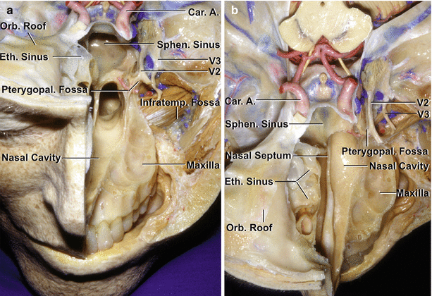
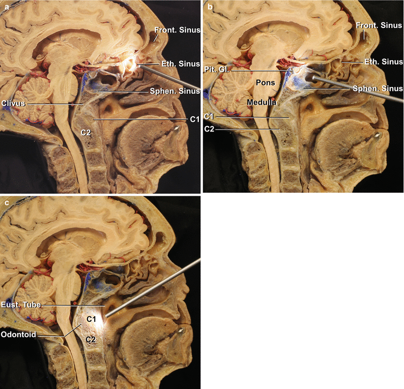
Fig. 1.1
Anterior and middle cranial base. (a) On the left side, the floor of the anterior fossa and the upper portion of the maxilla have been removed to expose the structures deep to the anterior and middle cranial fossa. The frontal, ethmoidal, and sphenoid sinuses and the nasal cavity lie below the medial part of the anterior cranial base. The orbit and maxilla are located below the lateral part of the anterior cranial base. The sphenoid sinus and sella are located in the medial part of the middle cranial base, and the infratemporal and pterygopalatine fossae are located below the lateral part of the middle cranial base. The carotid arteries pass upward on the medial part of the middle cranial base and are intimately related to the sphenoid and cavernous sinuses. The infratemporal fossa, which contains branches of the mandibular nerve, pterygoid muscles, pterygoid venous plexus, and maxillary artery, is located below the middle cranial base and greater sphenoid wing. The alveolar process of the maxilla, which encloses the roots of the upper teeth, has been preserved on the left side. The maxillary nerve enters the pterygopalatine fossa, which is located medial to the infratemporal fossa between the posterior wall of the maxilla and the pterygoid process of the sphenoid bone. (b) Superior view of the anterior and middle cranial base. The infratemporal fossa is located posterolateral to the maxilla. The right ethmoid air cells are exposed on the medial side of the right orbit. The nasal cavity extends upward between the ethmoid sinuses. (c) Oblique anterior view. The facial structures on the right side have been removed to expose the orbital apex located above the maxillary sinus. The wall of the right maxillary sinus forms the floor of the orbit, much of the lateral wall of the nasal cavity, and the anterior wall of the pterygopalatine and infratemporal fossa. On the left side, the mandibular nerve enters the infratemporal fossa. The maxillary nerve enters the pterygopalatine fossa, which is located in the lateral wall of the nasal cavity and contains the maxillary nerve, pterygopalatine ganglion, and terminal branches of the maxillary artery. (d) Anterior view. The orbital apex is located above the pterygopalatine fossa. The frontal branch of the ophthalmic nerve passes along the roof of the orbit, and the infraorbital branch of the maxillary nerve courses in the floor of the orbit. The posterior ethmoid air cells are located medial to the orbital apex. The vomer forms the posterior part of the nasal septum and attaches to the maxilla and palatine bones below and to the body of the sphenoid bone above. The sphenoid sinus is located in the middle cranial base below the sella turcica. The upper brainstem is seen in the posterior part of the exposure. A artery, Alv alveolar, Car carotid, Cart cartilage, CN cranial nerve, Eth ethmoid, Eust eustachian, Front frontal, Gang ganglion, Infraorb infraorbital, Infratemp infratemporal, M muscle, Max maxillary, N nerve, Nasolac nasolacrimal, Orb orbital, Pit pituitary, Pteryg pterygoid, Pterygopal pterygopalatine, Sphen Sphenoid, Sphenopal sphenopalatine (From Rhoton Jr AL. The anterior and middle cranial base. Neurosurgery. 2002;51(1 Suppl):S273–302; with permission)
Fig. 1.2
Specimen divided in the midsagittal plane to show the endoscopic endonasal approaches to the cranial base in midline through the frontal sinus, ethmoid sinus, sphenoid sinus, and nasopharynx. (a) Anterior cranial base approaches in midline through the frontal and ethmoid sinuses. (b) Approaches through the sphenoid sinus to the sellar region. (c) The endoscope is oriented downward to the nasopharynx for C1–C2 approaches. Eth ethmoid, Eust eustachian, Front frontal, Gl gland, Pit pituitary, Sphen sphenoid
The skull is divided into the cranium and the facial skeleton. The cranium is divided into the calvarium and the cranial base. The cranial base has an endocranial surface, which faces the brain, and an exocranial surface, which faces the nasal cavity and sinuses, orbits, pharynx, infratemporal and pterygopalatine fossae, and the parapharyngeal and intrapetrosal spaces (Fig. 1.3). Both surfaces are connected by canals, foramina, and fissures through which numerous neural and vascular structures pass. Both endocranial and exocranial cranial base surfaces are divided into anterior, middle, and posterior parts, each of which has central and paired lateral portions. On the endocranial side, the three parts correspond to the anterior, middle, and posterior cranial fossae (Figs. 1.3 and 1.4). On the endocranial side, the border between the anterior and middle cranial bases is the sphenoid ridge, which is joined medially by the chiasmatic sulcus, and the border between the middle and posterior cranial bases is the petrous ridges, which are joined by the dorsum sellae and posterior clinoid processes. On the exocranial side, the anterior and middle cranial bases are divided at the level of a transverse line extending through the pterygomaxillary fissures and the pterygopalatine fossae at the upper level and the posterior edge of the alveolar process of the maxilla at the lower level. Medially, this corresponds to the anterior part of the attachment of the vomer to the sphenoid bone. The middle and posterior cranial bases are separated by a transverse line crossing at or near the posterior border of the vomer-sphenoid junction, the foramen lacerum, carotid canal, jugular foramen, styloid process, and the mastoid tip.
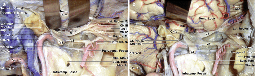
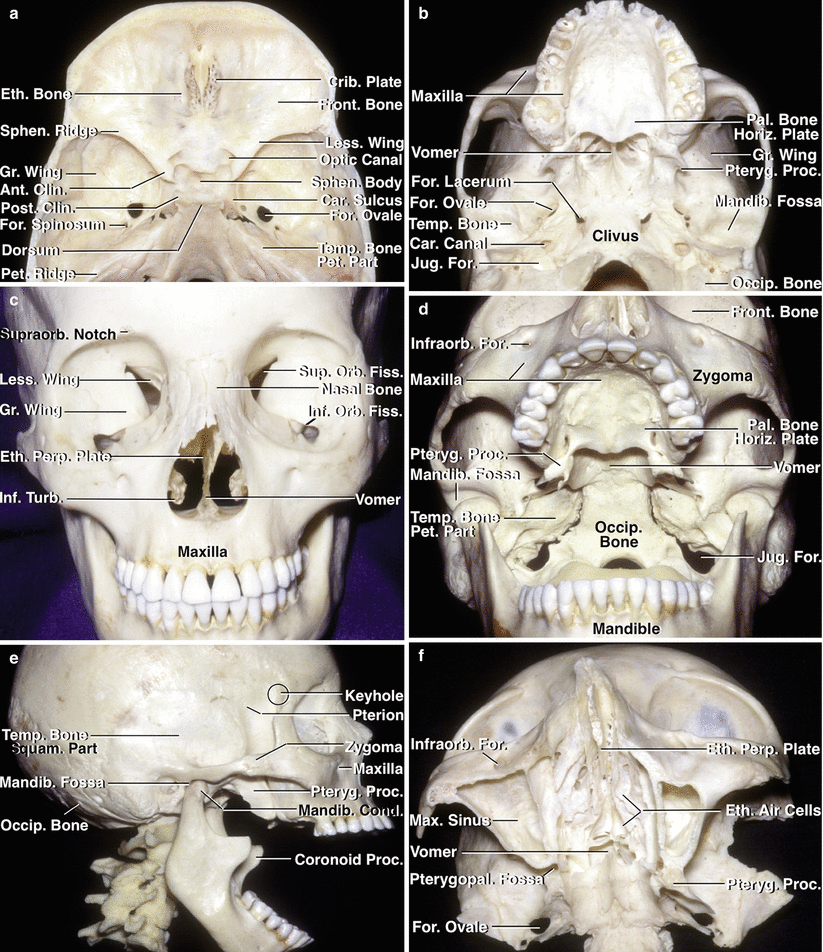
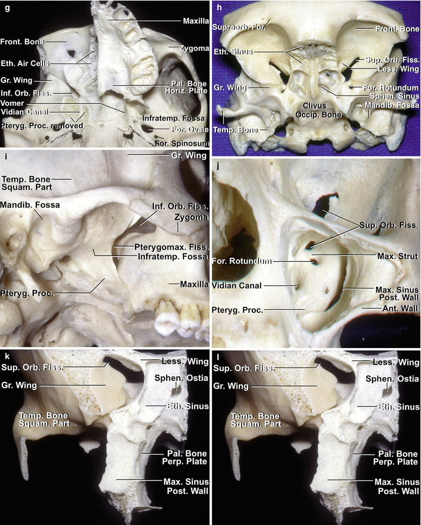
Fig. 1.3
Lateral view of the anterior, middle, and posterior cranial base. (a) The bone and structures lateral to the orbit, infratemporal, and pterygopalatine fossa and the parapharyngeal space and petrous part of the temporal bone have been removed to expose the structures below the anterior, middle, and posterior cranial base. The orbit and maxillary sinus are located below the anterior cranial base. The infratemporal and pterygopalatine fossae and the parapharyngeal space are located below the middle cranial base, and the suboccipital area is located below the temporal and occipital bones. The first trigeminal division is related to the upper part of the orbit. The second trigeminal branch is related to the lower part of the orbit and maxilla. The mandibular nerve exits the cranium through the foramen ovale and enters the infratemporal fossa. The pterygoid and levator and tensor veli palatini muscles have been removed to expose the eustachian tube and its opening into the nasal pharynx. The lateral part of the temporal bone has been removed to expose the cochlea, vestibule, and semicircular canals. The petrous carotid passes upward and turns medially below the cochlea. The sigmoid sinus turns downward under the semicircular canals and vestibule where the jugular bulb is located. The segment of the vertebral artery passing behind the atlanto-occipital joint is located below the posterior cranial base. (b) The dura has been opened to show the relationships of the frontal and temporal lobes and the cerebellum to the cranial base. The orbit is exposed below the frontal lobe. The pterygopalatine and infratemporal fossae and the temporal bone are located below the temporal lobe. The jugular bulb and internal jugular vein have been removed to show cranial nerves 9 through 12 exiting the jugular foramen. A artery, Car carotid, CN cranial nerve, Eust eustachian, Front frontal, Gr greater, Inf inferior, Infraorb infraorbital, Infratemp infratemporal, Int internal, Jug jugular, Lat lateral, M muscle, Max maxillary, N nerve, Ped peduncle, Pet petrosal, Pterygopal pterygopalatine, Rec rectus, Semicirc semicircular, Sphen sphenoid, Temp temporal, V vein, Vert vertebral (From Rhoton Jr AL. The anterior and middle cranial base. Neurosurgery. 2002;51(1 Suppl):S273–302.; with permission)
Fig. 1.4
Osseous relationships of the anterior and middle cranial base. (a) On the endocranial surface, the anterior and middle cranial base corresponds to the anterior and middle fossae. The anterior part of the cranial base is separated from the middle fossa by the sphenoid ridge and the chiasmatic sulcus. The middle cranial base is separated from the posterior cranial base by the dorsum sellae and the petrous ridges. The upper surface of the anterior cranial base is formed by the frontal bone, which roofs the orbit; the ethmoid bone, which is interposed between the frontal bones and is the site of the cribriform plate; and the lesser wing and anterior part of the body of the sphenoid, which forms the posterior part of the floor of the anterior fossa. The upper surface of the middle cranial base floor is formed by the greater sphenoid wing and posterior two thirds of the sphenoid body anteriorly and the upper surface of the temporal bone posteriorly. The posterior part of the cranial base is formed by the temporal and occipital bones. The cribriform plate, sella, and clivus are located in the medial part of the cranial base. The lateral part of the cranial base is located above the orbits, the pterygopalatine and infratemporal fossae, and the subtemporal and lateral part of the suboccipital areas. (b) Exocranial surface of the cranial base. This surface is more complicated than the endocranial surface. It is not demarcated into three well-defined fossae, as is the endocranial surface. The exocranial surface is formed by the maxilla, zygomatic, palatine, sphenoid, temporal, and occipital bones and the vomer. The maxilla, orbits, and nasal cavity are located below the anterior fossa. The anterior part of the hard palate is formed by the maxilla, and the posterior part is formed by the palatine bone. The anterior part of the zygomatic arch is formed by the zygoma and the posterior part by the squamosal part of the temporal bone. The mandibular fossa on the lower surface of the temporal squama is located below the posterior part of the middle fossa. The vomer attaches to the lower part of the body of the sphenoid and forms the posterior part of the nasal septum. (c) Anterior view. The orbital rim is formed by the frontal bone, zygoma, and maxilla. The roof of the orbit is formed by the frontal and sphenoid bones; the lateral wall by the greater sphenoid wing and the zygomatic bone; the floor by the maxilla, except for a small part of the posterior floor that is formed by the palatine bone; and the medial wall of the orbit that is formed by the maxilla, lacrimal, and ethmoid bones. The nasal bone is interposed above the anterior nasal aperture between the maxillae. The nasal cavity is located between the ethmoid bones and the maxillae and palatine bones above and the sphenoid pterygoid process below. It is roofed by the frontal and ethmoid bones, and the floor is formed by the maxillae and palatal bones. The osseous nasal septum is formed by the perpendicular ethmoid plate and the vomer. The inferior turbinate is a separate bone, and the middle and superior turbinates are appendages of the ethmoid bone. The orbit opens through the superior orbital fissure into the middle fossa and through the inferior orbital fissure into the pterygopalatine and infratemporal fossae. (d) Anteroinferior view of the cranial base. The anterior part of the hard palate is formed by the maxillae, and the posterior part is formed by the horizontal plate of the palatine bone. The vomer forms the posterior part of the nasal septum and divides the posterior nasal aperture in the midline. The infratemporal fossa is located below the greater sphenoid wing. The clivus is formed above by the body of the sphenoid bone and below by the basal part of the occipital bone. The petrous apex is interposed between the greater sphenoid wing and the clival part of the occipital bone. The mandibular condyles set in the mandibular fossa, located below the posterior part of the middle fossa on the inferior surface of the squamosal part of the temporal bone. (e) The cranial base is formed, in the lateral view, from anterior to posterior, by the maxilla and the frontal, zygomatic, sphenoid, temporal, and occipital bones. The zygomatic and frontal bones form the lateral part of the orbital rim. The pterion on the greater sphenoid wing marks the lateral end of the sphenoid ridge. The keyhole, a burr hole that exposes the dura of the anterior fossa and the periorbita in its depth, is located just above the frontozygomatic suture, behind the superior temporal line. The zygomatic arch is formed by the zygomatic bone and the squamosal part of the temporal bone. The condylar fossa, in which the mandibular condyle sits, is positioned above on the lower surface of the squamosal part of the temporal bone and posteriorly on the tympanic part of the temporal bone. The lower end of the pterygoid process unites with the posterior maxilla, but above, the process separates from the maxilla to create the pterygomaxillary fissure, which opens medially into the pterygopalatine fossa. (f) Inferior view of a cross section extending through the maxillae. The maxilla, which contains a large air-filled sinus, forms the anteromedial wall of the infratemporal fossa, the anterior wall of the pterygopalatine fossa, the lateral wall of the nasal cavity, the anterior portion of the hard palate, and much of the floor of the orbit. The pterygopalatine fossa is located between the pterygoid process and the posterior maxillary wall. The nasal septum is formed anteriorly and above by the perpendicular ethmoid plate and posteriorly and below by the vomer. (g) The right half of the maxilla and zygomatic arch has been removed. The inferior orbital fissure is located between the greater sphenoid wing and the maxilla. The right orbital roof and ethmoid air cells have been preserved. The right pterygoid process has been removed at its junction with the sphenoid body. The roof of the vidian canal, which extends through the base of the pterygoid process, has been preserved. (h) Anteroinferior view of the cranial base. The midline of the cranial base is formed, from anterior to posterior, by the frontal, ethmoid, sphenoid, and occipital bones. The roof of the orbit is formed by the frontal bone and lesser sphenoid wing. The ethmoid sinuses are located anterior to the sphenoid sinus between the orbits. (i) Lateral view of the pterygomaxillary fissure. The pterygomaxillary fissure is located between the posterior maxillary wall and the pterygoid process. The pterygomaxillary fissure opens from the infratemporal fossa into the pterygopalatine fossa. The mandibular fossa is formed above by the squamosal part of the temporal bone and posteriorly by the tympanic part of the temporal bone, which also forms the anterior and lower wall of the external auditory meatus. (j) Anterior view through the maxillary sinus. The anterior and posterior walls of the maxillary sinus have been removed to expose the pterygoid process, which forms the posterior wall of the pterygopalatine fossa. The lower part of the superior orbital fissure is seen through the upper part of the maxillary sinus. The foramen rotundum opens into the pterygopalatine fossa and is separated from the superior orbital fissure by the maxillary strut. The vidian canal opens through the pterygoid process below and medial to the foramen rotundum. (k) Anterior view of a cranium sectioned through the posterior part of the ethmoid and maxillary sinuses. The ethmoid sinuses are located anterior to the sphenoid body and sphenoid sinus. The part of the posterior wall of the maxilla forming the anterior wall of the pterygopalatine fossa has been preserved. The perpendicular plate of the palatine bone forms the medial wall of the pterygopalatine fossa. The ethmoid sinus overlaps the lateral margin of the sphenoid ostia. The superior orbital fissure is located between the lesser and greater sphenoid wings and sphenoid body. The infratemporal fossa is located below the greater wing of the sphenoid. The temporal fossa, which contains the temporalis muscle, is located between the greater wing and the zygomatic arch. (l) The posterior walls of the maxillary and ethmoid sinuses have been removed to expose the sphenoid sinus and pterygopalatine fossa. The lateral wing of the sphenoid sinus extends laterally into the pterygoid process below the foramen rotundum. Septa divide the sphenoid sinus. The vidian canal opens through the base of the pterygoid process into the pterygopalatine fossa. (m) The osseous cross section has been extended posteriorly to just in front of the superior orbital fissure. The optic strut extends from the base of the anterior clinoid to the sphenoid body and separates the optic canal from the superior orbital fissure. The foramen rotundum is located below the medial part of the superior orbital fissure. The vidian canal opens into the pterygopalatine fossa below and medial to the foramen rotundum. (n) Posterior view of the specimen in (k) showing the anterior part of the middle fossa from behind. The superior orbital fissure is positioned below the lesser sphenoid wing. The optic strut extends from the base of the anterior clinoid to the sphenoid body and separates the optic canal from the superior orbital fissure. The greater wing extends laterally to form part of the floor and anterior and lateral walls of the middle fossa. The medial and lateral pterygoid plates project backward from the pterygoid process. The horizontal plate of the palatine bone forms the posterior part of the hard palate. The posterior opening into the vidian canal is located above the medial pterygoid plate and extends forward through the pterygoid process at its junction with the sphenoid body. Ant anterior, Car carotid, Clin clinoid, Cond condyle, Crib cribriform, Eth ethmoid, Fiss fissure, For foramen, Front frontal, Gr greater, Horiz horizontal, Inf inferior, Infraorb infraorbital, Infratemp infratemporal, Jug jugular, Lat lateral, Less lesser, Mandib mandibular, Max maxillary, Med medial, Orb orbital, Occip occipital, Pal palatine, Perp perpendicular, Pet petrous, Post posterior, Proc process, Pteryg pterygoid, Pterygomax pterygomaxillary, Pterygopal pterygopalatine, Sphen sphenoid, Squam squamosal, Sup superior, Supraorb supraorbital, Temp temporal, Turb turbinate (From Rhoton Jr AL. The anterior and middle cranial base. Neurosurgery. 2002;51(1 Suppl):S273–302; with permission)
This chapter provides a concise review of the cranial base anatomy, including the anatomic basis of endoscopic endonasal approaches.
1.2 Anterior Cranial Base
1.2.1 Endocranial Surface
The anterior endocranial surface, formed by the ethmoid, sphenoid, and frontal bones, is divided into medial and lateral portions (Figs. 1.4 and 1.5). The medial part, covering the upper nasal cavity and the sphenoid sinus, is formed by the crista galli and the cribriform plate of the ethmoid bone anteriorly and the planum of the sphenoid body posteriorly. The lateral part, which covers the orbit and the optic canal, is formed by the frontal bone and the lesser wing of the sphenoid bone, which blends medially into the anterior clinoid process (Figs. 1.4 and 1.5). The foramen cecum in the midline serves as the site of passage of an emissary vein, and the cribriform plate is pierced by the filaments of the olfactory nerve. The optic canal transmits the optic nerve and the ophthalmic artery. The anterior cranial base faces the frontal lobes with the gyri recti medially and the orbital gyri laterally, along with the branches of the anterior cerebral arteries medially and middle cerebral arteries laterally.
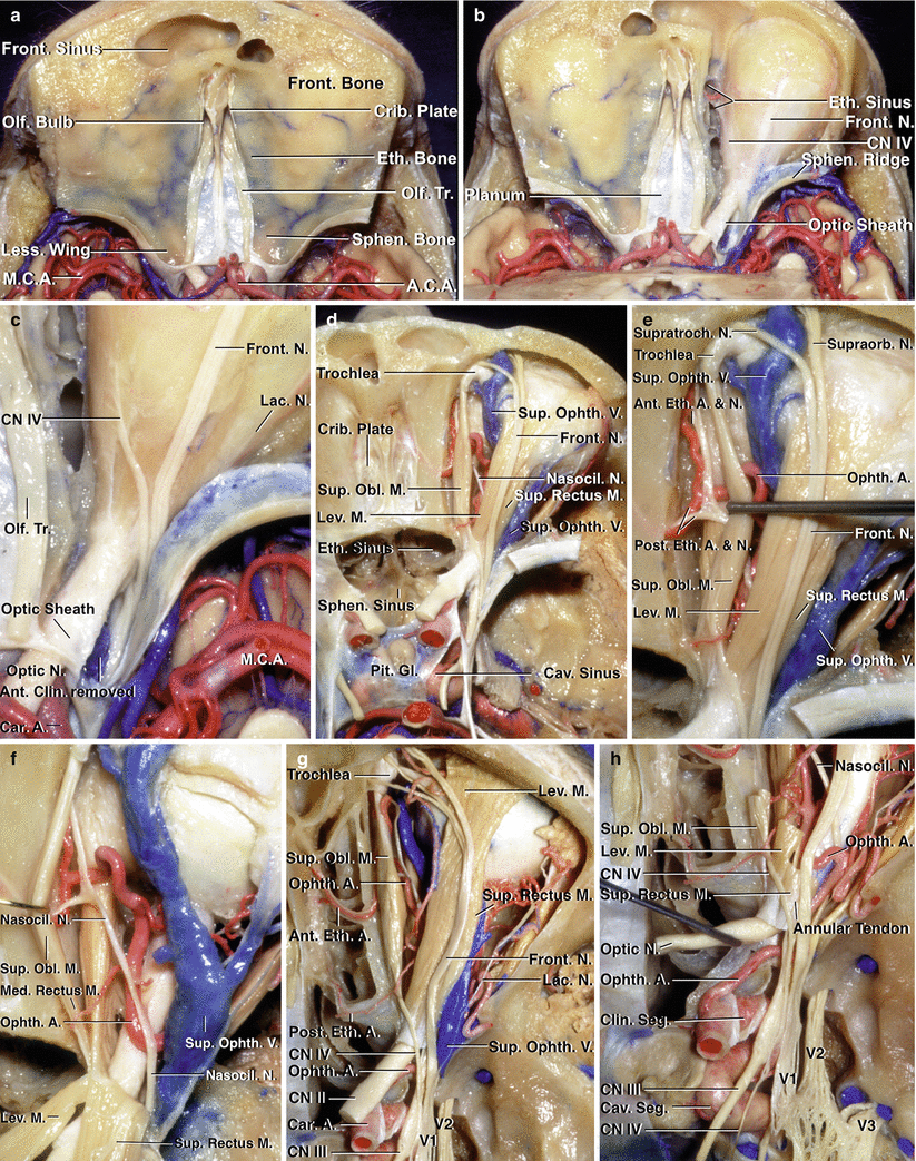
Fig. 1.5
Anterior fossa, orbit, and paranasal sinuses. (a) Superior view. The anterior cranial fossa is formed by the frontal, ethmoid, and sphenoid bones. The frontal bone splits anteriorly into two laminae, which enclose the frontal sinus. The ethmoid bones, which contain the ethmoid air cells and are the site of the crista galli and cribriform plate, are interposed between the frontal bones. Posteriorly, the frontal and ethmoid bones join the sphenoid bone, which encloses the sphenoid sinus and has the pituitary fossa on its upper surface. The olfactory bulbs and tracts have been preserved. (b) The roof of the right orbit has been removed to expose the periorbita. The right anterior clinoid process and roof of the optic canal have been removed to expose the optic nerve enclosed within the optic sheath as it passes through the optic canal to reach the orbital apex. (c) The frontal, trochlear, and lacrimal nerves can be seen through the periorbita. The trochlear nerve crosses above the orbital apex to reach the superior oblique muscle. (d) The orbital fat has been removed and the sphenoid sinus opened. The frontal branch of the ophthalmic nerve courses above the levator muscle. The ophthalmic artery, nasociliary nerve, and superior ophthalmic vein are located medially in the anterior part of the orbit and cross between the optic nerve and the superior rectus muscle to be situated on the lateral side of the optic nerve at the orbital apex. (e) Enlarged view. The superior oblique muscle has been retracted medially to expose the anterior and posterior ethmoidal branches of the ophthalmic artery and nasociliary nerve entering the anterior and posterior ethmoidal canal. The trochlea of the superior oblique muscle is attached to the superomedial margin of the orbit just behind the orbital rim. The frontal nerve divides into supraorbital and supratrochlear branches. (f) The levator and superior rectus muscles have been retracted posteriorly to expose the nasociliary nerve, ophthalmic artery, and superior ophthalmic vein passing above the optic nerve. (g) Superior view of the anterior fossa in another specimen. The nasal cavity, sphenoid sinus, and orbit have been unroofed. The dura has been removed from the roof and lateral wall of the cavernous sinus. The medial strip below the anterior cranial base is formed, from anterior to posterior, by the frontal, ethmoidal, and sphenoid sinuses. The orbital fat has been removed to expose the intraorbital structures. The frontal nerve courses above the levator muscle. The trochlear nerve passes above the annular tendon to reach the superior oblique muscle. The trochlea of the superior oblique muscle is attached in the superomedial part of the anterior orbit. The lacrimal nerve courses above the lateral rectus muscle. The ophthalmic artery and superior ophthalmic vein are seen in the interval between the levator and superior oblique muscles. The anterior and posterior ethmoidal branches of the ophthalmic artery course through the anterior and posterior ethmoidal canals. (h) Enlarged view of the cavernous sinus, superior orbital fissure, and orbital apex. The superior oblique, levator, and superior rectus muscles have been removed. The ophthalmic artery and nasociliary nerve enter the orbital apex on the lateral side of the optic nerve and cross between the optic nerve and superior rectus muscle to reach the medial part of the orbit. The optic nerve has been elevated to expose the ophthalmic artery, which courses through the optic canal on the lower side of the optic nerve and enters the orbital apex on the lateral side of the optic nerve. The ophthalmic artery then crosses medially between the optic nerve and superior rectus muscle, as does the nasociliary nerve. The maxillary nerve exits the foramen rotundum to enter the pterygopalatine fossa, and the mandibular nerve exits the foramen ovale to enter the infratemporal fossa. A artery, ACA anterior cerebral artery, Ant anterior, Car carotid, Cav cavernous, Clin clinoid, CN cranial nerve, Crib cribriform, Eth ethmoid, ethmoidal, Front frontal, Gl gland, Lac lacrimal, Less lesser, Lev levator, M muscle, Med medial, MCA middle cerebral artery, N nerve, Nasocil nasociliary, Ob oblique, Olf olfactory, Ophth ophthalmic, Pit pituitary, Post posterior, Seg segment, Sphen sphenoid, Sup superior, Supraorb supraorbital, Supratroch supratrochlear, Tr tract, V vein (From Rhoton Jr AL. The anterior and middle cranial base. Neurosurgery. 2002;51(1 Suppl):S273–302; with permission)
1.2.2 Exocranial Surface
On the exocranial side, the anterior cranial base is divided into a medial part related to the ethmoid and the sphenoid sinuses with the nasal cavity below and a lateral part that corresponds to the orbit and maxilla (Figs. 1.3, 1.6, 1.7, and 1.8).
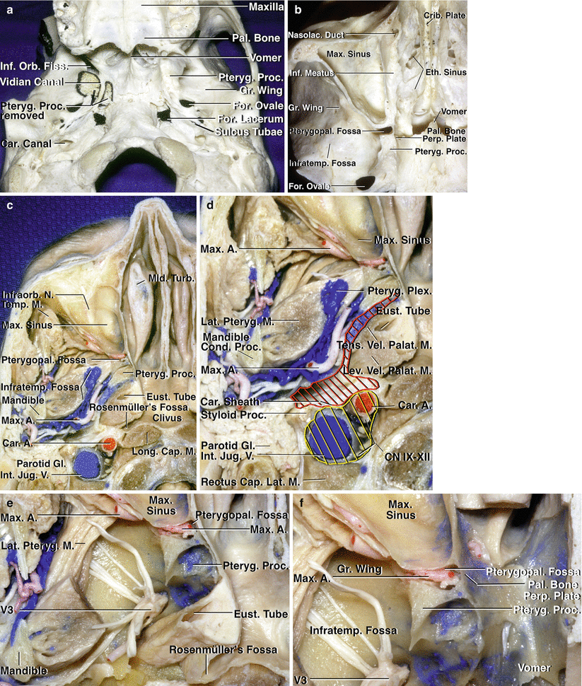
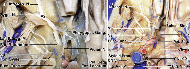
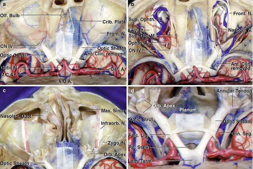
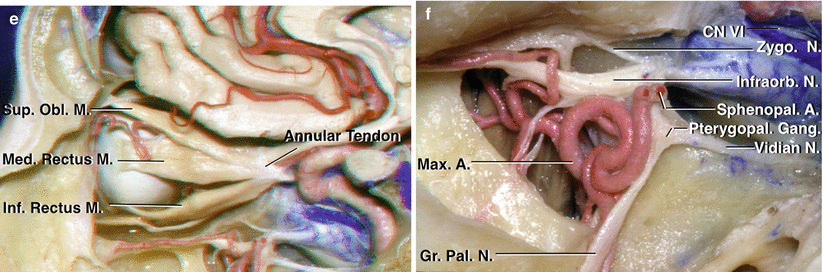
Fig. 1.6
Inferior view of the cranial base. (a) The right pterygoid process of a dry skull has been sectioned and removed at its junction with the greater wing and body of the sphenoid bone to expose the pterygopalatine fossa and the vidian canal. The vidian nerve, formed by the union of the superficial and deep petrosal nerves, courses in the vidian canal, which passes through the root of the pterygoid process. It opens posteriorly at the anterolateral margin of the foramen lacerum and anteriorly into the medial portion of the pterygopalatine fossa. The sulcus tubae, which is the attachment site of the cartilaginous part of the eustachian tube to the cranial base, is located on the extracranial surface of the sphenopetrosal fissure, anterolateral to the foramen lacerum and the carotid canal, and posteromedial to the foramina ovale and spinosum. The lateral part of the inferior orbital fissure opens into the infratemporal fossa located below the greater sphenoid wing, and the medial part opens into the pterygopalatine fossa located below the orbital apex between the maxilla and pterygoid process. The right zygomatic arch has been removed. (b) Inferior view of axial section of a cranium at the level of the maxillary sinus. The pterygopalatine fossa is located between the posterior wall of the maxillary sinus and the pterygoid process. The roof of the maxillary sinus forms the floor of the orbit. The infratemporal fossa is located below the greater wing of the sphenoid and opens medially into the pterygopalatine fossa. The medial wall of the pterygopalatine fossa is formed by the perpendicular plate of the palatine bone, which has an opening, the sphenopalatine foramen, through which branches of the maxillary artery and nerve reach the nasal cavity. The ethmoid air cells are located medial to the orbit. (c) Inferior views of an axial section of the cranial base. The infratemporal fossa is surrounded by the maxillary sinus anteriorly, the mandible laterally, the pterygoid process anteromedially, and the parapharyngeal space posteromedially. It contains the mandibular nerve and maxillary artery and their branches, the medial and lateral pterygoid muscles, and the pterygoid venous plexus. The posterior nasopharyngeal wall is separated from the lower clivus by the longus capitis, and the nasopharyngeal roof rests against the upper clivus and floor of the sphenoid sinus. (d) Enlarged view with highlighting of the pre- (red) and poststyloid (yellow) compartments of the parapharyngeal space. The styloid diaphragm, formed by the anterior part of the carotid sheath, separates the parapharyngeal space into pre- and poststyloid parts. The prestyloid compartment, a narrow fat-containing space between the medial pterygoid and tensor veli palatini muscles, separates the infratemporal fossa from the medially located lateral nasopharyngeal region containing the tensor and levator veli palatini and the eustachian tube. The poststyloid compartment, located behind the prestyloid part, contains the internal carotid artery, the internal jugular vein, and the cranial nerves 9 through 12. (e) Some of the lateral pterygoid muscle has been removed to expose the branches of the mandibular nerve in the infratemporal fossa. The lower part of the pterygoid process has been removed to expose the maxillary artery in the pterygopalatine fossa. The pharyngeal recess (fossa of Rosenmüller) projects laterally from the posterolateral corner of the nasopharynx below the foramen lacerum. (f) Enlarged view. The pterygopalatine fossa is located between the posterior maxillary wall anteriorly, the sphenoid pterygoid process posteriorly, the perpendicular plate of the palatine bone medially, and the infratemporal fossa laterally. The medial part of the eustachian tube has been removed. (g) The pterygoid process has been removed to expose the maxillary nerve passing through the foramen rotundum to enter the pterygopalatine fossa, where it gives rise to the infraorbital and zygomatic nerves and communicating rami to the pterygopalatine ganglion. The vidian nerve exits the vidian canal and joins the pterygopalatine ganglion. The terminal part of the petrous carotid is exposed above the foramen lacerum. (h) Enlarged view of the region of the carotid canal and jugular foramen. The bone below the carotid canal has been removed to expose the petrous carotid. The deep portion of the parotid gland has been removed to expose the facial nerve at the styloid foramen. The sigmoid sinus hooks downward from the posterior fossa and opens into the internal jugular vein. A portion of the occipital condyle has been removed to expose the hypoglossal nerve joining the nerves exiting the jugular foramen to pass downward in the carotid sheath. The styloid process and facial nerve at the stylomastoid foramen are located on the lateral side of the internal jugular vein. The right half of the floor of the sphenoid sinus has been removed to expose the sella. A artery, Cap capitis, Car carotid, CN cranial nerve, Cond condyle, Crib cribriform, Eth ethmoid, Eust eustachian, Fiss fissure, For foramen, Gang ganglion, Gl gland, Gr greater, Inf inferior, Infraorb infraorbital, Infratemp infratemporal, Int internal, Jug jugular, Lat lateral, lateralis, Lev levator, Long longus, M muscle, Max maxillary, Mid middle, N nerve, Nasolac nasolacrimal, Orb orbital, Pal palatine, Palat palatini, Perp perpendicular, Pet petrosal, petrous, Plex plexus, Proc process, Pteryg pterygoid, Pterygopal pterygopalatine, Seg segment, Sig sigmoid, Sphen sphenoid, Temp temporalis, Tens tensor, Turb turbinate, V vein, Vel veli, Zygo zygomatic (From Rhoton Jr AL. The anterior and middle cranial base. Neurosurgery. 2002;51(1 Suppl):S273–302; with permission)
Fig. 1.7
Superior view of the anterior cranial base. (a) Both orbits have been unroofed to expose the periorbita. The optic canals have been unroofed and the anterior clinoids removed to expose the optic nerves, which are enclosed in the optic sheath within the optic canal. The frontal, trochlear, and lacrimal nerves can be seen through the periorbita. The roof of the ethmoid sinuses and the olfactory bulbs sitting on the cribriform plate have been preserved. The anterior cerebral arteries course above the optic chiasm. (b) The intraorbital fat has been removed, and the levator and superior rectus muscles have been retracted laterally to expose both globes, ophthalmic arteries, superior ophthalmic veins, and nasociliary nerves. (c) The orbital contents have been removed to expose the lateral wall and floor of the orbit. The maxillary sinuses are exposed below the orbital floors. The maxillary nerves give rise to the infraorbital nerve, which courses along the floor of the orbit to reach the cheek and the zygomatic nerve, which courses along the lateral wall of the orbit to reach the malar eminence and temple. (d) Enlarged view. The optic nerves are enclosed within the optic sheath as they course through the optic canal. The annular tendon, from which the rectus muscles arise, surrounds the optic nerve and medial portion of the superior orbital fissure. Removal of the anterior clinoid exposes the clinoid segment of the carotid artery. The optic strut, which separates the optic canal and superior orbital fissure, has also been removed. The segment of anterior cerebral arteries passing above the chiasm has been removed to expose the lamina terminalis. The falciform dural fold extends across the optic nerve at the entrance into the optic canal. A artery, ACA anterior cerebral artery, Ant anterior, Car carotid, Clin clinoid, CN cranial nerve, Crib cribriform, Falc falciform, Front frontal, Infraorb infraorbital, Lac lacrimal, Lam lamina, Lev levator, Lig ligament, M muscle, Max maxillary, MCA middle cerebral artery, N nerve, Nasocil nasociliary, Nasolac nasolacrimal, Obl oblique, Olf olfactory, Ophth ophthalmic, Orb orbital, Pit pituitary, Seg segment, Sup superior, Term terminalis, Zygo zygomatic (From Rhoton Jr AL. The anterior and middle cranial base. Neurosurgery. 2002;51(1 Suppl):S273–302; with permission)
Fig. 1.8
Structures below the medial part of the anterior and middle cranial fossae. (a) Midsagittal section of the anterior and middle cranial base to the right of the nasal septum. The area below the medial part of the anterior cranial fossa is formed by the frontal and ethmoid sinuses and the nasal cavity. The nasal cavity is divided into the inferior, middle, and superior meatus and the sphenoethmoidal recess by the inferior, middle, and superior turbinates. The inferior meatus is located below the inferior turbinate, and the sphenoethmoidal recess, into which the sphenoid sinus opens, is located above the superior turbinate. The central part of the middle cranial base is formed by the body of the sphenoid bone, which contains the sphenoid sinus and sella with the pituitary gland. The cribriform plate is located in the roof of the nasal cavity. The nasopharynx and the opening of the eustachian tube are located below the sphenoid sinus. (b) Some of the mucosa has been removed from the turbinate. The inferior turbinate is a separate bone attached to the maxilla. The middle and superior turbinates are appendages of the ethmoid bone. The carotid artery courses along the lateral margin of the sphenoid sinus. The prominence within the sphenoid sinus, formed by the superior orbital fissure, is located anterior to the intracavernous carotid, and the prominence overlying the maxillary nerve is located below the intracavernous carotid. (c) The middle and superior turbinates have been removed to expose the ostia of the maxillary and frontal sinuses. Both open into the middle meatus below the middle turbinate. The nasolacrimal duct opens below the inferior turbinate. The Rosenmüller fossa is located behind the eustachian tube. (d) The medial wall of the maxillary sinus and the ethmoid air cells have been removed to expose the orbit. The optic nerve enters the orbit above the superior orbital fissure. The maxillary nerve exits the foramen rotundum to enter the pterygopalatine fossa. The vidian nerve passes through the vidian canal and enters the posterior margin of the sphenopalatine ganglion in the pterygopalatine fossa. The floor of the anterior cranial fossa forms much of the roof of the orbit and the maxillary sinus forms most of the floor of the orbit. The abducens nerve is seen below the intracavernous segment of the internal carotid artery. The pterygopalatine fossa is located anterior to the sphenoid sinus and below the orbital apex. (e) The intraorbital fat has been removed to expose the superior oblique and medial and inferior rectus muscles. (f) Enlarged view of the pterygopalatine fossa. The maxillary nerve exits the foramen rotundum to enter the pterygopalatine fossa, where it gives rise to the infraorbital, zygomatic, and palatine nerves and to communicating rami to the pterygopalatine ganglion. The vidian nerve exits the vidian canal to enter the pterygopalatine ganglion. The pterygopalatine fossa contains branches of the maxillary nerve, the junction of the vidian nerve with the pterygopalatine ganglion, and terminal branches of the maxillary artery. A artery, Car carotid, Cav cavernous, Crib cribriform, CN cranial nerve, Eust eustachian, Fiss fissure, Front frontal, Gang ganglion, Inf inferior, Infraorb infraorbital, Int internal, M muscle, Max maxillary, Med medial, Mid middle, N nerve, Obl oblique, Olf olfactory, Orb orbital, Palat palatine, Pterygopal pterygopalatine, Rec recess, Sphen sphenoid, Sphenoeth sphenoethmoidal, Sphenopal sphenopalatine, Sup superior, Turb turbinate, Zygo zygomatic (From Rhoton Jr AL. The anterior and middle cranial base. Neurosurgery. 2002;51(1 Suppl):S273–302; with permission)
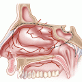
Detailed knowledge of the nasal anatomy is important in endonasal skull base procedures. The nose, a part of the upper respiratory tract, is divided into the external nose (which emerges from the face) and the internal nasal cavity, both divided into right and left halves by the midline nasal septum. The nasal cavity is a truncated pyramid that is wider at its base. It opens anteriorly onto the face through the external nose at the anterior nasal aperture, and it opens posteriorly into the nasopharynx by way of the posterior nasal apertures, also called choanas.
Nine bones form the structure of the nose and paranasal sinuses. The unpaired bones are the frontal, sphenoid, vomer, and ethmoid, and the paired bones are the nasals, palatines, maxillae, lacrimals, and inferior conchae (Fig. 1.9). The external nose is formed by the nasal bones anteriorly and superiorly, forming the dorsum of the nose and the frontal processes of the maxilla laterally. The frontal processes of the maxillae, which form the medial orbital rims, separate the orbit from the nose anteriorly. The superior (lateral) and inferior (lower or alar) nasal cartilages keep the air pathway open at the aditus or nasal vestibules and externally at the nostrils. Fibrofatty tissue and small cartilages form the lateral boundary of the nostrils. The posterior nasal aperture is formed by the anterior part of the sphenoid body above, the horizontal plate of the palatine inferiorly, the medial pterygoid plate laterally, and the posterior edge of the vomer medially, thereby dividing this aperture in two halves. The lateral wall of the nasal cavity can be divided into a superior part that forms the medial wall of the orbit and an inferior part that includes the medial aspect of the maxillary sinus. The superior part corresponding to the medial wall of the orbit is composed of the frontal process of the maxilla, the lacrimal bone, and the orbital plate of the ethmoid bone, whose air cells (ethmoid labyrinth) separate the orbit from the nasal cavity. The thin part of the ethmoid bone that faces the orbit is also called the lamina papyracea for its paper-thin fragility. The inferior part of the lateral wall of the nasal cavity is formed from anterior to posterior by the maxilla, the inferior concha or inferior turbinate bone, the perpendicular plate of the palatine bone, and the medial pterygoid plate. The palatine bone articulates posteriorly with the medial pterygoid plate of the sphenoid bone and forms the medial wall of the pterygopalatine fossa positioned between the pterygoid process of the sphenoid posteriorly and the posterior aspect of the maxilla anteriorly. The floor of the nasal cavity is composed from anterior to posterior by the palatine process of the maxilla and the horizontal plate of the palatine bone. The roof of the nasal cavity is formed from anterior to posterior by the nasal bones, the nasal spine of the frontal bone, the cribriform plate (ethmoid), and the sphenoid body. The nasal septum, with its osseous posterior part formed superiorly by the perpendicular plate of the ethmoid and, inferiorly by the vomer, with its anterior cartilaginous part formed by the quadrangular cartilage, divides the cavity into two halves (Figs. 1.9 and 1.10). There can be up to five osseous structures, three constant and two inconstant, that protrude from the lateral wall of the nasal cavity. The three constant protrusions are the superior, middle, and inferior turbinates (conchae), and the inconstant ones are the supreme turbinate (Santorini), and in 1 % of cases, there is a fifth turbinate superior to the supreme (Zuckerkandl) (Figs. 1.11 and 1.12). The supreme, superior, and middle turbinates are parts of the ethmoid bone, while a separate bone, the inferior concha, forms the inferior turbinate. The free spaces located lateral and below the superior, middle, and inferior turbinates are referred to as the superior, middle, and inferior meatus. The middle meatus, the most complex, is the site of two important structures that can be seen without further dissection. The first, located anteriorly, is an elongated, vertically oriented protrusion called the uncinate process, behind which is found the second bulging structure, the ethmoid bulla. The nasolacrimal groove and canal, the site of the lacrimal sac and nasolacrimal duct, respectively, descend just posterior to the origin of the middle nasal turbinate and end in the inferior meatus approximately 2 cm behind the nostril. The nasolacrimal canal is formed by the maxilla and the lacrimal bone and protrudes vertically just anterior to the middle meatus, creating a prominence. Farther posteriorly, approximately 1 cm behind the inferior turbinate, the eustachian tube opens into the lateral nasopharyngeal wall. The posterior ethmoid cells drain into the superior meatus; the anterior ethmoid cells, maxillary sinus, and frontal sinus drain into the middle meatus; and the nasolacrimal duct drains into the inferior meatus. The body of the sphenoid bone harbors the sphenoid sinus just below the planum sphenoidale, with the ostia located above the superior turbinate, and drains into the sphenoethmoidal recess. The ophthalmic, maxillary, and facial arteries, which form an anastomotic plexus in the nasal mucosa, provide the supply to the nasal cavity. The anterior and posterior ethmoidal arteries, branches of the ophthalmic artery, supply the ethmoid and frontal sinuses, the roof of the nose, and the superior part of the septum. The sphenopalatine branch of the maxillary artery supplies the mucosa of the conchae, meatus, and posteroinferior part of the nasal septum and is the principal supply to the nasal mucosa. Of great importance in endoscopic anatomy is the location of the sphenopalatine foramen where the sphenopalatine artery exits the pterygopalatine fossa and enters the nasal cavity (Fig. 1.9f). The sphenopalatine artery sends medial branches to the septum and lateral branches to the turbinates, meatus, and maxillary sinus. The number and distribution of these branches are highly variable. The medial branch, or posterior septal artery, supplies the nasoseptal flap used for skull base reconstruction. The sphenopalatine foramen is usually positioned immediately superior to the posterior aspect of the middle turbinate attachment and is formed anteriorly by the orbital process of the palatine bone, posteriorly by the sphenoid process of the palatine bone and superiorly by the sphenoid body (Fig. 1.9g). However, the location of the sphenopalatine foramen in the superior or middle meatus is variable. The greater palatine branch of the maxillary artery supplies the region of the inferior meatus. Its terminal part ascends through the incisive canal to anastomose on the septum with branches of the sphenopalatine and anterior ethmoidal arteries and with the septal branch of the superior labial artery. The mucosa of the maxillary sinus is supplied by the infraorbital artery and the superior, anterior, and posterior alveolar branches of the maxillary artery. The pharyngeal branch of the maxillary artery that runs through the palatovaginal canal supplies the sphenoidal sinus mucosa. The nasociliary branch of the ophthalmic nerve (V1) conveys sensation from the roof of the nasal cavity and upper septum by way of its anterior and posterior ethmoidal branches, which pass through the ethmoidal canals. The anterosuperior alveolar nerve branch of the maxillary nerve (V2) supplies part of the septum and floor near the anterior nasal spine and the anterior part of the lateral wall as high as the opening of the maxillary sinus. Branches of the greater palatine nerve supply the posterior three quarters of the lateral wall, roof, and floor, along with the inferior part of the nasal septum. Sympathetic postganglionic vasomotor fibers are distributed with the blood vessels. Parasympathetic fibers are carried through the vidian nerve, synapse in the pterygopalatine ganglion, and are distributed via branches of the maxillary nerve to the nasal mucosa.
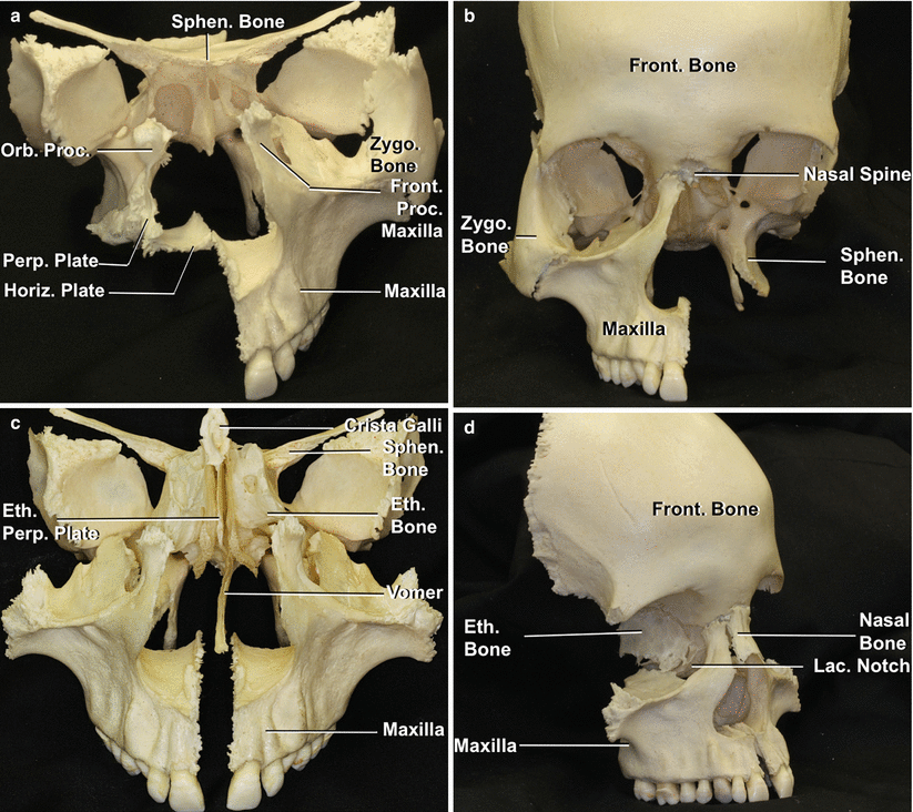
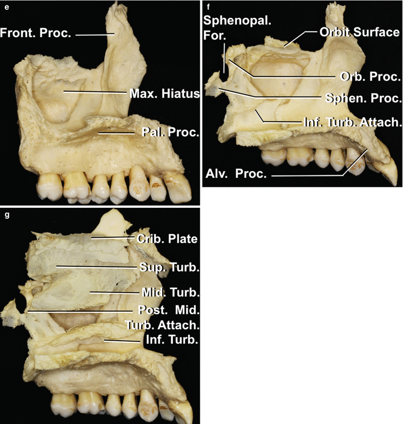
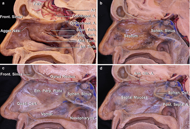
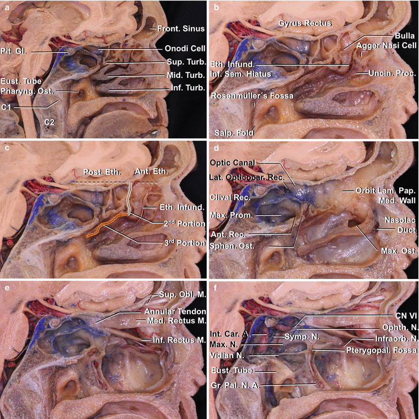
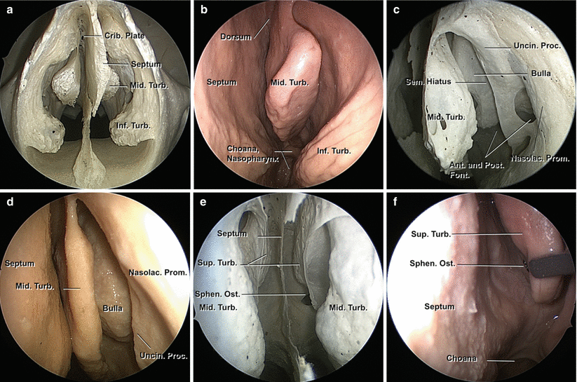
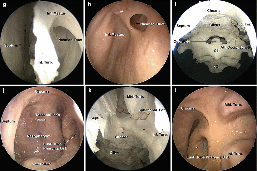
Fig. 1.9
Bones that form the structure of the nasal cavity. (a) Anterior view of the left maxilla, right palatine bone, and sphenoid bone. The hard palate, floor of the nasal cavity, is formed by the palatine process of the maxilla anteriorly and the horizontal plate of the palatine bone posteriorly. (b) The frontal bone articulates anteriorly with the frontal process of the maxilla, and its nasal spine forms part of the roof of the nasal cavity. (c) The ethmoid bone articulates posteriorly with the body of the sphenoid and forms part of the medial orbital wall, septum, and roof of the nose. (d) The roof of the nasal cavity is formed by the nasal bones, nasal spine of the frontal bones, cribriform plate of the ethmoid, and sphenoid (not shown in this figure). (e) Medial view of the maxilla; the maxillary sinus opens medially into the nasal cavity through the middle meatus. The frontal process of the maxilla articulates with the frontal, nasal, and lacrimal bones. The palatine process articulates with the contralateral mate and posteriorly with the palatine bone. (f) Articulation of the maxilla with the palatine bone. The orbital and the sphenoidal processes of the palatine bone limit the sphenopalatine foramen anteriorly and posteriorly. The sphenopalatine artery, terminal branch of the maxillary artery, exits the pterygopalatine fossa through this foramen and enters the nasal cavity. It gives medial or posterior septal branches for the septum and lateral branches for the turbinates, meatus, and maxillary sinus. (g) Articulation of the maxilla, palatine, inferior turbinate, and ethmoid bones, medial view. The ethmoid bone has been divided in midline and the perpendicular plate removed. The supreme (if present) superior and middle turbinates are part of the ethmoid bone, and the inferior turbinate is an independent bone. The posterior attachment of the middle turbinate is located below the sphenopalatine foramen. Alv alveolar, Attach attachment, Crib cribriform, For foramen, Front frontal, Horiz horizontal, Inf inferior, Lac lacrimal, Max maxillary, Mid middle, Orb Orbital, Pal palatine, Perp Perpendicular, Post posterior, Proc process, Sphen sphenoid, Sup superior, Sphenopal sphenopalatine, Turb turbinate, Zygo zygomatic
Fig. 1.10
Anatomic specimen divided in the midsagittal plane. (a) Right nasal cavity with the supreme, superior, middle, and inferior turbinates. The left hemisphere has been removed, keeping the A2 branches in midline. The orbitofrontal artery is the first branch of A2, lies in the olfactory sulcus in the frontobasal surface, and is the first branch encountered in an endoscopic craniofacial resection. The frontopolar artery arises superior to the orbitofrontal artery in midline. (b) The nasal septum mucosa. (c) The mucosa has been removed to show the structure of the septum with an anterior cartilaginous and posterior osseous portion formed by the perpendicular plate of the ethmoid bone superiorly and the vomer inferiorly. (d) The bone and cartilage have been removed and the periosteal-perichondrial surface of the septum dissected to show the main arterial supply: the posterior septal arteries, branches of the shenopalatine artery and of the ethmoidal arteries, and branches of the ophthalmic artery. A artery, Ant anterior, Cart cartilage, Eth ethmoid, Front frontal, Frontopol frontopolar, Inf inferior, Mid middle, Orbitofront orbitofrontal, Perp perpendicular, Post posterior, Quad quadrangular, Sphen sphenoid, Sup superior, Supr supreme, Turb turbinate
Fig. 1.11
Left side of a specimen divided in a sagittal plane passing through the left nasal cavity; dissection from medial to lateral. (a) Note that the ethmoid labyrinth in this specimen pneumatizes posteriorly up to the optic nerve (Onodi cell). (b) The uncinate process mucosa has been dissected to show its inferior attachment to the inferior turbinate, hiding the maxillary ostium behind. The middle turbinate has been resected, leaving its attachment to the lateral nasal wall. The ethmoid infundibulum is located between the bulla and the uncinate process and is a three-dimensional space. The two-dimensional plane between these two structures is called the inferior semilunar hiatus. The agger nasi cell is a pneumatization of the agger nasi region that is anterior to the middle turbinate attachment. (c) The second and third portions of the basal lamella of the middle turbinate attachment in the lateral nasal wall are shown in different colors. The first portion (not shown) attaches to the cranial base. The vertical second portion divides the ethmoid cells into anterior and posterior ethmoid cells according to the surgical classification. (d) When the uncinate process is resected, the maxillary ostium is visualized. The nasolacrimal duct has been opened. A complete ethmoidectomy shows the medial wall of the orbit. (e) Dissection of the orbit. (f) The lateral and posterior walls of the sphenoid sinus have been dissected to show the course of the internal carotid artery. A artery, Ant anterior, Car carotid, CN cranial nerve, Eth ethmoid, Eust eustachian, Front frontal, G gland, Gr greater, Inf inferior, Infund infundibulum, Int internal, Lam lamina, Lat lateral, M muscle, Max maxillary, Med medial, Mid middle, N nerve, Nasolac nasolacrimal, Obl oblique, Ophth ophthalmic, Opticocar opticocarotid, Orb orbital, Ost ostium, Pal palatine, Pap papyracea, Pharyng pharyngeal, Pit pituitary, Pos posterior, Proc process, Prom prominence, Rec recess, Salp salpingopharyngeal, Sem semilunar, Sup superior, Symp sympathetic, Turb turbinate, Uncin uncinate
Fig. 1.12
Images of the nasal cavity obtained with a 0-degree endoscope in a dry skull (left) and equivalent image in a formalin-fixed anatomic specimen (right). (a) General view introducing the endoscope through the piriform aperture, the septum separates the nasal cavity in midline, the inferior and middle turbinates can be identified. (b) The endoscope is introduced through the left nostril. The septum is located on the left side of the image, the middle and inferior turbinates as appendices protruding from the lateral wall of the nasal cavity. The choana or posterior nasal aperture opens into the nasopharynx. (c, d) Image of the middle meatus, anterior part. Below and lateral to the middle turbinate, the first prominence seen is the uncinate process and posteriorly the bulla ethmoidalis (anterior ethmoid cells). The inferior semilunar hiatus is the area between them and the three-dimensional space between them is the ethmoid infundibulum, common drainage of the anterior ethmoid cells, maxillary sinus, and in some cases the frontal sinus, depending on the uncinate process attachment. There is a craniocaudal prominence anterior to the middle turbinate, which covers the nasolacrimal duct. In the dry skull, the uncinate process is located between the anterior and posterior fontanelles that are closed by mucosa. The maxillary ostium is hidden by the uncinate process and is located between the uncinate process and the bulla ethmoidalis. (e, f) The sphenoid ostium is found posteriorly, usually medial to the superior turbinate and lateral to the septum. (g, h) View of the anterior part of the left inferior meatus; the nasolacrimal duct ostium is seen with the 45-degree endoscope. (i, j) View of the left choana with the 0-degree endoscope. The nasal cavity opens onto the nasopharynx through the posterior nasal apertures called choanae. The nasopharynx faces the middle clivus, inferior clivus, and craniocervical junction. Endoscopic endonasal procedures give access to pathologies up to the C2 body. The eustachian tube opens into the nasopharynx. (k, l) Equivalent views in a dry skull and an anatomic specimen of the posterior aspect of the left nasal cavity (45-degree endoscope). The sphenopalatine foramen is usually posterior and superior to the tail of the middle turbinate. The posterior septal arteries that supply the septum and thus the nasoseptal flap cross from lateral to medial coming from this foramen. Ant anterior, Atl atlanto, Crib cribriform, Eust eustachian, Font fontanelles, For foramen, Inf inferior, Jug jugular, Mid middle, Nasolac nasolacrimal, Occip occipital, Pharyng pharyngeal, Post posterior, Proc process, Prom prominence, Sem semilunar, Sphen sphenoid, Sphenopal sphenopalatine, Sup superior, Turb turbinate, Uncin uncinate
The orbital roof is formed by the lesser sphenoid wing and by the orbital plate of the frontal bone; the lateral wall is formed by the greater sphenoid wing and the zygomatic bone; the inferior wall is formed by the zygomatic, maxillary, and palatine bones; and the medial wall has been previously described as facing the nasal cavity and is formed by maxillary, lacrimal, ethmoid, and sphenoid bones. The main foramina of the region, besides the anterior and posterior ethmoidal foramina located in the superomedial orbital wall, are the supraorbital and supratrochlear notches or foramina, transmitting the arteries and nerves of the same name, and the optic canal, through which the optic nerve and ophthalmic artery pass (Figs. 1.4, 1.5, and 1.7). The superior orbital fissure is located between the lesser and greater wing of the sphenoid bone on the lateral side of the optic canal. The inferior orbital fissure, located between the greater sphenoid wing behind and the maxillary and palatine bones anteriorly, is closed by fibrous tissue and orbital muscle. Covered with periorbita and filled with a great amount of fat, the orbit is divided into an anterior space where the globe lies and a posterior space that shelters the nerves, vessels, and muscles behind the globe. The annular tendon of Zinn, a fibrous ring that surrounds the central part of the superior orbital fissure and the optic canal, gives attachment to the superior, medial, inferior, and lateral rectus muscles (Fig. 1.5). The superior oblique muscle attaches above the annular tendon, and the inferior oblique muscle arises from the inferomedial orbital wall just behind the rim. The oculomotor foramen, located inside the annular tendon and through which the oculomotor nerve passes, is located between the upper and lower attachment of the lateral rectus muscle. Just before passing through the superior orbital fissure and the oculomotor foramen in the annular tendon, the oculomotor nerve divides into an upper division supplying the superior rectus and levator muscles and a lower division to the medial and inferior rectus and inferior oblique muscles. The oculomotor nerve gives rise to the parasympathetic motor root to the ciliary ganglion, which lies lateral to the optic nerve. The abducens nerve passes through the oculomotor foramen and enters the medial surface of the lateral rectus muscle. The ophthalmic nerve divides just behind the annular tendon into lacrimal and frontal nerves, which pass outside the annular tendon and into the nasociliary nerve, which passes through the annular tendon. The ophthalmic nerve gives rise to the long ciliary nerves and the sensory root to the ciliary ganglion; the former conveys the sympathetic pupillomotor fibers and the latter conveys corneal sensation. The trochlear nerve passes above and outside the superomedial edge of the annular tendon. The optic nerve passes superior and medial from the globe to reach the optic canal and divides the retro-orbital space into medial and lateral parts. The main arterial supply to the orbit is from the ophthalmic artery and its branches. This artery courses below the optic nerve in the optic canal, crosses to the lateral side of the nerve at the orbital apex, and then courses from lateral to medial above the optic nerve. The main branches are the central retinal artery and the lacrimal, ciliary, ethmoidal, supraorbital, and dorsal nasal arteries plus numerous muscular branches. The main venous drainage of the orbit is through the superior and inferior ophthalmic veins that exit the orbit by passing outside the annular tendon and through the superior orbital fissure. The lacrimal gland, located in the superolateral part of the orbit, receives its sensory innervation from the lacrimal nerve and its parasympathetic and sympathetic innervation from the greater and deep petrosal nerves. The petrosal nerves join to form the vidian nerve, which enters the pterygopalatine ganglion; the latter sends branches to the zygomatic nerve that anastomose with the lacrimal nerve to reach the gland.
1.2.3 Endoscopic Endonasal Anatomy
The relationship of the anterior cranial base medially with the nasal cavity and paranasal sinuses delimits an area of access from the endonasal route. This area is defined by the frontal sinuses (formed by frontal and ethmoid bones) anteriorly, the crista galli and cribriform plate area (ethmoid bone) medially, the fovea ethmoidalis laterally, and the planum sphenoidale posteriorly. The fovea ethmoidalis is the roof of the ethmoid labyrinth, part of the frontal bone, which articulates with the rest of the inferiorly located ethmoid cells within the ethmoid bone (Figs. 1.4, 1.9, and 1.11).
A complete endonasal exposure of the medial aspect of the anterior cranial base may be performed with a Draf 3 procedure of the frontal sinus, complete resection of the ethmoid cells anteriorly and posteriorly, and resection of the middle turbinates bilaterally (Fig. 1.13). Both anterior and posterior ethmoidal arteries and nerves diverge medially from the ophthalmic artery in the orbit through the ethmoidal conducts. These may be totally covered by bone or hang into the fovea ethmoidalis, placing them at risk of inadvertent injury during surgery. Care must be taken during the approach to avoid tearing and retraction of these arteries into the orbit, where ongoing bleeding may cause intraorbital hematoma and even blindness. They travel to the lateral aspect of the cribriform plate, where they give off meningeal and nasal branches; the latter anastomose with the posterior septal branches from the sphenopalatine artery. The osseous canals of the ethmoidal foramina are usually formed by the frontal bone superiorly and the ethmoidal bone inferiorly. The posterior ethmoidal artery is usually smaller than the anterior one or may be absent in 30 % of the cases. The distance between the anterior lacrimal crest of the maxilla and the anterior ethmoidal foramen is 22–24 mm, between the anterior and posterior ethmoidal foramina is 12–15 mm, and from the posterior to the optic canal is 3–7 mm. Removal of the fovea ethmoidalis bilaterally, cribriform plate, and planum allows exposure of the dura mater, which when opened gives access to the gyrus rectus, part of the orbital gyri with the orbitofrontal artery bilaterally (level of the frontal sinuses and ethmoid cells), and both olfactory nerves (level of the cribriform plate). The anterior communicating artery and the initial A2 segments can be encountered superior to the chiasm after drilling the planum (Fig. 1.10a).
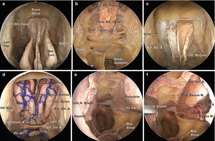
Fig. 1.13
Endoscopic endonasal dissection of the anterior cranial base and orbit. (a) Bilateral ethmoidectomy and bilateral opening of the frontal sinus (Draf 3). (b) A complete ethmoidectomy and superior septectomy have been performed, and the sphenoid sinus has been opened. Note the anterior and posterior ethmoidal canals. (c) The bone of the anterior cranial fossa surrounding the right olfactory fossa has been drilled to expose the dura mater. (d) The dura mater of the anterior skull base has been resected and the falx divided anteriorly so that the olfactory bulbs, gyri recti, and part of the orbital gyri are exposed from orbit to orbit in an endoscopic craniofacial resection. The pituitary gland and both parasellar segments of the internal carotid arteries are dissected posteriorly. (e, f) Endoscopic endonasal approach to the left orbit. (e) To approach the inferior and medial walls of the orbit, the orbit, medial maxillary antrostomy, and unilateral ethmoid labyrinthectomy with lamina papyracea removal need to be performed. (f) For intraconal pathologies, the periorbit needs to be incised, and the approaches are commonly directed between the medial and inferior rectus muscles. A artery, Ant anterior, Car carotid, Eth ethmoid, Front frontal, Gl gland, Inf inferior, Int internal, Lam lamina, M muscle, Max maxillary, Med medial, Mid middle, N nerve, Olf olfactory, Orb orbital, Orbitofront orbitofrontal, Pap papyracea, Pit pituitary, Post posterior, Sept septum, Sphen sphenoid, Tuberc tuberculum, Turb turbinate
The orbit can be approached medially by removing the lamina papyracea of the ethmoid bone and inferiorly through the superior wall of the maxillary sinus (Fig. 1.13).
1.3 Middle Cranial Base
1.3.1 Endocranial Surface
The endocranial surface of the middle cranial base, formed by the sphenoid and temporal bones, has medial and lateral parts (Figs. 1.3, 1.4, and 1.14). The medial part is formed by the body of the sphenoid bone, the site of the tuberculum sellae, pituitary fossa, middle and posterior clinoid processes, the carotid sulcus, and the dorsum sellae (Fig. 1.4). The lateral part is formed by the lesser and greater sphenoid wings, with the superior orbital fissure between them (Fig. 1.4). The lesser wing is connected to the body of the sphenoid bone by an anterior root, which forms the roof of the optic canal, and by a posterior root, also called the optic strut, which forms the floor of the optic canal and separates the optic canal from the superior orbital fissure (Fig. 1.4). The greater wing forms the largest part of the endocranial surface of the middle fossa, with the squamosal and the petrosal parts of the temporal bone completing this surface. The superior orbital fissure transmits the oculomotor, trochlear, ophthalmic, and abducens nerves; a recurrent meningeal artery; and the superior and inferior ophthalmic veins. The maxillary and mandibular nerves pass through the foramen rotundum and ovale, both located in the greater wing of the sphenoid bone. The not infrequently occurring sphenoidal emissary foramen, located anteromedial to the foramen spinosum, gives passage to a vein connecting the cavernous sinus and the pterygoid venous plexus. The upper surface of the petrous bone is grooved along the course of the greater and lesser petrosal nerves (Fig. 1.14). The carotid canal extends upward and medially and provides passage to the internal carotid artery and carotid sympathetic nerves in their course to the cavernous sinus. The posterior trigeminal root reaches the middle fossa and the impression on the upper surface of the petrous bone where the Meckel cave and the semilunar ganglion sit. The roof of the carotid canal opens below the trigeminal ganglion near the distal end of the carotid canal (Figs. 1.14 and 1.15). The arcuate eminence approximates the position of the superior semicircular canal. A thin lamina of bone, the tegmen tympani, provides a roof for the area above the middle ear and auditory ossicles on the anterolateral side of the arcuate eminence. The internal auditory canal can be identified below the floor of the middle fossa by drilling along a line approximately 60° medial to the arcuate eminence, near the middle portion of the angle between the greater petrosal nerve and the arcuate eminence (Fig. 1.14). The petrous apex, medial to the internal acoustic meatus, is free of important structures.
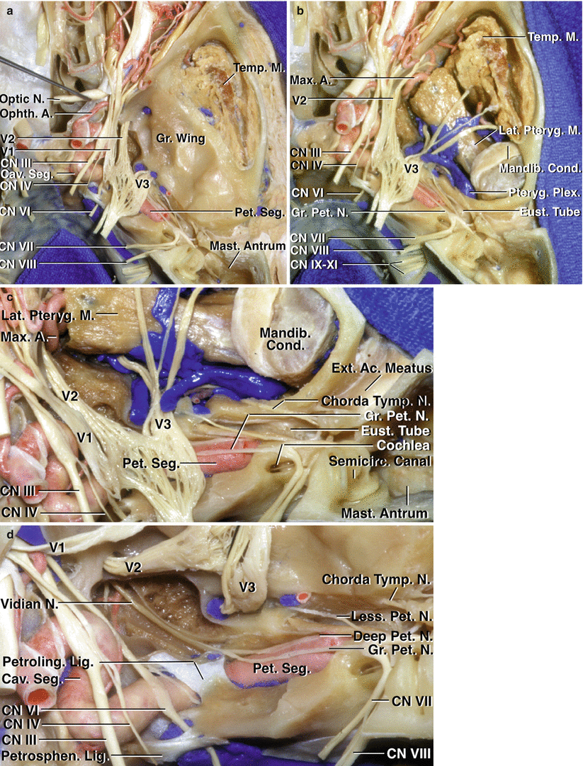
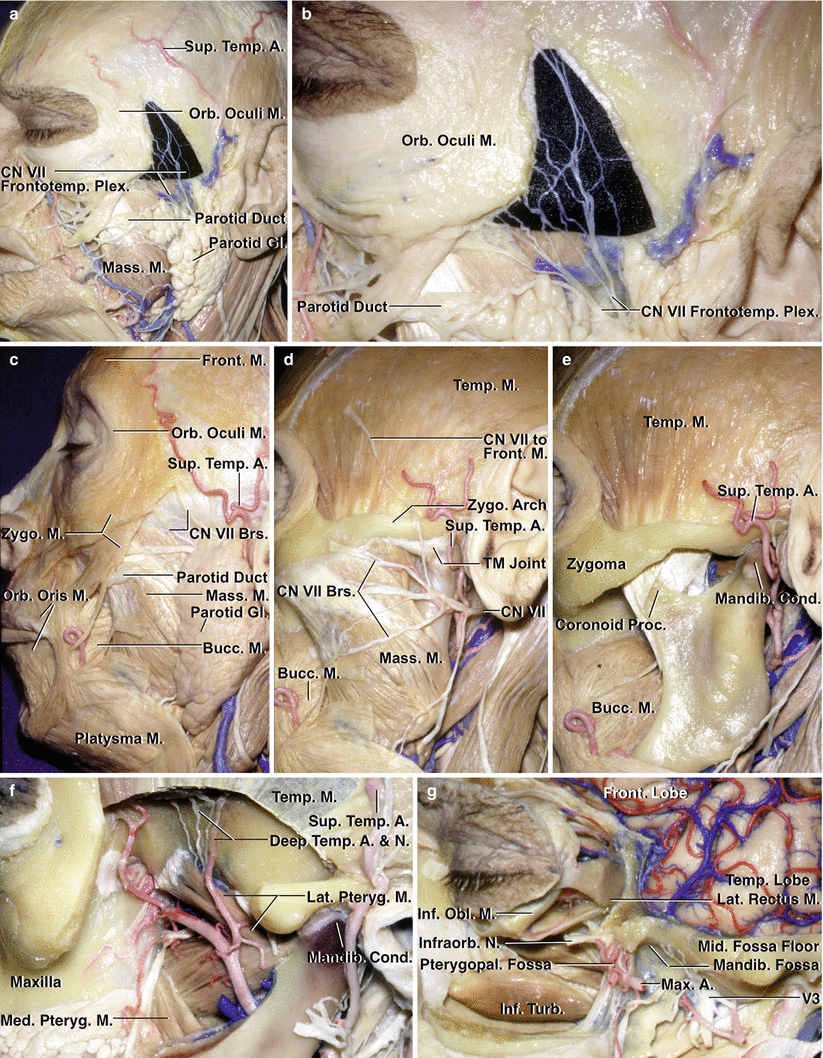
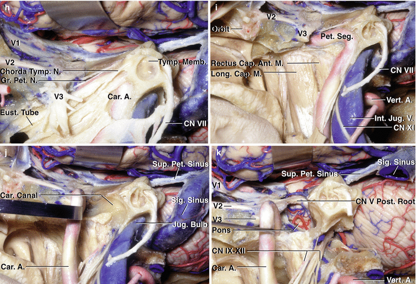
Fig. 1.14
Superior endocranial view of middle cranial base. (a) The floor of the middle fossa has been preserved. The anterior part of the floor of the middle fossa is formed by the greater sphenoid wing, which roofs the infratemporal fossa, and the posterior part of the floor is formed by the upper surface of the temporal bone. The internal acoustic meatus, mastoid antrum, and tympanic cavities have been unroofed. The dural roof and lateral wall of the cavernous sinus have been removed. The petrous segment of the internal carotid artery is exposed lateral to the trigeminal nerve. The temporalis muscle is exposed in the temporal fossa lateral to the greater sphenoid wing. (b) The floor of the middle fossa has been removed to show the relationships below the floor. The temporalis muscle descends medial to the zygomatic arch in the temporal fossa to insert on the coronoid process of the mandible. The infratemporal fossa is located medial to the temporal fossa, below the greater sphenoid wing, and contains the pterygoid muscles and venous plexus and branches of the mandibular nerve and maxillary artery. The mandibular condyle is located below the posterior part of the middle fossa floor, which is formed by the temporal bone. (c) Enlarged view of the posterior part of the area below the middle fossa floor. The roof of the temporal bone, which forms the posterior part of the floor of the middle fossa, has been opened to expose the mastoid antrum, eustachian tube, semicircular canals, cochlea, the nerves in the internal acoustic meatus, and the mandibular condyle. (d) The trigeminal nerve has been reflected forward. The abducens nerve passes below the petrosphenoid ligament and through the Dorello canal. The petrous segment of the carotid passes below the petrolingual ligament to enter the cavernous sinus. The greater petrosal nerve is joined by the deep petrosal branch of the carotid sympathetic plexus to form the vidian nerve, which passes forward in the vidian canal, which has been unroofed. The lesser petrosal nerve arises from the tympanic branch of the glossopharyngeal nerve, which passes across the promontory in the tympanic nerve plexus and regroups to cross the floor of the middle fossa, exiting the cranium to provide parasympathetic innervation through the otic ganglion to the parotid gland. The tensor tympani muscle and eustachian tube are layered, with the former above the latter, along and separated from the anterior surface of the petrous carotid by a thin layer of bone. A artery, Ac acoustic, Cav cavernous, CN cranial nerve, Cond condyle, Eust eustachian, Ext external, Gr greater, Lat lateral, Less lesser, Lig ligament, M muscle, Mandib mandibular, Mast mastoid, Max maxillary, N nerve, Ophth ophthalmic, Petroling petrolingual, Pet petrosal, petrous, Petrosphen petrosphenoid, Plex plexus, Pteryg pterygoid, Seg segment, Semicirc semicircular, Temp temporalis, Tymp tympani (From Rhoton Jr AL. The anterior and middle cranial base. Neurosurgery. 2002;51(1 Suppl):S273–302; with permission)
Fig. 1.15


Stepwise lateral exposure of the middle cranial fossa. (a) The branches of the facial nerve, which form a fine plexus in the fat pad overlying the temporalis fascia and are directed to the orbicularis oculi and frontalis muscle, have been dissected free, and a small piece of black material has been placed deep to their fine branches to highlight this neural network in the fat pad. (b) Enlarged view of the facial nerve plexus innervating the orbicularis oculi and frontalis muscle. (c) Lateral view of the structures superficial to the anterior and middle cranial base. The frontotemporal and zygomatic branches of the facial nerve are exposed anterior to the parotid gland. The orbicularis oculi surrounds the orbit, and the frontalis muscle extends upward from the superior orbital rim. The levators of the lip and zygomaticus muscles are located in front of the maxilla. The orbicularis oris surrounds the mouth, and the buccinator muscle surrounds the oral cavity deep to the masseter muscle. The parotid duct crosses the masseter muscle. The superficial temporal artery divides into anterior and posterior branches. The parotid gland has been removed to show the branches of the facial nerve. (d) The parotid gland has been removed to expose the facial nerve exiting the stylomastoid foramen. The facial nerve branch to the frontalis muscle has been preserved in the dissection and has been laid back against the temporalis muscle to show it crossing the zygomatic arch in its course to the forehead. The superficial temporal artery passes deep to the facial nerve in front of the ear. (e) The masseter muscle has been removed to expose the temporalis muscle inserting on the coronoid process. The buccinator muscle, which surrounds the oral cavity, is situated on the deep side of the masseter muscle. (f) The coronoid process and the lower part of the temporalis muscle have been removed to expose the deep temporal branches of both the maxillary artery and mandibular nerve passing upward along the greater sphenoid wing and temporal squama to enter the deep side of the temporalis muscle. The lateral pterygoid muscles extend backward from the pterygoid process and greater wing of the sphenoid to insert along the mandibular condyle and temporomandibular joint. (g) A craniotomy has been done to expose the floor of the middle fossa, and the lateral wall of the orbit has been removed to expose the extraocular muscles. The mandibular condyle has been removed and the pterygoid muscles reflected to expose the mandibular nerve at the foramen ovale. The pterygopalatine fossa is located behind the maxilla. The floor of the orbit and the upper part of the maxilla have been removed to expose the nasal cavity. (h) Enlarged view following resection of the floor of the middle fossa and the external auditory canal to expose the tympanic membrane and the mandibular nerve below the foramen ovale. The mastoid segment of the facial nerve has been preserved. The greater petrosal nerve crosses above the petrous carotid. The tensor tympani muscle and eustachian tube are layered along the anterior margin of the petrous carotid. (i) The eustachian tube and tensor tympani have been resected to expose the upper cervical and petrous carotid. The nasopharyngeal mucosa has been opened to expose the longus capitis and rectus capitis anterior muscles. (j) The carotid artery has been reflected forward out of the carotid canal. This exposes the petrous apex in front of the jugular foramen on the medial side of the internal carotid artery. (k) The petrous apex has been drilled and the dura opened below the trigeminal nerve to expose the upper anterior part of the posterior cranial fossa. Segments of the internal jugular vein and jugular bulb have been resected to expose the 9th through 12th cranial nerves below the jugular foramen and hypoglossal canal. A artery, Ant anterior, Brs branches, Bucc buccinator, Cap capitis, Car carotid, CN cranial nerve, Cond condyle, Eust eustachian, Front frontal, frontalis, Frontotemp frontotemporal, Gl gland, Gr greater, Inf inferior, Infraorb infraorbital, Int internal, Jug jugular, Lat lateral, Long longus, M muscle, Mandib mandibular, Mass masseter, Max maxillary, Med medial, Memb membrane, Mid middle, N nerve, Obl oblique, Orb orbital, Pet petrosal, petrous, Plex plexus, Post posterior, Proc process, Pteryg pterygoid, Pterygopal pterygopalatine, Rec rectus, Seg segment, Sig sigmoid, Sup superior, Temp temporal, temporalis, Tymp tympani, tympanic, V vein, TM temporomandibular, Turb turbinate, Vert vertebral, Zygo zygomatic (From Rhoton Jr AL. The anterior and middle cranial base. Neurosurgery. 2002;51(Suppl):S273–302; with permission)
Related posts:


Stay updated, free articles. Join our Telegram channel
Full access? Get Clinical Tree
