and Ziv Gil2
(1)
Division of Otolaryngology Head and Neck Surgery and Maxillofacial Surgery, Tel Aviv Sourasky Medical Center, Tel Aviv, Israel
(2)
The Head and Neck Center Department of Otolaryngology Head and Neck Surgery, Rambam Healthcare Campus, Haifa, Israel
Keywords
MeningitisCerebrospinal fluid leakTension pneumocephalusWound infectionOsteonecrosisDiplopia and telecanthus13.1 Introduction
Since the developments and popularization of modern skull base approaches through the twentieth century, surgical extirpation of tumors has become the mainstay of treatment for both benign and malignant diseases. Open and endoscopic approaches allow intradural and extradural tumor resection, and they can be performed in a single procedure that also allows precise reconstruction of the dura. Despite their technical reproducibility, however, these procedures still involve high risks for postoperative complications and mortality. Recent data show that over the last four decades, advancements in imaging modalities and refinements in surgical and reconstructive methods have allowed an increasing number of patients with skull base cancers to undergo curative surgical resections. Better understanding of skull base anatomy, the increasing experience of surgeons, and modifications of surgical techniques allow successful resections of complex skull base tumors involving multiple craniofacial compartments. As a result of improved preoperative imaging assessment, technical advances in tumor resection, new reconstruction methods, and the fruitful cooperation of multidisciplinary clinical teams, we have witnessed a decline in the rate of complications associated with skull base surgery.
The purpose of this chapter is to review the complications associated with skull base surgery in general and anterior skull base tumor resection specifically.
13.2 Risk of Complications after Anterior Skull Base Tumor Resection
Patients who undergo surgery for the removal of skull base tumors have a risk of severe complications. Factors that may increase such risks are comorbidity, dural invasion, and aggressive tumors requiring wide resections. Traditionally, the rate of complications after craniofacial resection was nearly 50 %. However, analysis of recent studies has revealed a significant reduction in the complication to 30 % (Fig. 13.1). Analyzing the types of complications revealed that while the rates of intracranial, systemic, and ocular complications remained stable over the last 30 years, the incidence of wound complications declined by more than 20 %. In addition, although the overall complication rate decreased by 28 % for younger patients in the later time period, it decreased by only 14 % for those over 50 years old. Similarly, among patients with significant comorbidities, there was a 4 % reduction in complications in the later period, compared with a 23 % reduction in healthy patients. In addition to the reduction in the risk of complications, the risk of postoperative mortality also declined by 1 % during the last three decades, despite the higher-risk population.
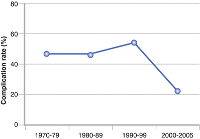
Fig. 13.1
Change in complication rate after craniofacial resection during the last decade as recorded at Memorial Sloan Kettering Cancer Center
There is a significant variability in the reported risk of complications after anterior skull base surgery. The large variability in mortality (0–8 %) and in the incidence of intracranial (4–23 %), systemic (5–10 %), and orbital (4–6 %) complications can be largely explained by differences in the study period, patient characteristics, the surgeon’s experience, and the definition of complications.
A summary of the data from several of the reports that described the type and frequency of complications after open approaches is shown in Table 13.1. Although during the beginning of endonasal resection the risk of complications from cerebrospinal fluid (CSF) leak was around 30 %, development of vascularized local flaps has contributed to a significant reduction in the rate of CSF leaks, which is now between 5 % and 10 %. Table 13.2 shows the rate of complications after endonasal skull base resections.
Table 13.1
Morbidity after open anterior skull base resection of tumors
Author | Subjects, n | Mortality (%) | Overall complications (%) | Wound (%) | Intracranial (%) | Systemic (%) | Orbital (%) |
|---|---|---|---|---|---|---|---|
Ketcham et al. (1985) | 89 | 3 | 54 | 20 | 13.5 | – | – |
Janecka et al. (1994) | 183 | 2 | 33 | 2.2 | 4.4 | – | – |
Kraus et al. (1994) | 85 | 2 | 39 | 24.7 | 17.6 | 5.9 | 4 |
Dias et al. (1999) | 104 | 7.6 | 48.6 | 38.5 | 23 | 10.6 | 5.8 |
Donald et al. (1999) | 107 | 5.6 | 50.5 | 22.4 | 15.9 | – | – |
Solero et al. (2000) | 168 | 4.7 | 29.7 | 4.8 | 20 | – | 1.8 |
Patel et al. (2003) (International Study) | 1,193 | 4.7 | 36.3 | 19.8 | 16.2 | 4.8 | 1.7 |
Gil et al. (2009) | 156 | 3.3 | 33 | 7.5 | 13 | 5.8 | 3.3 |
Mean | 2,089 | 4.1 | 40.5 | 17.5 | 15.5 | 7 | 3 |
Table 13.2
Morbidity after endoscopic anterior skull base resection of tumors
Author | Subjects, n | Mortality (%) | Overall (%) | Wound (%) | Intracranial (%) | Systemic (%) | Orbital (%) |
|---|---|---|---|---|---|---|---|
de Divitiis et al. (2008) | 11 | 0 | 30 | 0 | 27 | 1 | 2.7 |
Dehdashti et al. (2009) | 22 | 0 | 27 | 0 | 22.5 | 0 | 4.5 |
Stamm et al. (2008) | 7 | – | – | – | 29 | – | – |
Cavallo et al. (2007) | 21 | 0 | 14.5 | 5 | 9.5 | – | – |
Tabaee et al. (2007) | 127 | – | – | – | 8.7 | – | – |
Zanation et al. (2009) | 70 | – | – | – | 5.7 | – | – |
Villaret et al. (2010)
Related posts:Stay updated, free articles. Join our Telegram channel
Full access? Get Clinical Tree
 Get Clinical Tree app for offline access

|