Tenosynovitis Disorders of the Upper Extremity
Mary C. Burns
Michael W. Neumeister
Tendonopathies of the upper extremity are extremely common disorders encompassing a large variety of conditions from tendon strains to ruptures or lacerations. The most common tendinopathy is tenosynovitis or inflammation of the tendon sheaths. Inflammation may arise from a number of causes as categorized in Table 79.1. It is important to understand that inflammation is a continuous process that involves a cascade of events leading to variable clinical symptoms and signs. Acute inflammation involves a vascular response, fluid exudate and swelling, cellular exudate with phagocytosis, and alterations in tissue composition. The exudates associated with inflammation differ from normal interstitial fluid by having a greater protein content and higher specific gravity (over 1.020).1
The fate of acute inflammatory conditions, however, depends upon the treatment and cessation of the inciting caustic agent or activity. Upon cessation, the inflammation may resolve completely leaving normal tissues behind. Alternatively, the acute inflammatory response may result in a number of pathologic states, including (A) organization of the exudate leading to fibrosis, (B) tissue destruction and secondary healing by repair/scarring, (C) chronic inflammation, and (D) in the case of infection, suppuration.
LATERAL EPICONDYLITIS
Lateral epicondylitis, or tennis elbow, is a common condition of the lateral elbow. First described in 1873 by Runge, lateral epicondylitis is the direct result of strain at the fascia and origin of the extensor muscles of the forearm.2 Active extension against resistance, such as occurs when one hits a tennis ball with a back hand shot (hence the term “tennis elbow”), presumably results in microtrauma to the origin of the extensor carpi radialis brevis (ECRB), the extensor digitorum communis (EDC), and the extensor carpi ulnaris. Although the specific etiology is unknown,3 it is postulated that this disease results from micro-tears at the origin of the common extensor muscle mass. Many reports believe that these small tears are caused by overuse, repetitive or cumulative injury, and strain of the common extensor origin at the lateral epicondyle. Continued use of the upper extremity results in continued strain and re-injury to the muscle origins.4,5 This can lead to degeneration and possible avascularity at the muscle origin, which in turn, theoretically, leads to chronic inflammation causing the injured areas to remain weak and painful. Ironically, however, inflammation is not a characteristic histologic finding of lateral epicondylitis, but fibrosis and mucinoid degeneration of the origin of the extensor muscles have been documented.6 Table 79.2 highlights the histologic changes observed at the origin of the extensor muscles of the forearm.
TABLE 79.1 ETIOLOGY OF INFLAMMATION OF TENDONS OR TENDON SHEATHS | ||||||||||||||||||||
|---|---|---|---|---|---|---|---|---|---|---|---|---|---|---|---|---|---|---|---|---|
| ||||||||||||||||||||
Patients generally present with pain and localized tenderness of the extensor origin and lateral epicondyle of the humerus (Figure 79.1). The onset may be acute or insidious, and it is more common in males between 40 and 50 years of age. Generally, there is increased pain with wrist extension or with gripping. Activities requiring full elbow extension and forearm pronation aggravate the discomfort that may radiate down the posterior forearm or proximally over the lateral upper arm above the elbow. The pain may also be accentuated by extreme wrist flexion from the passive stretch of the ECRB muscle or by active contraction of the wrist extensors.
During the initial examination of the patient, the surgeon should also evaluate the range of motion (ROM) of the elbow, assess crepitus of the radiohumeral joint, observe for bursitis and osteochondritis of the capitellum, and rule out radial tunnel or posterior interosseous nerve entrapment.
There is limited value in obtaining further diagnostic studies for lateral epicondylitis. One may consider plain radiographs if degenerative elbow changes are considered or identified on physical exam. Similarly, magnetic resonance imaging scans may identify articular pathology or masses, but generally these tests are not indicated for clinically confirmed lateral epicondylitis. Nerve conduction and electromyogram studies may be useful if radial tunnel or posterior interosseous nerve entrapment is suspected. These nerve entrapments are not uncommonly found in patients with lateral epicondylitis. Injecting a local anesthetic agent around the extensor origin and lateral epicondyle confirms the diagnosis but the diagnosis is usually made at physical exam. The lateral epicondyle and the proximal radius are the origins of the extensor muscles of the forearm. The extensor carpi radialis longus (ECRL) has its origin just above the lateral epicondyle on the humerus and the intermuscular septum. The ECRB originates from the lateral epicondyle (common extensor origin) and the lateral ligament of the elbow.
TABLE 79.2 HISTOLOGIC FEATURES AT THE LATERAL EPICONDYLE IN PATIENTS WITH LATERAL EPICONDYLITIS | |||||||
|---|---|---|---|---|---|---|---|
|
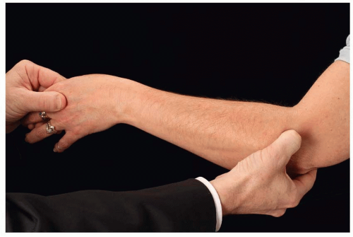 FIGURE 79.1. Location of pain in “tennis elbow.” Lateral epicondylitis is characterized by point tenderness over the lateral epicondyle. The patients often complain of weakness, pain on wrist extension, and lateral elbow pain. |
The natural history of lateral epicondylitis is variable. Many authors believe that over time (1 to 2 years), the condition burns itself out and therefore no specific treatment may be warranted. The level of discomfort, however, dictates the treatment. If the condition is mild, and the patient can tolerate benign neglect, improvement with anti-inflammatory medication is probable. In general, patients are offered physical therapy, splinting, and oral anti-inflammatory medication. The physical therapy regimen includes stretching the extensor muscle origin and gradual loading. Counterforce braces or forearm support bands are common splints utilized for lateral epicondylitis (Figure 79.2). The counterforce brace essentially changes the origin of the muscles to a more distal site. The brace acts to decrease the force of muscle contraction by inhibiting muscle expansion, thereby reducing tension at the origin of the muscle. This essentially off-loads the muscle origin, prevents repetitive trauma at this site, and allows healing. Most therapists will have patients wear the counterforce brace throughout the day, particularly when performing activities utilizing the extensor muscles. The brace can be removed during periods of inactivity and when they are sleeping. The brace is typically applied approximately 2 cm distal to the lateral epicondyle and should fit with a comfortable amount of pressure. Maximum contraction of the wrist extensors is avoided. A brace that is applied too tightly may result in swelling of the distal forearm and hand, compression of the posterior interosseous nerve, and pain. When lateral epicondylitis is severe, complete immobilization may be necessary. A wrist cock-up splint or even a long-arm splint with the elbow at 90° of flexion may be warranted. The Richard wrist cock-up splint maintains the wrist in extension to offload the extensors. Orthoses have been described in various positions ranging from neutral to 45° of wrist extension. The rigid wrist orthosis, however, is fraught with noncompliance and is therefore limited to only the more severe cases of lateral epicondylitis.
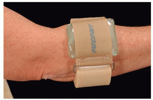 FIGURE 79.2. Nonsurgical treatment for lateral epicondylitis. A static band at the origin of the ECRB off-loads the epicondyle. |
Steroid injections have been utilized for the treatment for lateral epicondylitis. The duration of the effects of the steroid injection, however, is variable.7 Most patients respond favorably within the first 6 weeks but recurrence of the pain is common. The steroid is injected at the site of the maximum tenderness at the lateral epicondyle. Typically, 1 cc of dexamethasone or Kenalog is injected into this area. No significant difference has been demonstrated between different steroid preparations. Most surgeons wait for a period of 2 to 3 months before re-injection at this site and limit the number of injections to 3 or 4, mostly because of the secondary effects of the steroids on the overlying soft tissue. The steroid injections can result in significant atrophy of the fat and overlying skin and may also result in hypo-pigmentation of the area.
Some authors have tried platelet-rich plasma or even the patient’s own blood injected into the lateral epicondyle. Gossen et al. compared corticosteroid with platelet-rich plasma injections in patients with refractory lateral epicondylitis for at least 6 months.7 The long-term outcomes were similar in both groups following the injections but it was generally felt that the corticosteroid injection was more effective in reducing the pain in the short term. Autologous blood injections were thought to initiate an inflammatory response at the lateral epicondyle with subsequent fibrosis that improved the condition by decreasing strain at the origin. Edwards and Calandruccio injected autologous blood in 28 patients and demonstrated an 80% reduction in pain over 9 months.7 Missra and Delco injected buffered platelet-rich plasma into patients with lateral epicondylitis with similar findings comparing corticosteroid and the platelet-rich plasma.7
Botulinum toxin A has been injected into extensor muscles to induce paralysis as a means of off-loading the muscle’s origin.8 Other topical agents have been used for lateral epicondylitis, including topical nitrates, steroid creams, and salicylate creams; however, no randomized controlled studies have been performed with these agents.17
Surgery For Lateral Epicondylitis
The surgical treatment for lateral epicondylitis is restricted to those patients who have failed conservative measures and have been treated for 6 months to a year or are significantly disabled as a result of the discomfort.
A 2 to 3 cm incision is made over the lateral epicondyle and the extensor fascia is incised. Any degenerative muscle or granulation tissue is debrided. The prominent lateral epicondyle is shaved with an osteotome. Care is taken not to disrupt the collateral ligament of the elbow. Hemostasis is secured and the fascia is repaired. The wounds are typically closed with a resorbable sutures and a bulky dressing is applied.
Variations to the procedure include resecting the sensory nerves to the lateral epicondyle, transposing the anconeus muscle over the shaved epicondyle. To denervate the lateral epicondyle, the incision is usually extended proximally to identify the arcade of nerves that innervate the lateral epicondyle in the elbow. The nerves are resected and allowed to retract proximally or are buried in the brachialis muscle.8
DE QUERVAIN’S TENOSYNOVITIS
The anatomy of the first dorsal compartment is variable. There may be multiple slips of the abductor pollicis longus (APL) tendon which may insert in a variety of locations around the basilar joint, including the thumb metacarpal, the trapezium, the volar carpal ligament, the opponens pollicis, and abductor pollicis brevis (APB) (Figure 79.3). The extensor pollicis brevis (EPB) tendon is also housed within the first dorsal compartment. The EPB tendon, however, may travel in its own separate compartment within the first dorsal compartment along with the APL tendon. This septation of the EPB tendon seems to increase the probability of acquiring de Quervain’s tenosynovitis, and it may also increase the probability that conservative measures will not be successful and that surgical decompression is warranted. The treatment of de Quervain’s tenosynovitis is usually conservative at the first presentation. Nonsteroidal anti-inflammatories, corticosteroid injections, and off-loading of the APL and EPB tendon are the mainstay of treatment in de Quervain’s tenosynovitis. Failing all nonsteroidal anti-inflammatories, most patients should receive a trial of corticosteroid injections into and around the first dorsal compartment. This steroid is injected around the radial styloid, which is the site of greatest constriction. Most studies quote steroid injections’ success rates of 50% to 80%.9,10
Related posts:


Stay updated, free articles. Join our Telegram channel

Full access? Get Clinical Tree


