Technique for Dorsal Augmentation Using Costal Cartilage Graft
Dean M. Toriumi
DEFINITION
Low nasal dorsum is when the bridge of the nose demonstrates inadequate height/projection.
A low dorsum can appear wide and flat on frontal view with inadequate lateral wall shadowing.
Asian patients tend to have inadequate dorsal height and frequently are in need of dorsal augmentation.
Inadequate dorsal height may be directly related to excessive nasal tip projection. The relationship between dorsal height and tip projection frequently requires creating balance between these two parameters.
ANATOMY
The anatomy of the low dorsum can vary from patient to patient.
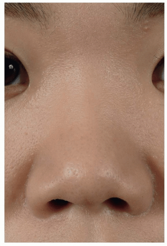
In the Asian patient, the nasal dorsum can be low and wide (FIG 1).
Asian patients tend to have a low radix (low nasal starting point) as well.
Asian patients tend to have very small and thin septal cartilage that provides insufficient stock for dorsal augmentation.
Some patients have a congenitally low dorsum.
Patients that suffered from trauma can present with a low dorsum or saddle nose deformity.
Most patients with a saddle nose deformity have a deficiency in the middle nasal vault usually with a normal bony nasal vault. Many of these patients may actually have a dorsal convexity above the saddled area.
PATIENT HISTORY AND PHYSICAL FINDINGS
Any evidence of previous trauma or surgery should be elicited in the history.
Any prior placement of alloplastic implants should be elicited in the history.
Physical exam should reveal any septal deviations or fractures.
Asian patients who have a low dorsum also tend to have an underprojected nasal tip.
All patients should be queried as to any previous injectable fillers placed into their nose.
SURGICAL MANAGEMENT
Anesthesia
It is preferable to perform these surgeries under general anesthesia. With the patient intubated, the airway is protected from blood contacting the vocal cords and potentially causing laryngospasm. A protected airway is particularly important if any work is planned on the nasal septum or turbinates.
Local anesthetic agent (1% lidocaine with 1:100 000 epinephrine) is injected into the nose to provide hemostasis. Injections are made along the septum, along the marginal incisions, over the nasal dorsum and middle vault, and in the nasal tip area. At least 10 minutes should pass before the procedure is initiated to allow the full vasoconstrictive effect to set in.
 FIG 1 • Asian patient with low dorsum showing lack of lateral wall shadowing and flat appearance to nasal dorsum. |
TECHNIQUES
▪ Harvesting Costal Cartilage for Dorsal Augmentation
Costal cartilage can be harvested from relatively small incisions with low morbidity. Costal cartilage of adequate length and width can be harvested through an 11-mm chest incision.1,2
When less experienced, a larger incision should be used to safely harvest the costal cartilage.
Consider using a 3-cm incision until you are comfortable with the chest wall anatomy and have significant experience harvesting costal cartilage.
Avoid cutting the muscle layer.
It is preferred to separate the muscle fibers bluntly.
This allows the muscle layer to be tightly resutured at the end of the operation to provide good support to the area.
Costal perichondrium should be harvested from the surface of the rib. Costal perichondrium can be used for camouflage or as a soft tissue graft.
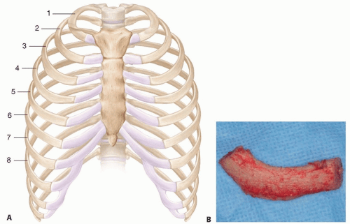
The best rib to harvest for dorsal augmentation is typically the 7th rib. The 7th rib is longer and straighter and provides the best cartilage stock for major dorsal augmentation3,4 (TECH FIG 1A,B).

TECH FIG 1 • A. Chest wall showing rib anatomy. The 7th rib is longer and straighter than the 5th and 6th ribs. The 6th rib has a genu where the rib passes below the breast. B. Harvested rib.
The 5th rib is curved and not appropriate for large dorsal augmentations.
The 6th rib is relatively straight but has a genu that could shorten the straight segment that can be used for the dorsal graft.
The 7th rib is also more superficial than the 6th rib, so it is easier to harvest through a smaller incision.
The 7th rib cartilage is sharply dissected away from the 6th and 8th ribs using a Freer elevator and lifted off of the underlying perichondrium.
Typically, a 3.5- to 4.5-cm segment of costal cartilage should be harvested.
Perform a Valsalva maneuver after the rib is harvested to see if there is a tear in the pleura.
If so, a red rubber catheter can be placed during closure, and then the chest can be expanded and the catheter removed after the closure is completed.
The 7th rib tends to lie below the level of the diaphragm and therefore lessens the likelihood of a pneumothorax. As you move medially on the 7th rib, it may move above the diaphragm.
▪ Preparing and Affixing the Dorsal Graft
The harvested costal cartilage should be initially carved into three separate segments (TECH FIG 2A), which are then assessed for the most ideal curvature.
If a very thick dorsal graft is needed, the rib should be carved differently by taking thinner peripheral segments to provide a thicker dorsal graft (TECH FIG 2B). In this case, the middle segment is typically used for the dorsal graft.
If the central core of the costal cartilage has a soft (pearlike) texture, this increases the likelihood of warping of the cartilage. In this case, it is preferable to use the outer core of the rib for the dorsal graft.
If this cartilage must be used, it is recommended to laminate the cartilage by suturing two thinner layers of cartilage together to create one dorsal graft. The lamination acts to decrease the likelihood of warping.

TECH FIG 2 • A. Younger patient with the rib being carved into three separate segments. Note the bending of the cartilage. B. For larger dorsal grafts, the central segment of the rib can be made thicker to allow for a thicker dorsal graft.
Stay updated, free articles. Join our Telegram channel

Full access? Get Clinical Tree

 Get Clinical Tree app for offline access


