Targeted Muscle Reinnervation (TMR) is a reproducible procedure for effective prevention and treatment of neuropathic pain. Recent studies have demonstrated that TMR is more successful in treating neuroma pain than the standard method of neuroma excision and burying into muscle. The fundamental concept of TMR is that providing severed nerve endings a place to go and something to do facilitates normal healing. Future studies will continue to support the use of TMR for any region of the body where an injured nerve may develop a painful neuroma.
Key points
- •
Targeted muscle reinnervation (TMR) is a reproducible technique for effective treatment and prevention of neuropathic pain.
- •
The fundamental concept of TMR is to give severed nerve endings a place to go and something to do, which facilitates healing of these severed peripheral nerves.
- •
Methods to encourage normal healing of severed peripheral nerves instead of burying the nerve ending in surrounding tissue, will lead to improved outcomes of neuroma treatment.
- •
Neuropathic pain is a surgically treatable condition.
Introduction
Chronic pain after surgery leads to significant disability worldwide. Although there are numerous established causes of chronic postsurgical pain, neuropathic pain due to peripheral nerve injury is a known contributor. In some proportion of peripheral nerve injuries, symptomatic neuromas—disorganized axons encased in scar—form and may be exquisitely painful to light touch, pressure, vibration, and extreme temperatures or even at rest. Many management strategies have been proposed, yet symptomatic neuromas remain a challenge. Pharmacologic therapy with antidepressants, anticonvulsants, opioids, and topical anesthetics have variable efficacy and places patients at risk for opioid dependence. Behavioral strategies include desensitization protocols, cognitive behavioral therapy, and group psychotherapy. Interventional methods for neuropathic pain include minimally invasive treatments of the injured nerve with radiofrequency ablation, perineural injection with alcohol or corticosteroids, regional blocks, spinal cord stimulation, transcutaneous electrical nerve stimulation, and direct operative handling of the injured nerves.
Whenever possible, nerve injuries should be repaired. When this is not feasible, such as in the case of an absent distal nerve stump or in the case of a failed reconstruction, direct operative handling of the injured nerve is preferred. For decades, the standard surgical approach for symptomatic neuroma has been neuroma excision and relocation to a more favorable position, commonly buried into bone or muscle, with the hope that the recurrent neuroma would be hidden and asymptomatic. , In contrast, more recent strategies are based on the concept of providing severed axons an end organ—somewhere to go and something to do. One such technique is targeted muscle reinnervation (TMR), a nerve transfer procedure that treats pain via direct nerve to nerve healing.
First performed by the senior author G.A. Dumanian in 2002, TMR is defined as the coaptation of cut peripheral nerves to a newly divided nearby motor nerve branch. The fascicles of the proximal nerve grow into motor end plates, and likely into proprioceptors and other sensory end organs, to reinnervate the target muscle. , TMR has evolved from a technique to improve intuitive prosthesis control in amputees to a technique to treat and prevent neuropathic pain from peripheral neuroma. A recent randomized clinical trial in the treatment of chronic postamputation pain demonstrated that healing a nerve ending with TMR had better patient-reported outcomes for residual limb pain and phantom limb pain at 18 months of follow-up, compared with neuroma excision and muscle burying. Furthermore, a cohort study of patients who underwent TMR at the time of amputation compared with unselected amputees from the general population demonstrated significantly fewer TMR patients with moderate to severe residual limb and phantom limb pain by multiple measures. Additional clinical data have shown that pain outcomes from nonamputees with neuroma pain who were treated with TMR are better than those who were treated with standard neuroma excision and hiding. There is growing acceptance for TMR as an effective surgical strategy for management of neuropathic pain. TMR may be performed by any surgeon with knowledge of peripheral nerve anatomy and nerve dissection experience.
Pathophysiology
Traumatic neuromas are regions of nerve swelling/inflammation that can occur in any part of the body after nerve injury. Nerve injury can be direct—transection during dissection, or indirect—stretched or crushed during retraction. The physiologic response to axonal injury is axon sprouting and regeneration from the proximal segment and wallerian degeneration of the distal segment. Seddon’s classification distinguishes types of nerve injury in terms of neuropraxia, axonotmesis, and neurotmesis. Sunderland further grades nerve injury based on expected recovery. With intact or repaired perineurium and epineurium, sprouting axons find a distal target and eventually prune excess axons. For Sunderland grade 4 or greater, however, sprouting axons fail to connect to a distal segment or target organ and become encased in scar, thus forming neuromas, specifically neuroma-in-continuity in axonotmesis or end neuroma in neurotmesis ( Table 1 ).
| Seddon (1947) | Sunderland (1951) | Mackinnon (1988) | Summary |
|---|---|---|---|
| Neuropraxia | 1 | Local conduction block with possible segmental demyelination. No injury to axons, so no regeneration. Remyelination and complete recovery by 12 wk postinjury. | |
| Axonotmesis | 2 | Axonal injury occurs. Intact endoneurium, perineurium, and epineurium. Distal segment undergoes wallerian degeneration. Proximal nerve fibers regenerate at 1mm/day. Full recovery possible unless distance of injury from motor end plate far and prolonged denervation of the receptor prevents motor recovery. | |
| 3 | Axonal injury occurs. Wallerian degeneration combined with fibrosis of endoneurium. Perineurium and epineurium intact. Incomplete recovery likely, due to scar within endoneurium hinders regenerating fibers interaction with correct end organs. | ||
| 4 | Nerve-in-continuity but injury causes complete scar block. Only epineurium intact. Recovery only possible if scar is surgically removed and nerve repaired or grafted. | ||
| Neurotmesis | 5 | Nerve completely divided and must be surgically repaired before regeneration can occur. | |
| 6 | Mixed nerve injury. Combination of any of the types of injuries 1–5, and different levels of injury occur at different regions of the nerve. |
Within the neuroma bulb, the authors’ data have shown that the axons are sensory, with no motor staining found. The model is that the sensory nerves continue to seek a distal nerve attempting to heal and reinnervate, whereas without Schwann cells and other trophic factors, the motor axons recede back toward the spine. This is consistent with clinical peripheral nerve surgery that motor outcomes tend to be less successful than sensory recoveries for mixed nerve repairs. Furthermore, it has been the authors’ experience that cut pure motor nerves do not form neuromas, a key to TMR.
Histologically, TMR restores axon count, size, and myelination in a rabbit amputation model. In the authors’ clinical experience, reestablishment of muscle function can be detected clinically in more than 95% of nerve transfers. Electromyography after transhumeral TMR has demonstrated physiologic synaptic inputs to reinnervated muscles. What is unclear is the fascicles that fail to find a distal target after nerve transfer due to obvious mismatch in size and fascicle numbers between donor and recipient nerves. This size mismatch undoubtedly led to the slow adoption of TMR as a surgical technique. It is hypothesized that the large denervated block of muscle near the nerve coaptation site is a source of additional end organs to soak up escaping axons.
Presentation and diagnosis
A typical patient presenting with a symptomatic neuroma may describe the pain as electric, shooting, or burning. The pain may be associated with light touch or pressure or changes in weather and temperature or be without any provocation at all. Neuropathic pain may affect sleep quality, such as the common complaint of night waking in carpal tunnel syndrome; mood; ambulation; and, for the amputee, prosthetic use. Patients may present with high opioid tolerances. For the amputee, postamputation pain may include residual limb (or stump) pain, phantom limb pain, back pain, and hip pain. , To clarify distinctions between pain syndromes, residual limb pain is defined as pain local to the residual limb, often due to a neuroma ; phantom limb pain is defined as unpleasant or painful sensations perceived in the missing limb, thought to be a complex interplay between the neuroma and several levels of the central nervous system ; and chronic pain is defined as pain lasting greater than 6 months and causing physical debilitation and decreased quality of life. A simple history for pain includes an assessment of intensity on an 11-point numerical rating scale (0 is no pain and 10 is the worst pain imaginable) and an assessment of frequency.
For the patient who presents with intolerable symptomatic neuroma, the clinician must address the nerve injury. Symptomatic neuromas by definition are associated with injury to a mixed or sensory nerve. The clinical diagnosis is made as a combination of the chief complaint as chronic pain postamputation/surgery/injury and a focused physical examination. The clinician asks patients to point to a location on their extremity where the pain is the greatest, and the patients can quickly identify this painful spot. There likely is a Tinel sign when this spot is tapped by the clinician, or patients may experience severe acute pain when this region is palpated. The nerve leading to this neuroma often is excitable to palpation along its length. Imaging typically is not necessary or recommended for the work-up of intolerable neuropathic pain.
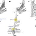
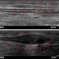
Injections with local anesthesia may be performed to confirm that treatment of the injured nerve will lead to a change in symptoms. An inability to improve symptoms temporarily with an anesthetic injection should give both surgeon and patient pause before beginning surgery. A few patients have centralization of their pain, with a complete lack of pain reduction after neuroma treatment. Fortunately, these completely centralized pain patients are infrequent but may be more common as the time from amputation or injury increases over decades. Diagnostic nerve blocks are even more important as the surgeon attempts to localize neuromas of the head and neck, trunk, or in nonamputees. Injections can be done for thin patients in the office, but heavier patients may require interventional radiology for an ultrasound-guided procedure.
Surgical technique
Targeted Muscle Reinnervation for the Amputee (ie, Treatment of End Neuroma)
The procedural method of TMR has been well described in the literature for the various levels of upper and lower extremity amputation. , The fundamental technical steps of TMR are (1) nerve identification and preparation to healthy fascicles, (2) recipient motor nerve identification, and (3) tension-free coaptation. Common nerve transfers are listed in Tables 2–8 for convenience.
| Nerve | Primary Muscle Target |
|---|---|
| Common peroneal, possible sparing of motor nerve to anterior tibial | Lateral gastrocnemius |
| Tibial | Soleus |
| Medial sural | Soleus, or medial gastrocnemius |
| Lateral sural | Flexor hallucis longus |
| Nerve | Primary Muscle Target |
|---|---|
| Deep peroneal | Tibialis anterior |
| Superficial peroneal | Peroneus longus |
| Tibial | Soleus, or flexor digitorum longus |
| Medial sural a | Medial gastrocnemius |
| Lateral sural a | Lateral gastrocnemius |
Related posts:


Stay updated, free articles. Join our Telegram channel

Full access? Get Clinical Tree


