One-stage septorhinoplasty has become a surgical standard of care because many surgeons in the mid-twentieth century recognized that septal surgery played an essential role in the management of the crooked nose and therefore combined septoplasty and rhinoplasty into a single operation. Definitive predictable correction of the crooked nose is one of the most exigent aspects of this operation. The surgeon should methodically analyze the anatomy and aesthetics of a patient’s nose, as a unique structure and as part of the overall face, and must have an understanding of the interrelationships of the structural components of the nose and of the dynamics of change that result from altering these various structures. This article discusses the general principles and the surgical details of septorhinoplasty.
Many surgeons consider mastery of septorhinoplasty to be one of the most difficult surgical challenges. Definitive predictable correction of the crooked nose is one of the most exigent (taxing) aspects of this operation; the literature is replete with references that describe how difficult it is to achieve long-term optimal results. The surgeon must have the talent to methodically analyze the anatomy and aesthetics of a patient’s nose as a unique structure and as part of the overall face, and must have an understanding of the interrelationships of the structural components of the nose and of the dynamics of change that result by altering these various structures. The surgeon must also have the surgical skill to appropriately change the structural framework and modify the soft tissue components of the nose to create the desired outcome, taking into consideration the forces of healing that can aid and work against the surgeon’s goals.
An awareness of the historical development of the septorhinoplasty operation and the principles that drove changes in and refinement of techniques over the years allows the surgeon to apply these concepts to surgical treatment of the crooked nose, thereby increasing the chances of success. Up until 1890, surgeons attempted to correct deviations of the nasal septum by fracturing the septum and maintaining it in a midline position by intranasal tubes left in place over a period of months. Although this technique was generally successful for bony deviations, cartilaginous deflections were more resistant to the surgeon’s efforts to restore their midline positions. The cartilage, being more elastic than the bone, was not easily fractured and tended to return to its original position. This tendency to return to its normal position persisted decades later, even when surgeons cross-hatched the cartilage, attempting to weaken its integrity and memory.
Morris Asch was a well-known pioneer nasal surgeon. He developed forceps specifically for the purpose of achieving reduction of the cartilaginous segment of the nasal septum. He described the technique and tools for closed septal redisplacement, a technique that relied on blunt force to disrupt the intrinsic memory and attachments of the cartilaginous septum. Asch recognized the danger of forceful and uncontrolled fracture of the perpendicular plate of the ethmoid or vomer, advocating his techniques for correction of cartilaginous deviations only; he did not address the factors responsible for the cartilaginous deformities and thus his patients did not have lasting improvement. Because of this lack of efficacy, his techniques were abandoned around the turn of the twentieth century.
As there was no single technique that produced consistent results, various procedures were described by surgeons of the time, many of which seem to violate the fundamental surgical principles of present day surgery. Krieg apparently understood that cartilage memory was often the cause of surgical failure, and described a technique in which the entire deflected cartilaginous segment along with the overlying mucosa was simply resected. This left a large perforation; the resultant crusting and bleeding could have been expected to produce more obstructive symptoms. Eventually, Krieg recommended removal of only the deviated portion of the cartilage with mucosal preservation, the precursor to the submucous resection procedure that became popular among surgeons decades later.
In 1912, Otto Freer authored what can be considered a landmark paper of the times, The Anatomy of Deflections of the Nasal Septum , in which he painstakingly described various types of septal deformity. He wrote, “Of far more surgical consequence than the external form of deflections is their internal structure, my knowledge of which, taught me by my submucous resections by my open or flap method, virtual dissections on the living, is here set forth. The insight into the distorted anatomy of the deflected septum so gained has also shown me how deflections come to be and the propriety, denied by me in my earlier experience, of grouping them into traumatic deflections and those due to faulty growth.” It is evident that Freer recognized that the shape of the nose largely depends on the shape of the septum, but he advocated submucosal resection of the entire septal cartilage, seemingly not realizing the role that the septum played in the overall structural support of the nose. Many other surgeons such as Samuel Fomon and colleagues felt that the only physiologically important structure in the nose was the mucosa, and when saddling occurred as a result of the removal of the entire septal cartilage, the blame was placed on cicatricial forces of healing rather than on the removal of the cartilage itself.
George Killian, a contemporary of Freer, recognized the structural importance of the dorsal and caudal portion of the septal cartilage, preserving it in his operations, thereby maintaining support. Other surgeons, such as Jacques Joseph, Maurice Cottle, and Jack Sheen, further refined surgical techniques, emphasizing preservation and realignment, which is now the basis of modern septorhinoplasty procedures. The 1 stage septorhinoplasty has become a surgical standard of care, because many surgeons in the mid-twentieth century recognized that septal surgery played an essential role in the management of the crooked nose and combined septoplasty and rhinoplasty into 1 operation.
Etiology
Crooked noses are characterized by the deviation of the cartilaginous lower two-thirds of the nose in relationship to the bony upper one-third; the nose may take on a sinusoidal appearance. However, many variations can exist and asymmetry can result from any or all of the nasal thirds being off of the midline. Intrinsic and extrinsic forces produce nasal deviation. There is almost always a major septal deformity in a patient with a severely deviated nose.
An accurate diagnosis is requisite in the formulation of an appropriate surgical plan. Successful correction of the crooked nose requires an understanding of the developmental anatomy of the osseocartilaginous structures of the nose, the nasal function and physiology, and the surgical techniques, which can restore form, function, and achieve facial balance. The nasal bones, paired lateral cartilages, nasal septum, and turbinates must be addressed as an integrated whole, although arguably the key to successful repair (and the cause of most surgical failures) in most crooked noses is the nasal septum.
The nasal septum, seemingly a simple, unimportant structure, is actually complex. Studies have shown that in the ethmoideoseptal synchondrosis, postnatal endochondral ossification contributes to the growth of the ethmoid bone, the body of which is a derivative of the basicranial cartilage primordium. Unique in bone histology, there is a syndesmosis between the cartilaginous septum and the membranous vomer bone where new cartilage is formed in the perichondrium without endochondral ossification. Vertical growth depends on a complex interplay of resorption, new cartilage formation, and forces exerted on the apposing structures.
In the absence of identified trauma, patients with a deviated septum or twisted nose often wonder when and how septal deformity developed; often the surgeon may not have a reliable answer. In a study by Gray, the incidence of septal deformity was investigated in 2380 Caucasian infants at birth, in 2112 adult skulls of 5 ethnic groups, and in 918 animals representing a variety of other mammalian species. Forty-two percent of infant septa were straight, 27% were deviated, and 31% were kinked. A similar ratio was found in the adult skulls. Gray observed that the varying degrees of septal deformity occur at a constant rate at birth and in adults, varying only slightly by ethnic group. Based on these observations, Gray concluded that septal deformity is of two kinds, which may occur independently or together: deformity of the quadrangular cartilage caused by direct trauma or pressure at any age; and combined septal deformity involving the cartilaginous and bony septum, caused by compression across the maxilla from pressures that occur during pregnancy or parturition.
Evaluation of 93 consecutive patients who underwent septoplasty by one of the authors (BG) revealed 6 basic types of septal deformity: 40% of the patients had a septal tilt deformity, 32% had a C-shaped anteroposterior deviation, 14% had localized deviations or large spurs, 9% had S-shaped anteroposterior deformities, 4% had C-shaped cephalocaudal deformities, and 1% had an S-shaped anteroposterior deformity. Recognition of the type of deformity is important in surgical planning, because various surgical procedures are necessary to deal with the different types of septal deviation.
In addition to the septum, the nasal bones can also contribute to a crooked nose deformity. The nasal bones are the most commonly fractured bones of the face and fractures of the nasal bones are one of the most frequent causes of a crooked nose. Progressive nasal obstruction after such an injury is not unusual, and loss of support and scarring can lead to decreased airflow and an asymmetric appearance. Internal and external valve collapse may occur.
The patient can sometimes can identify the specific injury that caused the crooked nose and, in these cases, knowledge about the mechanism of injury, force of impact, and vector of impact can be useful in understanding how the deformity developed and in surgical planning. When no specific injury is identified, childhood trauma, birth trauma, or intrauterine forces may be the cause of the crooked nose. Disturbance of the growth centers may result in asymmetric growth of the osseocartilaginous nose through childhood and especially during adolescence. Because in childhood the nose is composed primarily of cartilage, injuries to it may go unrecognized. The nasal bones are smaller and more compliant and tend to absorb energy applied as external trauma instead of fracturing.
When a nasal fracture occurs as a result of a low impact lateral force, 1 of the nasal bones may be depressed without harming the septum, resulting in a nose that appears deviated to the opposite side, but which in reality is not. Surgery that replaces the nasal bone in its native position corrects the asymmetry and illusion of crookedness, and restores baseline airflow. Likelihood of success in these cases is high.
As the magnitude of the force increases and the force vector becomes more oriented from a frontal direction, the nasal bones tend to splay and the nasal septum, acting as the shock absorber of the nose, can also fracture. The surgery becomes more difficult and the likelihood of success decreases.
Although developmental disturbance and trauma are the most common causes of a twisted nose, the surgeon must exclude other potential pathologies. Autoimmune, immunologic, and connective tissue diseases can result in damage and resorption of cartilage with resulting loss of structural support, scarring, and twisting. The use of drugs such as cocaine and nasal steroids, or mass lesions such as polyps or neoplasms can destroy normal structures, altering the appearance and symmetry of the nose. Although uncommon, a careful history and examination can exclude such unusual causes of a deviated nose.
If a patient has undergone previous surgery, whether done by a novice or an expert, and whether poorly done or executed extremely well, the process of healing, aging, and gravity can affect the appearance of the nose causing it to appear asymmetric and crooked.
Evaluation of the patient
A complete history and physical examination are necessary in any patient in whom surgical correction of the twisted nose is being considered. Although the nasal surgeon’s primary focus is the nose and breathing, all other relevant data needs to be uncovered before offering surgery, including information about the patient’s cardiovascular health, a personal or family history of diabetes or glucose intolerance, history of tobacco and alcohol use, history of easy bleeding or bruising, and history of the patient’s psychological or psychiatric issues.
The surgeon should determine when the nose became crooked, and should know if gradual twisting occurred as the patient matured from a child to an adolescent and then to an adult, or if there was a more immediate and sudden cause of the deformity, such as with a facial or nasal trauma or from a previous surgery. The surgeon should understand if the patient’s expectations are limited to improvement in breathing and straightening the nose, or if other aesthetic goals need to be addressed at the same time. The patient should be questioned about associated symptoms such as epistaxis, rhinitis, and congestion and their response to various pharmacologic interventions. The physician should ask whether there is a seasonal nature to the patient’s complaints of congestion, whether it seems to be worse at various times of the day, and whether the patient routinely uses nasal steroid sprays or sympathomimetics (which can cause rebound). The patient should be asked whether breathing is more impaired during exertion or exercise and whether deep breathing through the nose exacerbates the symptoms.
The patient’s general physical examination is usually performed by the primary care physician, however it is essential that the rhinoplasty surgeon examines not only the external and internal nose but also the nose in relation to the entire face. This skill must become second nature and the surgeon must be able to identify subtle nuances of facial form and symmetry. Even if the patient only desires surgical correction of a crooked nose, balance and symmetry of the facial aesthetic subunits must be evaluated separately and in relationship to the nose.
A systematic examination of the nose should occur in each patient contemplating rhinoplasty. Beginning with the upper third of the nose, the surgeon should examine the nose, the position and depth of the nasion, the position, length, and symmetry of the nasal bones, and the thickness of the skin overlying the bones. As the examination moves caudally, the surgeon determines whether there seems to be deficiency, collapse, or disarticulation of the upper lateral cartilages. Examination should determine if there is deviation of the entire nose or if it is primarily cartilaginous in nature.
Thereafter, the tip is examined and the surgeon notes whether it appears to be in the midline or not. Tip support, rotation, projection, bulbosity, symmetry, width of the lateral crura of the lower lateral cartilages, nostril shape, and the columella are assessed. The tip should be visualized at rest and with respiration, and should be palpated and visually examined. The caudal septum and the position of the anterior and posterior septal angles in relationship to the tip and the nasal spine should be assessed.
Intranasal examination includes a thorough examination of the septum and the turbinates before and after decongestion, and the response of the turbinates to the decongestion is noted. Anterior rhinoscopy alone is rarely sufficient to appreciate the subtle anatomy of the various forms that a septal deviation can assume, and in the case of a severely deviated septum, seeing beyond the crooked septum is often impossible without using a fiberoptic device for visualization. Spurs and alterations from midline should be identified, as should the presence of septal deviations, absence of cartilage from a previous nasal surgery, and the contribution of the dorsal septum to the outward appearance of the nasal dorsum. Examination should address whether or not the angle between the upper lateral cartilages and the septum approaches the ideal value of 9° to 15° degrees and if there is collapse on inspiration. Narrowing of the nasal valve can cause airway obstruction. The size and color of the turbinates should be noted and their potential role in obstruction must be determined. The Cottle maneuver, retracting the cheek skin laterally to see if airflow is improved, is often described as a way to determine whether valve collapse plays a role in airway obstruction; however, using a small curette or small cotton-tipped application to gently lift the nose in the area of the internal and external nasal valves is a much more precise technique for determining whether valve support is lacking. Deep inspiration with alar collapse indicates a weak external valve.
Photodocumention is essential and serves several functions. It allows the surgeon to precisely record visual abnormalities that can never be fully described in narrative fashion. It is a tool that aids in diagnosis and planning. With the help of digital manipulation, it may assist the patient in understanding what their nose may look like after the procedure and it may assist the surgeon in making sure the patient’s expectations mirror the surgeon’s plans. Photographs document the progress of healing and recovery and can be ultimately used as a tool for surgeon self-assessment and teaching. In cases of trauma or assault, photographs may play a medico-legal role.
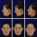
Life-sized photographs are useful in surgical planning for any rhinoplasty, but can be particularly useful in the patient with a crooked nose ( Fig. 1 ). Standard rhinoplasty views are essential, but views from above can be useful in visualizing and recording the deformity in a patient with a twisted nose ( Fig. 2 ). Furthermore, for the frontal view, an overhead light source is preferred to illumination by a flash from the camera or from flash umbrellas, because an overhead light source will accentuate the visibility of the deviation seen in the photographs, as it more closely duplicates the way sunlight or overhead lighting falls on the nose and casts shadows.
Radiographic imaging is usually not needed except when patients also suffer from allergy and/or paranasal sinus disease, computed tomography scanning can identify whether inflammation of the mucosal lining of the sinuses is present or not. Furthermore, in patients with facies that seem to have some degree of abnormal growth and development, imaging can identify anatomic variations of the facial bones including variations of the nasal bones and turbinates ( Fig. 3 ).
Evaluation of the patient
A complete history and physical examination are necessary in any patient in whom surgical correction of the twisted nose is being considered. Although the nasal surgeon’s primary focus is the nose and breathing, all other relevant data needs to be uncovered before offering surgery, including information about the patient’s cardiovascular health, a personal or family history of diabetes or glucose intolerance, history of tobacco and alcohol use, history of easy bleeding or bruising, and history of the patient’s psychological or psychiatric issues.
The surgeon should determine when the nose became crooked, and should know if gradual twisting occurred as the patient matured from a child to an adolescent and then to an adult, or if there was a more immediate and sudden cause of the deformity, such as with a facial or nasal trauma or from a previous surgery. The surgeon should understand if the patient’s expectations are limited to improvement in breathing and straightening the nose, or if other aesthetic goals need to be addressed at the same time. The patient should be questioned about associated symptoms such as epistaxis, rhinitis, and congestion and their response to various pharmacologic interventions. The physician should ask whether there is a seasonal nature to the patient’s complaints of congestion, whether it seems to be worse at various times of the day, and whether the patient routinely uses nasal steroid sprays or sympathomimetics (which can cause rebound). The patient should be asked whether breathing is more impaired during exertion or exercise and whether deep breathing through the nose exacerbates the symptoms.
The patient’s general physical examination is usually performed by the primary care physician, however it is essential that the rhinoplasty surgeon examines not only the external and internal nose but also the nose in relation to the entire face. This skill must become second nature and the surgeon must be able to identify subtle nuances of facial form and symmetry. Even if the patient only desires surgical correction of a crooked nose, balance and symmetry of the facial aesthetic subunits must be evaluated separately and in relationship to the nose.
A systematic examination of the nose should occur in each patient contemplating rhinoplasty. Beginning with the upper third of the nose, the surgeon should examine the nose, the position and depth of the nasion, the position, length, and symmetry of the nasal bones, and the thickness of the skin overlying the bones. As the examination moves caudally, the surgeon determines whether there seems to be deficiency, collapse, or disarticulation of the upper lateral cartilages. Examination should determine if there is deviation of the entire nose or if it is primarily cartilaginous in nature.
Thereafter, the tip is examined and the surgeon notes whether it appears to be in the midline or not. Tip support, rotation, projection, bulbosity, symmetry, width of the lateral crura of the lower lateral cartilages, nostril shape, and the columella are assessed. The tip should be visualized at rest and with respiration, and should be palpated and visually examined. The caudal septum and the position of the anterior and posterior septal angles in relationship to the tip and the nasal spine should be assessed.
Intranasal examination includes a thorough examination of the septum and the turbinates before and after decongestion, and the response of the turbinates to the decongestion is noted. Anterior rhinoscopy alone is rarely sufficient to appreciate the subtle anatomy of the various forms that a septal deviation can assume, and in the case of a severely deviated septum, seeing beyond the crooked septum is often impossible without using a fiberoptic device for visualization. Spurs and alterations from midline should be identified, as should the presence of septal deviations, absence of cartilage from a previous nasal surgery, and the contribution of the dorsal septum to the outward appearance of the nasal dorsum. Examination should address whether or not the angle between the upper lateral cartilages and the septum approaches the ideal value of 9° to 15° degrees and if there is collapse on inspiration. Narrowing of the nasal valve can cause airway obstruction. The size and color of the turbinates should be noted and their potential role in obstruction must be determined. The Cottle maneuver, retracting the cheek skin laterally to see if airflow is improved, is often described as a way to determine whether valve collapse plays a role in airway obstruction; however, using a small curette or small cotton-tipped application to gently lift the nose in the area of the internal and external nasal valves is a much more precise technique for determining whether valve support is lacking. Deep inspiration with alar collapse indicates a weak external valve.
Photodocumention is essential and serves several functions. It allows the surgeon to precisely record visual abnormalities that can never be fully described in narrative fashion. It is a tool that aids in diagnosis and planning. With the help of digital manipulation, it may assist the patient in understanding what their nose may look like after the procedure and it may assist the surgeon in making sure the patient’s expectations mirror the surgeon’s plans. Photographs document the progress of healing and recovery and can be ultimately used as a tool for surgeon self-assessment and teaching. In cases of trauma or assault, photographs may play a medico-legal role.
Life-sized photographs are useful in surgical planning for any rhinoplasty, but can be particularly useful in the patient with a crooked nose ( Fig. 1 ). Standard rhinoplasty views are essential, but views from above can be useful in visualizing and recording the deformity in a patient with a twisted nose ( Fig. 2 ). Furthermore, for the frontal view, an overhead light source is preferred to illumination by a flash from the camera or from flash umbrellas, because an overhead light source will accentuate the visibility of the deviation seen in the photographs, as it more closely duplicates the way sunlight or overhead lighting falls on the nose and casts shadows.