Over the last 2 decades, many of the difficulties in shaping primary tips and rebuilding destroyed secondary tips have been solved through the use of tip sutures and grafts. Dorsal grafts, which are a highly visible determinant of the nasal profile and contour, have become the greatest challenge in rhinoplasty surgery. This article reviews the author’s different approaches to dorsal grafts using fascia and diced cartilage, either separately or in combination.
Over the last 2 decades, many of the difficulties in shaping primary tips and rebuilding destroyed secondary tips have been solved through the use of tip sutures and grafts. Dorsal grafts have become the single greatest challenge in rhinoplasty for many reasons. Dorsal grafts are a highly visible determinant of the nasal profile and contour, as well as being technically demanding with a high complication rate. Due to these challenges, most surgeons try to avoid using a dorsal graft whenever possible, sometimes to the detriment of the final result. Over the last 7 years, the author’s approach to dorsal grafts has dramatically altered because of surgical advances using fascia and diced cartilage, either separately or combined. Thus, a new era in rhinoplasty has emerged with the advent of “The Designer Dorsum.”
Overview
Ever since rhinoplasty began, a wide variety of dorsal grafts have been used with respect to composition and indications. Early grafts were composed of virtually any material available, including ivory, gold, paraffin, duck bone, and glass. More recently, modern chemistry has brought in silicone, Gore-Tex (W.L. Gore & Associates Inc, Flagstaff, AZ, USA), Medpor (Porex Surgical Inc, Newnan, GA, USA), and a plethora of new fillers. The problem lies in the selection of the best material for an individual patient. In deciding which material to use, the critical factors are as follows: recipient site requirements, technical difficulty, resistance to infection, and both short- and long-term complication rates. When these factors are taken into consideration, autogenous grafts are clearly superior to alloplastic ones. Even so, the most common autogenous grafts used for dorsal grafts had technical problems rather than viability or compatibility difficulties, which forced surgeons to develop newer techniques.
Septal Cartilage
Septal cartilage is the gold standard of autogenous grafts in rhinoplasty, but its use as a dorsal graft has declined for 2 reasons. In many primary cases among Asians and also most secondary cases, septal material for a full-length dorsal graft (35 × 8 mm) is not available. Other challenges are the requisite height of the dorsal graft, and whether one stacks or bends the septal cartilage. Visibility is a major factor, at least short-term in thin skin patients, and long-term in patients with even an average skin envelope.
Conchal Cartilage
As championed by Sheen and Sheen, auricular cartilage has been used extensively in nasal surgery. When it is used as a dorsal graft, surgeons have adopted the method of either suturing or layering to overcome its intrinsic curvature. Despite excellent results early on, the limitations of the conchal cartilage have become disappointingly apparent to patient and surgeon with time. The layered conchal graft requires dove-tail techniques to achieve length, and stacking to obtain height. Conchal grafts can be sutured transversely to increase their height, but their lateral junction with the underlying dorsum is always visible. Also, as the skin envelope tightens the intrinsic irregularity and asymmetry of the concha become more visible.
Rib Cartilage
Costal cartilage has been the material of choice for nasal reconstruction and repair of complex secondary rhinoplasty deformities. The primary challenge is the prevention of warping of solid costal cartilage grafts. The basic principle is to do “balanced cuts” to offset contracting forces once the peripheral cartilage has been excised during the shaping process. Although conceptually simple, warping will occur. Gunter and colleagues inserted a K-wire into the center of the graft to prevent warping. Although probably effective, the reality is that the K-wire has a 10% morbidity ranging from extrusion to infection. Additional challenges of solid dorsal grafts include technical factors (misalignment, radix fullness, abnormal shape) and long-term issues (warping, visibility through thinned-out skin). Due to its inevitable absorption, cadaver costal cartilage is not recommended except in extenuating situations (older patient, ethnicity).
Designer dorsal grafts
If all the classic autogenous techniques have major limitations, what is the solution? The answer is to have a highly flexible technique that can be designed to fit a specific defect, rather than inserting the same type of dorsal graft into a wide range of deformities. In the last 7 years, the author has used a combination of fascia and cartilage to fabricate a wide range of dorsal grafts. These grafts can be classified in several ways. Based on composition, there are 4 types: (1) fascia, (2) diced cartilage (DC), (3) diced cartilage beneath fascia (DC + F), and (4) diced cartilage encased in fascia (DC − F). The shape of the grafts can vary widely to fit the defect with respect to length (partial or full-length) and thickness (1–8 mm, uniform thickness or tapered). Dermis grafts are used when the overlying skin envelope is damaged. Each of the specific grafts is discussed in detail.
Fascia (F)
Fascia is the ultimate material for concealment under thin skin and subtle augmentations in the 1- to 2-mm range. The donor material of choice is deep temporal fascia for 4 reasons: (1) the fascia is ideal in thickness and pliability; (2) the donor site is hidden in the hair; (3) survival and resistance to infection is a virtual certainty; and (4) surgical morbidity is minimal. All alternative sources have limitations. Superficial temporal fascia is too thin, whereas tensor fascia lata leaves a visible scar. Rectus fascia, removed during a rib harvest, is too thick and noncompliant. Cadaver fascia (Tutoplast [IOP Inc, Costa Mesa, CA, USA]) is acceptable, but introduces the problems of cost, and the uncertainty of nonviable material.
Perichondrium, removed during a rib harvest, is considered by some to be an equivalent to fascia but is different as regards thickness, pliability, and perhaps, postoperative thickening.
Applications
The most common application of fascia is reducing dorsal visibility under thin skin in primary and secondary cases, providing concealment and preventing “shrink wrappage” of thin skin. However, it is not a substitute to a smooth underlying osseocartilaginous dorsum. Actual augmentation depends on the number of layers used: 1 layer (0.5 mm), 2 layers (1.0 mm), or 3 layers (1.5 mm). The fascia is pinned to a silicone block and folded as necessary. The free edge is sutured with a running 4-0 plain catgut suture. Two 4-0 plain catgut sutures on small straight needles (ST-1) are placed at the cephalic end, and then used to guide the graft into the dorsal pocket. The graft is then smoothed, and the caudal end is sutured at the anterior septal angle.
Problems
Fascia grafts have had surprisingly few problems. Displacement, visibility, and overgrafting are not issues. However, additional swelling is an important expectation associated with fascia. Also, the patient should “compress” the graft and not massage it, because shearing leads to more fluid accumulation, not less. Rather, the concern should be directed more toward insufficiency Donor site problems are minimal, and include the occasional scar revision (2 in 428 cases) or hematoma (1 in 428 cases).
Diced Cartilage (DC)
Since the 1930s, the viability and effectiveness of DC grafts have been validated in facial and cranial reconstruction. The fundamental concept is that cartilage can be cleanly cut into small pieces and placed into the recipient site, where fibrous tissue creates a semi rigid larger graft. Because the grafts are cleanly cut, the pieces survive. The cartilage is not morselized, bruised, or crushed because this would lead to unpredictable survival. Dicing is usually done using #11 blades to cut it into tiny pieces less than 0.5 mm in length. The pieces should be a fine “paste” and small enough to spurt out of a tuberculin syringe. The grafts are placed into the recipient bed using either an elevator for small areas or a tuberculin syringe for larger areas. It is important that the recipient area is not overgrafted—what you see at the end of the operation is what you get postoperatively.
Applications
It is important to distinguish how one uses DC. Intrinsically, it is used as a filler material rather than a “structured graft.” The author uses DC either as a volume or site filler. As a volume filler, DC is placed on the sides of dorsal rib grafts or in peripyriform depressions to alleviate the pyriform contracture that occurs in the cocaine nose. As a site filler, DC can be placed in the infralobule to create a round tip or hide bifidity, but it does not create tip definition. It can be extremely useful for filling dorsal depressions as a closed approach, especially in revision cases.
Problems
If one avoids overgrafting, there are few problems with DC. Absorption is not an issue, but visibility can occur in a thin-skin nose. As regards dorsal corrections, the biggest challenge is decision making. One should never confuse a limited “filler concealment” with secondary rhinoplasty. Many surgeons and dermatologists show results of fillers placed in secondary cases emphasizing what can be done, but do not acknowledge the associated problems or limited benefits. Because DC does not have the risks of absorption or infection, it can be conceptualized as a “permanent autogenous filler” that has its advantages and disadvantages. However, the results would be limited improvement, rather than a major functional and aesthetic change that a complete secondary rhinoplasty can provide. The typical patient would probably be the facial rejuvenation recipient, who would accept an incidental, incomplete improvement in his or her nasal appearance.
Diced Cartilage and Fascia (DC + F)
DC + F grafts have the same standard harvest and preparation as fascia and DC grafts. Once the graft materials are ready, a 2-stage insertion is done. The fascia is inserted first, then the DC is placed underneath until the desired contour is achieved. Essentially, the fascia keeps the DC from being visible.
Applications
DC + F grafts provide a true major volume graft in the radix, and a fine type of dorsal augmentation. The author uses DC + F grafts to correct deep hypoplastic radix area deficiencies. Because the purpose of the fascia is to conceal rather than augment, the amount used is less than that of the standard radix fascia graft. The fascia is inserted and then elevated with an Aufricht retractor; this is followed by placement of the DC against the bare bone that has been stripped of its periosteum to promote fusion with the cartilage. The area is not overcorrected, and is filled precisely. DC + F grafts for the dorsum are done for subtle contour improvements, and not for major augmentations. A single- or double-layer fascia graft is inserted and elevated, and small amounts of DC are placed as “contour fill.” Because the cartilage can be easily displaced, this maneuver is often the last step before closure.
Problems
The most common problems are either technical or judgmental errors. In the radix area, one can misjudge the amount of cartilage in either direction or dissect the pocket too laterally, either of which can result in a bulge. The primary risk is displacement and visibility of cartilage pieces over the dorsum. In selected cases, these pieces can be fragmented using a #16 needle in the examination room.
Diced Cartilage in Fascia (DC − F)
When major augmentations are required, the DC is placed in a fascial sleeve. This sleeve acts as a container but also promotes vascularity, and minimizes visibility under thin skin.
The “construct” graft is made to measure on the back table and then placed carefully into the recipient bed, where its volume can be adjusted in situ.
Applications
The range of applications is dramatic. When a combined radix/upper dorsum graft is required, the fascia is pinned to a silastic block, the DC is placed on top, and the fascial edges are sutured together to create a “bean bag” graft. In the recipient site, placing additional DC(s) deep to the fascia against bare bone can increase the volume of the graft. The graft can be reduced in size by “milking out” cartilage from the graft through a small separation of the suture line using the suction cannula. The dramatic flexibility of these grafts is shown in the range of dimensions for dorsal grafts, with thickness being tapered to uniform thickness, height from 2 to 8 mm, and any desired length. Custom-made dorsal grafts are easily created using the DC − F technique. The steps are as follows:
- 1.
A large sheet of deep temporal fascia (4 × 2 cm) is folded transversely and pinned to a silastic block to create a “tube.” The free longitudinal edge is sutured with a running 4-0 chromic suture.
- 2.
The cephalic end is closed with 4-0 plain sutures on small straight needles, which are left in place to facilitate percutaneous placement.
- 3.
The DC is then placed from the caudal end. When a uniform graft is required, the author uses a tuberculin syringe filled with compressed DC. The syringe is placed against the closed cephalic end, and the DC is injected as the syringe is removed. The nondominant hand is used to mold the graft to create the desired contour. When a tapered graft is required, the volume of DC is carefully controlled to fit the recipient site defect.
- 4.
The graft is guided into the recipient site using the percutaneous sutures and elevation of the skin envelope with the Aufricht retractor. The graft is carefully placed over the dorsum, and carefully inspected for edges and volume. Any excess volume is milked out of the caudal end.
- 5.
Once the surgeon is satisfied, the graft is sutured, closed, and fixed to the dorsum near the anterior septal angle.
Problems
As long as one avoids using DC − F grafts for structural support, graft problems are minor, and their frequency diminishes as one advances on the learning curve. A common error is to place the graft too high and blunt the radix. Kelly avoided this by using a distinct fascial radix graft above the DC − F graft.
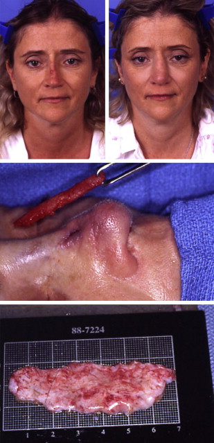
Dermis
When the skin envelope is severely thinned out or attenuated, then a dermal graft is the solution. The objective is not to achieve major augmentation, but to thicken and normalize the skin envelope. These cases are divided into 2 groups: the infected nasal implant, and the multiply operated nose with a crucified skin envelope. For the latter, the first stage is to insert dermis to allow safe elevation of the skin envelope at the second stage of reconstruction ( Fig. 1 ). In contrast to Sheen and Sheen, who worried about dermis grafts surviving a possible infection, the author routinely uses dermis when low-grade infection is present because of an extruding alloplastic implant. The implant is removed, the capsule excised, and the wound profusely irrigated with antibiotic solution. The dermis graft is excised from the suprapubic area, often using a Cesarean section scar. The area (as big as 14 × 4 cm) is de-epithelialized first, and then removed at the subdermal plane leaving the fat behind. The graft is turned over, and any hair follicles present are carefully removed. The dermis is guided into the wound with percutaneous sutures. In most cases, the dermis is placed in multiple layers and even “stuffed” into the partially closed incision. The nose is overgrafted because the amount of survival is unpredictable. Thus, until now, dermis grafts have been part of the solution, and not of the problem.