In this review, the complications of rhinoplasty are examined in terms of their timing of presentation. An algorithmic approach to postoperative problems is discussed. Complications can frequently be avoided by meticulous technique, recognition of pitfalls, and early attention to perioperative morbidity. Reoperative rates can be minimized with good patient education and proper command of the postoperative situation, so that unnecessary procedures are not undertaken.
The aesthetic surgeon who wishes to reshape the nose to improve function and aesthetics must be thoroughly familiar with the consequences of these operations. There are numerous complicated maneuvers that are performed during the rhinoplasty sequence; each one can contribute to a successful outcome or an unwanted problem.
Patient education and the setting of appropriate expectations is the first step in decreased morbidity and reoperation rates in rhinoplasty. Education must focus on the goals and limitations of the procedure. A prepared patient will understand the difference between postoperative morbidity and a true problem.
The preoperative evaluation must be precise; anatomic analysis guides surgical goals. Previous medical history is important, because the perioperative use of medications such as steroids and vasoconstrictors is not without complication and interaction. Routine follow-up appointments must be scheduled to ensure patient confidence and to intercept early signs of complications. The patient should be told to purchase a bottle of oxymetazoline nasal (Afrin) or over-the-counter oxymetazoline spray and have it ready at the bedside postoperatively to facilitate early outpatient management of bleeding. An important part of patient education is preparation for this management of bleeding. Optimal postoperative care is facilitated by having the patients’ contact information and keeping in touch with them in the early postoperative period.
In this review, the complications of rhinoplasty are examined in terms of their timing of presentation. An algorithmic approach to postoperative problems is discussed.
Intraoperative complications
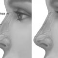
Intraoperative injuries to the nasal tissues are uncommon and manageable when encountered. Fracture of the septum or the L-strut left after septoplasty is uncommon, but possible where extensive septal harvest has weakened its structural integrity. A recent review demonstrates that this occurs in about 1% of cases. This history and the following findings on physical examination warn the surgeon of this possibility: (1) secondary cases where a previous hump reduction was performed, (2) short nasal bones with a dorsal hump, (3) the severely deviated nose with a deviated septum, and (4) severe fracture or comminution of the nasal bones or septum. Significant hump removal and septoplasty with osteotomies can lead to instability; the surgeon should be vigilant in cases where these maneuvers are required. When septal fracture is recognized, continued exposure must be cautiously exercised, as intact mucoperichondrium helps in stabilization of the fracture segments. Ultimately, fractures can be treated through several methods: (1) suture fixation of the septal parts, using the mucoperiosteum as support, (2) direct suture fixation of spreader grafts to the dorsal strut, (3) direct suture fixation to the dorsal osseocartilaginous junction, (4) percutaneous Kirschner wire (K-wire) fixation of the nasal bones to the L-strut, or any combination of sutures, grafts, and K-wires needed to maintain central structural stability.
Cribriform plate fracture can occur in patients with a history of trauma. In these patients, force should be used judiciously when dissecting the septum and performing dorsal osteotomies. Osteotomies can also result in skin tears if the osteotome is not thoughtfully guided during tapping. Also, an aggressive lateral osteotomy can enter the maxillary sinus or the lacrimal drainage system and cause several problems, such as enophthalmos, silent sinus syndrome, or mucocele formation.
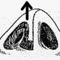
Nasal bones can collapse because of overaggressive infracturing or misplacement of an osteotomy. Failure to correct collapse results in a poor aesthetic appearance with visible asymmetry and palpable deformity. Adjacent internal valve collapse negatively affects the airway. If this is noted, an elevator should be used to replace the bones to their appropriate position, much as one would perform a closed reduction of a nasal fracture. Internal and external splinting should be used postoperatively.
Immediate postoperative complications
Bleeding
Bleeding is the most common immediate complication. A thorough preoperative history will reveal any medications and herbal supplements that may put the patient at risk for postoperative bleeding. Aspirin should be discontinued 10 days before the procedure, as should any herbal supplements. Several clinical pearls have been useful in decreasing bleeding in our hands: (1) appropriate preoperative injection using local anesthetic mixed with epinephrine into the subcutaneous tissue and topical Afrin or cocaine on the mucosa, with adequate time given for the onset of these drugs; (2) dissection in the correct tissue planes, that is, maintaining dissection in the submucoperichondrial plane, where appropriate. If dissection is too superficial, it will reach the nasal superficial musculoaponeurotic system (SMAS), which is well vascularized, resulting in bleeding and subsequent irritation of the nasal soft tissue.
Besides the use of bipolar cautery for hemostasis, bleeding can be minimized by not packing the nose at the end of the operation, as this can lead to some bleeding with removal. Goldwyn retrospectively reviewed 780 patients who underwent elective rhinoplasty, and demonstrated that there was a 3.6% incidence of excessive bleeding and a 0.9% incidence of severe bleeding. Bleeding was managed successfully by repacking the nose in the short term, cauterizing a bleeding site, or suturing an open area in the incision or at the columella-septum junction. No specific factors were statistically associated with the incidence of bleeding. However, other surgeons have demonstrated a significant benefit to packing the nose. Guyuron and colleagues demonstrated in a randomized prospective study that packing the nose was superior to a septal quilting stitch-only, in terms of airway improvement, residual deviation, and drip pads used. Guyuron and both of the senior authors of this article (J. G. and S. S.) prefer soft Silastic Doyle splints when splinting is deemed necessary.
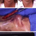
Mild epistaxis is easily managed with head elevation and digital pressure; for this reason, it is not of concern to the plastic surgeon. However, bleeding can be greatly distressing to the patient, and for this reason, the patient should be able to get in touch with the surgeon with great ease. The surgeon should have an algorithm for the situation whereby the patient calls emergently with the complaint of postoperative bleeding, as this will occur in a busy rhinoplasty practice. Early light epistaxis can be managed by either oxymetazoline (Afrin, Schering-Plough HealthCare Products Inc, Memphis, TN, USA), or ice with digital pressure, or a combination of both. The key is to allay the patient’s fears and inquire as to the amount and color of the blood. Color is key, as patients consider even a drop of blood to be a great deal. If the blood is bright red, and bleeding is persistent despite ice and digital pressure at the nasal root, or oxymetazoline twice into each nostril and elevation of the head, the patient is instructed to come into the emergency room for immediate evaluation. In the emergency room, the head should continue to be elevated, and the patient’s blood pressure should be checked. The bottom line in this situation is that the plastic surgeon must intervene and stop the bleeding. The bleeding source should be observed; this observation is facilitated by removing all clots. Quick Relief (Biolife, L.L.C., Sarasota, FL, USA) may be considered for minor epistaxis located more anteriorly. Epinephrine-soaked cottonoids should be inserted to minimize bleeding so that a thorough and direct examination can be performed. If anterior packing does not work, then posterior packing with a Foley or Rhino Rocket (Shippert Medical Technologies Corporation, Centennial, CO, USA) is the next step ( Fig. 1 ). The Rhino Rocket is inserted and maintained for 24 to 48 hours and should be coated with Bactroban ointment (GlaxoSmithKline PLC, Brentford, Middlesex, UK) to ease its slide into the nostril. Ice packs should be applied, and the nose should be elevated. Activity must be limited. The patient should be educated concerning optimal care for the period that the Rhino Rocket is in place: this includes saline spray to the ends of the rockets every 2 hours while awake to keep them moist; 1 to 3 Afrin sprays every 12 hours in each nostril; and broad-spectrum oral antibiotics to prevent the development of toxic shock syndrome. When the rockets are pulled, they should be pulled a little bit at a time to prevent rebleeding. The surgeon should pull just hard enough to move the rocket a millimeter, and then wait 10 minutes; advance about 1 cm every 10 minutes until it is completely removed. Once it is out, 3 puffs of Afrin should be administered in each nostril. Afrin should be continued for 3 weeks postoperatively.