Introduction
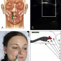
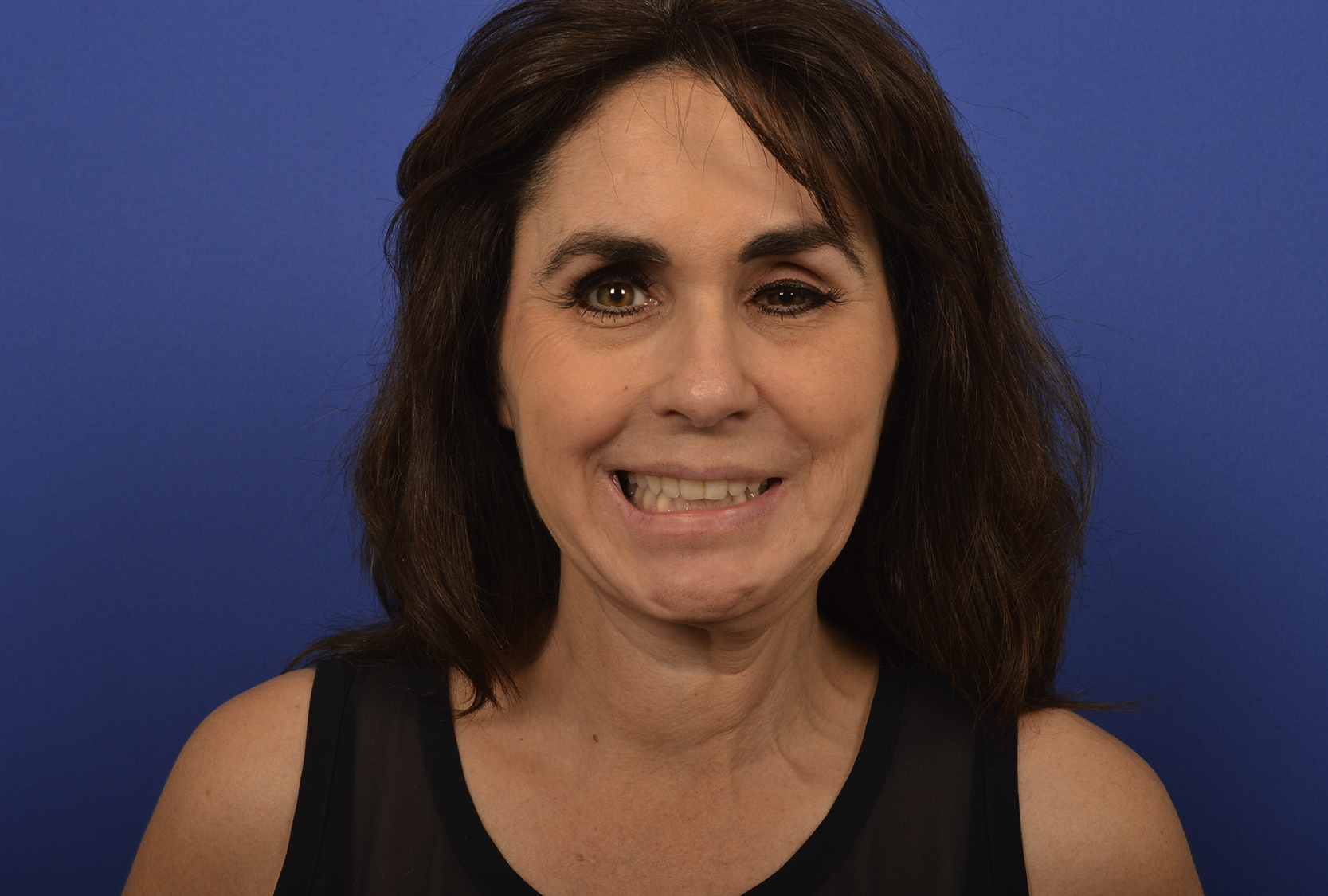
Features of a natural smile include spontaneity, symmetric simultaneous superolateral excursion of the oral commissure, appropriate orientation of the nasolabial fold, and symmetrical upper and lower teeth show. , These components are compromised in patients with facial palsy and almost all facial reanimation options do not obtain a significant number of these key features of a normal smile. The only way to attain these components is to maintain the entire neural pathway of facial expression. Unlike complete flaccid facial paralysis, patients with post–facial paralysis with synkinesis (PFPS) have functional smile muscles with completely intact neural input ( Fig. 11.1 )
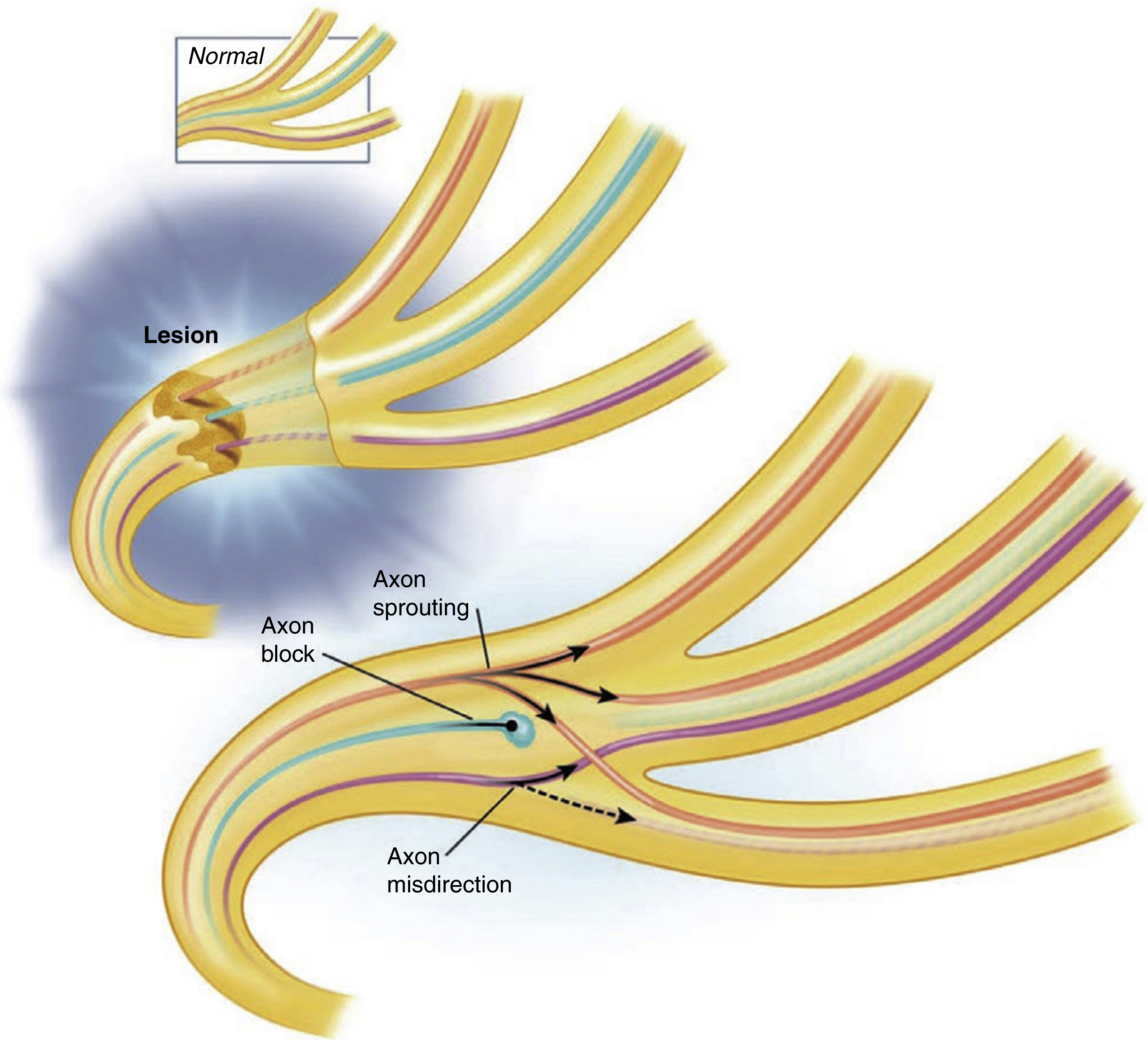
Synkinetic patients have smile dysfunction due to overactivity and uncoordinated movement of counterproductive muscles such as platysma, buccinators, depressor anguli oris (DAO), and orbicularis oris. The most supported theory of PFPS is aberrant facial nerve regeneration where proximal axons reroute, sprout, and/or degenerate, leading to abnormal reinnervation of both correct and inappropriate muscles ( Fig. 11.2 ).
The aim of treatment is always to improve cosmesis and spontaneous smile function as well as functional deficits such as oral incompetence and cervicofacial tightness. There are a number of surgical and nonsurgical management approaches used at present to treat synkinesis, including neuromuscular retraining, chemodenervation, and surgery. These options are complementary and synergistic and in the authors’ experience, together yield the optimal outcome for individuals with PFPS. , This chapter will focus on surgical options for smile reanimation as other sections of the book discuss nonsurgical and eyelid management.
Masseteric-Facial Nerve Transfer
Masseteric-facial nerve transfer for reinnervation of patients with an irreversible complete facial paralysis of less than 2-years duration has gained popularity ( Fig. 11.3 ). “Supercharging” of the facial nerve with the masseteric nerve has also been proposed for individuals with post-paralysis with synkinesis to increase the voluntary excursion of oral commissure.
Although this approach can theoretically serve a role in improving the oral commissure excursion, the masseteric nerve does not provide a spontaneous and involuntary smile mechanism for patients with synkinesis.
Furthermore, the underlying factors that lead to synkinesis—specifically the overactivity of buccinator, DAO, platysma, and orbicularis—are not addressed with this technique.
Static Sling
Static support of facial structures with tensor fascia lata is perhaps one of the most commonly used nondynamic reanimation techniques for facial paralysis ( Figs 11.4 and 11. 5 ).
Although this approach can be a useful tool in patients with long-standing flaccid paralysis, its utilization for synkinesis is less common. In fact, for patients with severe synkinesis who have contracted musculature, static sling can cause increased tightness and less mobility of the zygomatic major. In the senior author’s (BA) experience, this technique can improve function and symmetry in patients with flaccid paralysis and non-synkinetic partial paralysis who have ptotic oral commissure and effaced nasolabial folds.
Gracilis Free Functional Muscle Transfer
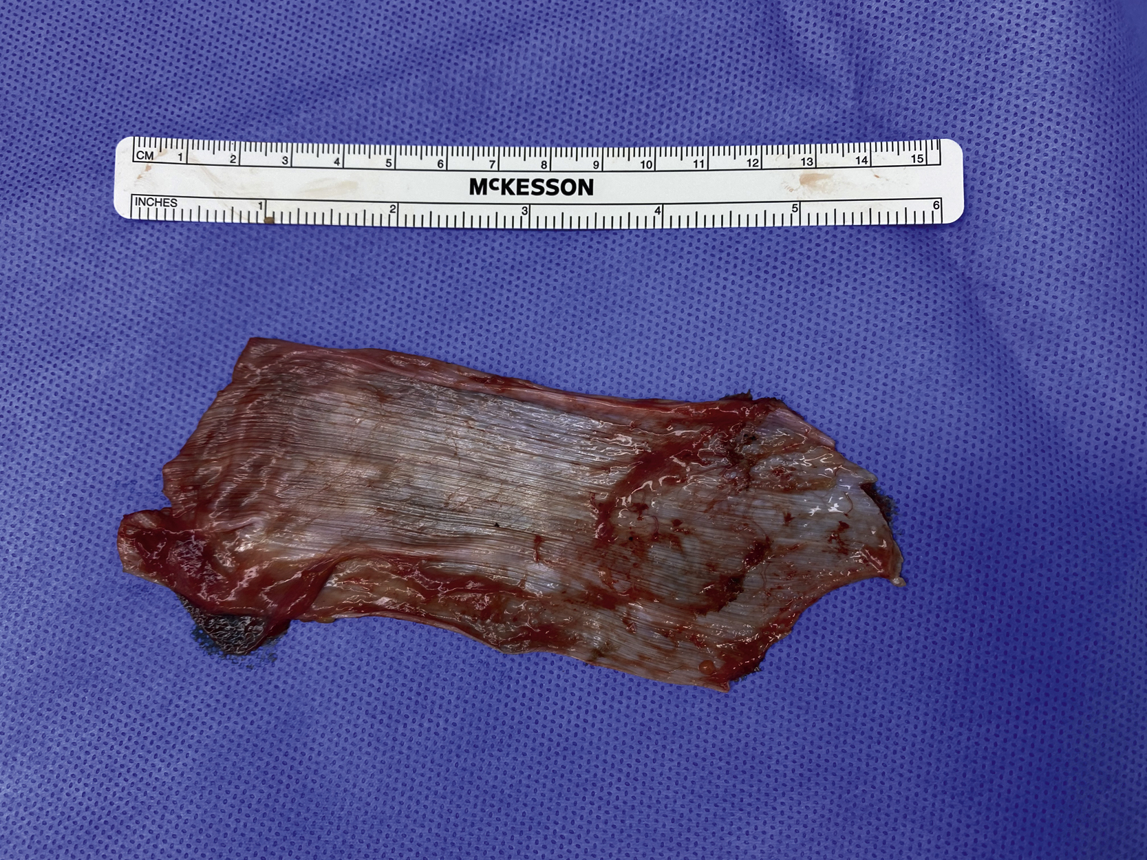
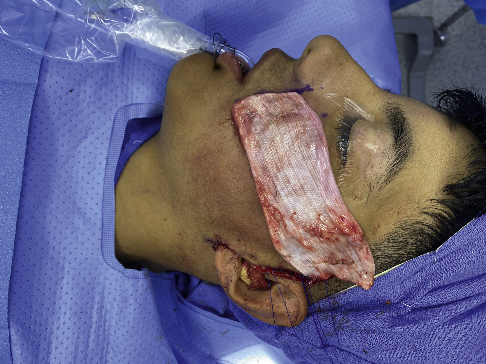
Since the 2000s, the gracilis free functional muscle transfer has become one of the most advanced facial reanimation techniques for pediatric and adult patients with unilateral or bilateral facial paralysis ( Figs 11.6 and 11.7 ). The gracilis muscle is most commonly innervated by a contralateral sural nerve graft and/or ipsilateral masseteric nerve.
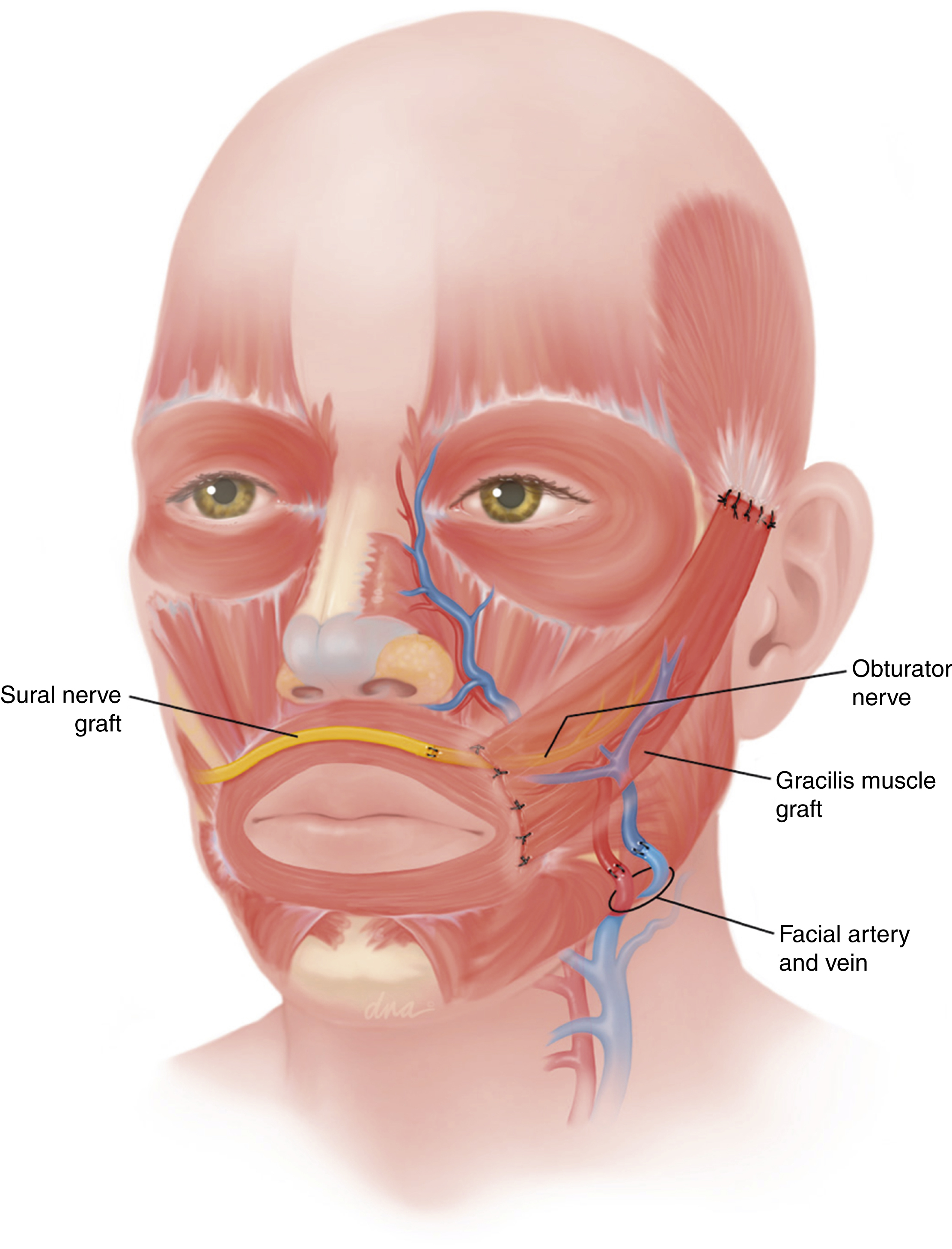
Although cross face nerve grafts improve spontaneity, studies have shown that the masseteric nerve can enhance excursion of the muscle. , Given its popularity and excellent overall outcomes in patients with flaccid paralysis that lack any functional smile muscles, the utilization of the gracilis muscle has also become extremely prevalent for patients with synkinesis.
As discussed previously, patients with synkinesis have active zygomatic muscles and the main factors that cause smile dysfunction are overactivity and uncoordinated movement of counterproductive muscles such as the DAO, platysma, and buccinator muscles. Gracilis muscle transfer does not truly address the underlying issues. Furthermore, this approach requires multiple procedures and extensive recovery. As a result, gracilis muscle transfer should only be considered if other options do not result in a satisfactory outcome.
Modified Selective Neurectomy
Traditional surgical treatment of synkinesis has focused primarily on augmenting and increasing the power of the elevators of the lip and oral commissure. Although there are various dynamic surgical procedures that focus on the upward excursion of oral commissure in PFPS such as masseteric-facial nerve transfer, orthodromic temporalis tendon transfer and gracilis free functional muscle transfer, they do not address the underlying factors that lead to smile dysfunction in this patient population. As described earlier in the text, the underlying issue with PFPS is not the lack of power of the zygomatic muscles but rather the overactive and untimely activation of counterproductive facial muscles such as the DAO, buccinator, platysma, and orbicularis oris. As a result, we are not dealing with a problem of weakness but rather a problem of overactivity. As a result, if we can reduce the untimely activity of the DAO, buccinator, platysma, and orbicularis oris muscles while preserving the normal function of the zygomatic major/minor, depressor labii inferioris (DLI), and levator labii alaeque nasii, we would be able to obtain a natural well-timed smile.
Although neurotoxins are effective in reducing muscle activity, they are not specific and if injected in the region of the oral commissure, can impact both the zygomatic muscles as well the counterproductive muscles. That is the reason neurotoxins have had great success in treating the narrowing of the eyelid aperture and platysma tightness where there are no other muscles in the vicinity.
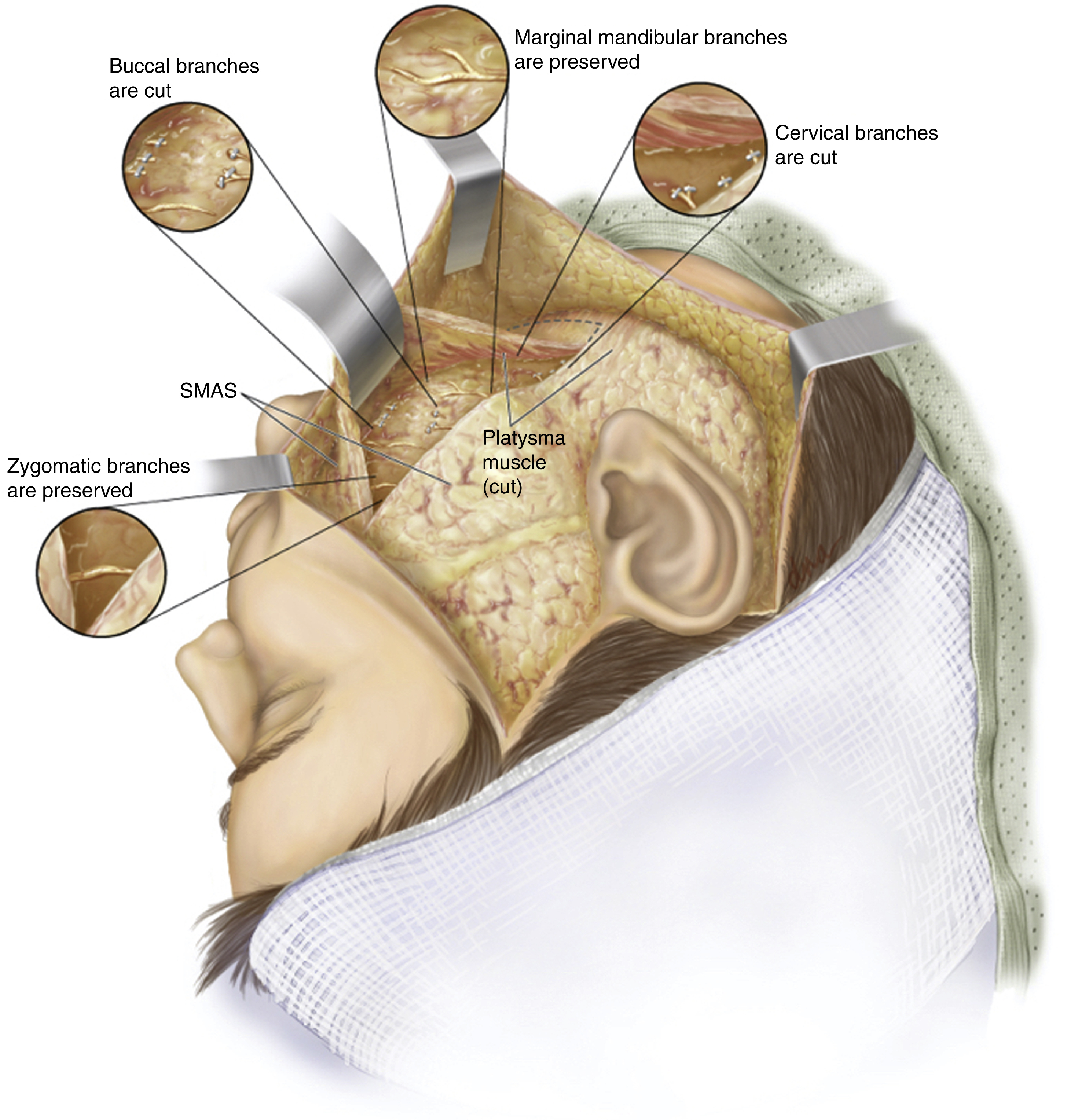
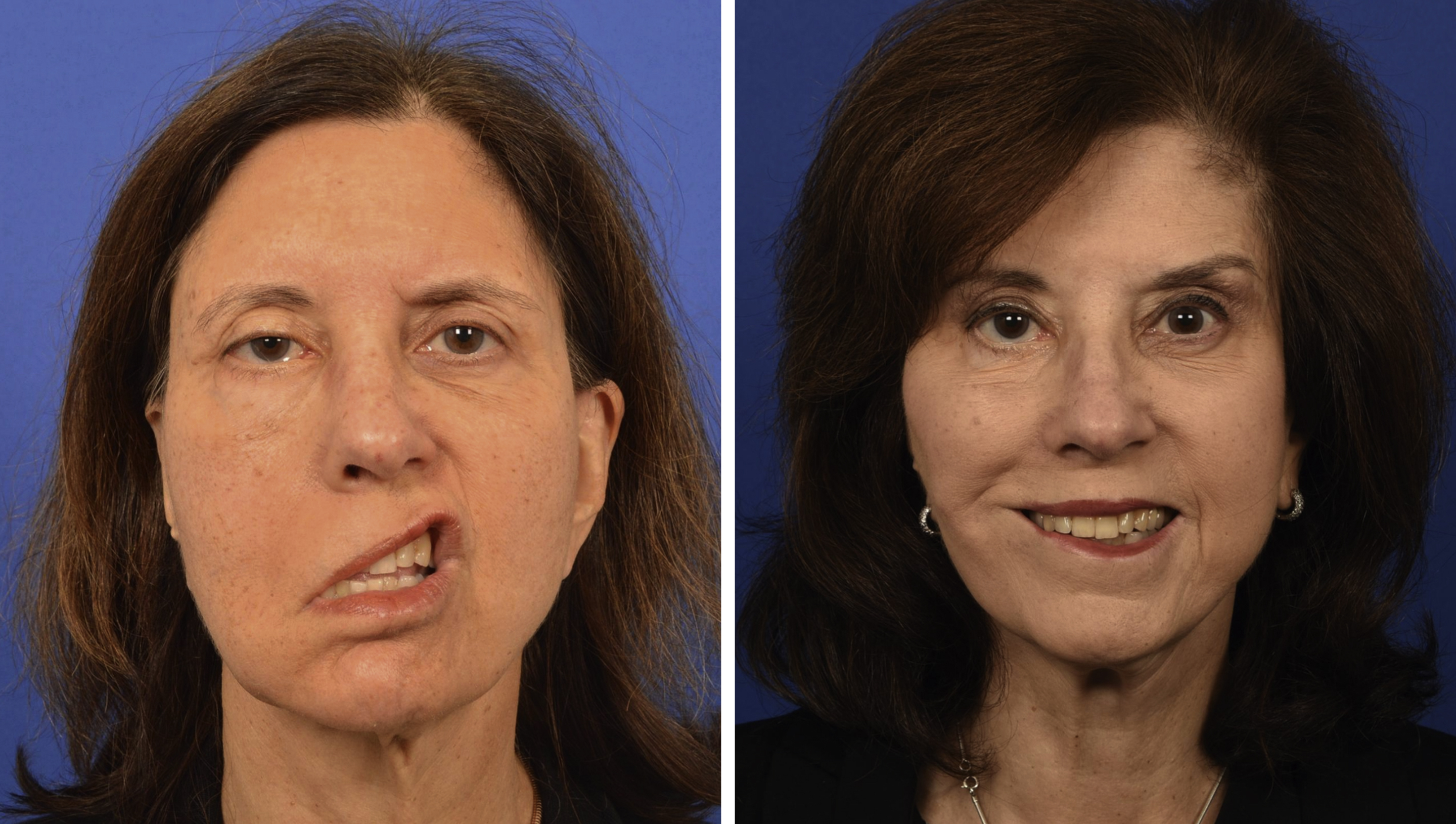
Modified selective neurectomy, pioneered by the senior author (BA), addresses this unresolved problem by selectively transecting the distal nerve branches to the DAO, platysma, buccinator, and orbicularis oris while preserving the innervation to zygomaticus major/minor, levator labii alaeque nasii, levator labii superioris, and DLI, thereby reducing the overactivity of the counterproductive muscles and unlocking the activity of key smile muscles ( Figs 11.8 and 11.9 ). This approach maintains the entire neural pathway for smile mechanism and is the only approach that addresses the underlying issues related to PFPS. As a result, the modified selective neurectomy achieves results rarely seen in traditional facial reanimation procedures by producing a well-timed, natural, spontaneous, and symmetric smile.