This article introduces and discusses several biophysical and cellular modalities that are being tested or used in clinical practice to optimize wound bed preparation, effect soft tissue coverage, and improve the quality of the inevitable and resultant scar. Among these promising technologies is the use of electrical stimulation to mimic a physiologic current of injury in an effort to accelerate re-epithelialization and the wound healing process. Over the past several years an on-site individualized regenerative medicine kit has become commercially available (ReCell, Avita Medical), utilizing well-established laboratory techniques of cell separation without the need for cell cultivation in an effort to expand and promote wound coverage and end result.
- •
Desiccated wounds and wounds exposed to air exhibit a reduced electrical potential. Electrical stimulation mimics the current of injury, in effect restarting or accelerating re-epithelialization and the wound healing process. The use of wireless microcurrent stimulation (WMCS) uses the current-carrying capacity of charged gas molecules, oxygen and nitrogen, to donate electrons to the wound field, transferring a charge to the wound via the air without having to touch or pierce either the wound or surrounding skin.
- •
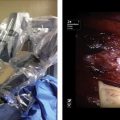
The ReCell (Avita Medical, Cambridge, UK) kit is based on well-established laboratory techniques of trypsinization, cell separation, and filtration. Because no cell cultivation is necessary with this technique, the product is available for use in the operating room during the skin transplantation operation (on-site techniques and 1-step individualized regenerative medicine).
Introduction
Burn and reconstructive plastic surgeons today are faced with an ever-expanding range of life-threatening and complex wound types, each with its own specific pathophysiological features, qualities, and presentations. As experience and expertise evolve, so have new technologies. In this article, the authors introduce and discuss several modalities that are being tested or used in clinical practice to optimize wound bed preparation, effect soft tissue coverage, and improve the quality of the inevitable and resultant scar. Several of these treatment modalities have only recently been introduced to clinical practice and, as such, long-term results may not be available. Every effort has been made to accurately present and reflect these limitations.
Wound bed preparation
Wireless Microcurrent Stimulation
Although first reported more than 150 years ago, the application of electrical field therapy to facilitate wound healing has, over the past decade, experienced a resurgence of interest. Based on several newer clinical reports and the development of noninvasive and better-engineered hardware, this technology may show promise in wound healing and is currently being tested in Europe. This technology and its potential clinical application are founded on the appreciation of existent electrical currents in both intact as well as wounded skin. Skin, depending on which area of the body is tested, possesses an electrical field of between 10 mV and 60 mV. Cell membranes possess a membrane potential, a reflection of the electrical potential difference or voltage across the membrane. Cells within intact skin are negatively charged interiorly whereas the exterior of the cell, the extracellular space, is positively charged. The difference in charge arises because cell membranes possess active and passive pumps that move sodium ions out of the cell in exchange for potassium ions, which are pumped into the cell. For the skin, this results in the epidermis being negatively charged relative to the deeper tissues, which carry a positive charge. When wounded and the epidermal barriers are disrupted, the skin battery is short-circuited and an outward current of approximately 30 μA/cm 2 is generated and can be measured at the wound edge. Since the early 1990s, many reports have described this effect at both the tissue aand cellular levels and applications for wound healing have been proposed.
Electrical currents stimulate several cell activities (eg, DNA synthesis, cell proliferation, cell migration, synthesis of the extracellular matrix, collagen, expression of growth factors, and receptors) depending on the cell type. When wounding occurs, a weak negatively charged electric field establishes between the skin and underlying tissues, termed the current of injury . It is thought that this current persists until the skin defect is repaired and that the healing process is interrupted if the current ceases. Desiccated wounds and wounds that are exposed to air exhibit a reduced electrical potential. Electrical stimulation mimics the current of injury, in effect restarting or accelerating re-epithelialization and the wound healing process.
Electrical stimulation in wound healing is defined as the use of an electrical current to transfer energy to a wound. Studies of stimulation current effects on wound healing using a porcine model identified direct current stimulation as most effective in wound area reduction. Until recently, potential clinical application of these currents required using invasive transcutaneous methods of transferring current through wet electrolytic contact with the external skin surface and the wound bed, 2 electrodes having been required to complete the electric circuit.
A newer method is available that uses the current-carrying capacity of charged gas molecules, oxygen and nitrogen, to donate electrons to the wound field ( Fig. 1 ). This method is known as wireless microcurrent stimulation (WMCS). In so doing, a charge can be transferred to the wound via the air without having to touch or pierce either the wound or surrounding skin, resulting in the current of injury measurable by an amp meter. If a weak direct current electric field is applied to the atmosphere, the naturally produced N 2 + and O 2 − move, the former in the direction of the field and the latter in the opposite direction. If the field strength exceeds a certain value, which at atmospheric pressure is approximately 3 × 10 6 V m −1 (3 MV/m, the breakdown field strength), a few free electrons can be accelerated to high velocities and energies, so that they can knock electrons from the oxygen and nitrogen molecules, creating more positive and negative molecules. If an O 2 − molecule lands on a conductive surface (like the skin), it gives up its negative charge, ceases to exist as an O 2 − molecule, and turns into an oxygen molecule and a few water molecules, a process termed plate-out . In this process the plated-out O 2 − induces a current in the wound cells and fluids whereas O 2 − never enters the body. In so doing, this novel approach creates a current of injury via a noninvasive electrical field that can potentially cover the total surface area of the wound (see Fig. 1 ).
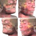
In the authors’ burn center in Germany, WMCS is currently applied in an effort to accelerate the healing of burns wounds. A 1.5-μA current is applied as the present therapeutic basis, and all treatments are performed in sessions of 60 minutes/day per target area ( Fig. 2 ). The return path is connected by either a wrist or ankle strap. Preliminarily the authors have found this modality easy to apply and the noninvasive, noncontact mode of application likely minimizes the risk of bacterial contamination and local trauma. Early results with this technology seem to show measurable improvement in the rate of early re-epithelialization in partial-thickness injuries; further study is under way.
Extracorporeal Shock Waves for Burn Wound Healing
Over the past several years there has been an increasing body of literature addressing the use of extracorporeal shock waves (ESWs) for healing of a variety of wound types, cellulite, and Dupuytren contracture. Several of these articles describe its use in a subset of burn wounds. In a recent article, the University of Barcelona burn unit prospectively examined its use in the treatment of 15 patients who suffered burn wounds deemed at least deep partial thickness in depth as determined by laser Doppler assessment. Wounds were treated in the emergency department on day 3 and again on day 5 postinjury. Although 1 patient was lost to follow-up and 2 patients required skin grafting, the rest healed well without surgery. Low pain scores were noted during treatment and there was a 5% incidence of hypertrophic scarring. Ottomann and colleagues investigated the effect of ESWs on wound healing in partial-thickness wounds and donor sites. In a prospective randomized trial, a group from Berlin compared time to heal after SWT with 100 impulses/cm 2 at 0.1 mJ/mm 2 to donor sites. The investigators noticed a significantly faster (2 days) healing. In a phase II trial with 50 patients with partial-thickness wounds a similar effect with significantly accelerated wound healing was noticed. The investigators concluded that a larger phase III trial is warranted.
ESW therapy (ESWT) is based on acoustic wave energy source. The exact mechanism of action of ESWT is at present ill defined. Many hypotheses with regard to mechanisms of action have been reported, ranging from its modulation of free oxygen radical, to decreasing apoptotic rates, and to increasing the expression of heat shock proteins as well as promotion of various growth factors, including transforming growth factor (TGF)-β1 and insulinlike growth factor 1. The latter two effects, the promotion of transforming growth factor (TGF)-β1 and insulin like growth factor 1 perhaps explain the low incidence of problem scar formation noted. Experimentally ESWT has been shown to stimulate angiogenesis and increase perfusion to ischemic tissues by increasing vascular endothelial growth factor while diminishing early proinflammatory immune response to deep burn injury in mice. Although promising, this form of therapy is in its infancy. Shock wave therapy is currently being evaluated by the Food and Drug Administration and is in phase III trials for diabetic foot ulcers.
Laser for Wound Bed Preparation
Laser (light amplification by stimulated emission of radiation) modalities have been promoted over the past decades as a method to effect controllable excisional débridement of various wound types with the potential additional benefit of decreasing bacterial burden in the wound. Early attempts using carbon dioxide (CO 2 ) ablative lasers for burn wound excision and débridement proved slow and costly and incurred a measure of additional thermal injury, significantly limiting its adoption for general use in burn care. A more positive experience, however, was published in 1999 by Dr Robert L. Sheridan and colleagues, who described their experience with the use of laser ablation in the treatment of 21 children who suffered full-thickness burns. The full-thickness wounds were ablated using continuous-wave CO 2 laser system and immediately autografted. The results were compared with controls. Endpoints included engraftment at 7 days and serial Vancouver assessments of scarring. They identified no problems with bleeding and engraftment rates averaged 94.7% ± 3.5% in the control sites and 94.7% ± 3.3% in the study sites ( P = 1.0). No significant difference was noted in Vancouver Scar Scale scores at an average follow-up of 32.0 ± 5.2 weeks.
Recent publications using animal models of cutaneous chemical injury have also demonstrated efficacy by delivery energy patterns, which incur less residual or surrounding thermal damage. Most promising among them is probably the erbium:yttrium-aluminum-garnet (Er:YAG) laser. Reported benefits are of improved control of depth of excision and/or débridement, allowing differential control to the papillary or upper reticular layers of skin. Accelerated rates of wound healing are suggested but ill defined in several animal studies and thought to result from decreases in bioburden in the wound as well as potential mitogenic effect of low-energy laser stimulation. A clinical case report was published in 2003 by Reynolds N et al in the journal, Burns , describes a single case of mixed partial-thickness and full-thickness burns to the foot successfully managed with the use of an Er:YAG laser. It seems likely that with increasing availability and evolving experience, newer forms of laser energy delivery systems may bring this technology to the forefront of clinical practice for use in acute burn wound management.
Hydrotherapy with Carbon Dioxide for Wound Healing
Balneotherapy, derived from the Latin balneum (bath), has been advocated for the management of various diseases and wound types since at least biblical times. Near continuous hydrotherapy over days was emphasized by Passavant and von Hebra in the mid-nineteenth century for burn wound treatment and in many centers today hydrotherapy remains a mainstay in burn treatment, with the caveat that meticulous sanitary attention to equipment and microbial surveillance are maintained to prevent infectious colonization and cross-contamination. Hydrotherapy is generally used during the initial cleansing of a patient and facilitates a minimally traumatic débridement of loose necrotic tissue during the wound healing process while affording a measure of pain relief resulting from warm water immersion. Additional therapeutic effects can be expected by the addition of different salts or CO 2 in high concentrations to the water.
Various studies have demonstrated a positive effect of CO 2 on microcirculatory parameters of the skin. Karagülle and colleagues reported that immersion into mineral water containing 3500 mg/L CO 2 for 16 minutes increased skin microcirculatory values by 2-fold. Pain threshold levels were unaffected by CO 2 compared with controls.
Further investigations revealed that thermoneutral baths with a CO 2 concentration of greater than 500 mg/L led to a vasodilatation in the skin resulting from precapillary arteriolar dilatation with subsequently improved transcutaneous partial oxygen pressure. A prolonged improvement of the cutaneous microcirculation was recorded for up to 60 minutes post-treatment. A similar effect on the microcirculation of the skin is also observed in dry CO 2 bath where skin areas or extremities are immersed in high concentrations of CO 2 gas (without water). The authors have demonstrated a positive prolonged effect on cutaneous blood flow as measured by laser Doppler imaging. Although the application of CO 2 hydrotherapy has previously been used for the treatment of critically decreased limb perfusion and complex regional pain syndrome, Werner and colleagues investigated the effect of CO 2 on chronic wounds and demonstrated improved healing. Recently, the authors have begun to use this technique in an effort to improve burn wound healing. In the authors’ center, a patient’s wound is immersed into warm water saturated with 500 mg/L CO 2 for 30 minutes. Extremity wounds can be immersed in water containing CO 2 or, alternatively, invested occlusively in a bag, which is then filled with CO 2 gas for 30 minutes ( Fig. 3 ). Caution must be taken with cardiopulmonary unstable patients, and monitoring of cardiopulmonary function is strongly recommended for these patients. The authors hypothesize that the positive effect on microcirculation improves the overall healing process and improves re-epithelialization in skin-grafted wounds, which manifested some measure of graft loss. Preliminary observational evidence suggests that wounds can be improved with this technique. Controlled randomized studies are necessary, however, to demonstrate the positive effect on re-epithelialization of partial-thickness wounds as well as potential salvage of the zone of stasis. In any case, the authors currently consider hydrotherapy with additional CO 2 a helpful adjunct for wound healing.
Wound bed preparation
Wireless Microcurrent Stimulation
Although first reported more than 150 years ago, the application of electrical field therapy to facilitate wound healing has, over the past decade, experienced a resurgence of interest. Based on several newer clinical reports and the development of noninvasive and better-engineered hardware, this technology may show promise in wound healing and is currently being tested in Europe. This technology and its potential clinical application are founded on the appreciation of existent electrical currents in both intact as well as wounded skin. Skin, depending on which area of the body is tested, possesses an electrical field of between 10 mV and 60 mV. Cell membranes possess a membrane potential, a reflection of the electrical potential difference or voltage across the membrane. Cells within intact skin are negatively charged interiorly whereas the exterior of the cell, the extracellular space, is positively charged. The difference in charge arises because cell membranes possess active and passive pumps that move sodium ions out of the cell in exchange for potassium ions, which are pumped into the cell. For the skin, this results in the epidermis being negatively charged relative to the deeper tissues, which carry a positive charge. When wounded and the epidermal barriers are disrupted, the skin battery is short-circuited and an outward current of approximately 30 μA/cm 2 is generated and can be measured at the wound edge. Since the early 1990s, many reports have described this effect at both the tissue aand cellular levels and applications for wound healing have been proposed.
Electrical currents stimulate several cell activities (eg, DNA synthesis, cell proliferation, cell migration, synthesis of the extracellular matrix, collagen, expression of growth factors, and receptors) depending on the cell type. When wounding occurs, a weak negatively charged electric field establishes between the skin and underlying tissues, termed the current of injury . It is thought that this current persists until the skin defect is repaired and that the healing process is interrupted if the current ceases. Desiccated wounds and wounds that are exposed to air exhibit a reduced electrical potential. Electrical stimulation mimics the current of injury, in effect restarting or accelerating re-epithelialization and the wound healing process.
Electrical stimulation in wound healing is defined as the use of an electrical current to transfer energy to a wound. Studies of stimulation current effects on wound healing using a porcine model identified direct current stimulation as most effective in wound area reduction. Until recently, potential clinical application of these currents required using invasive transcutaneous methods of transferring current through wet electrolytic contact with the external skin surface and the wound bed, 2 electrodes having been required to complete the electric circuit.
A newer method is available that uses the current-carrying capacity of charged gas molecules, oxygen and nitrogen, to donate electrons to the wound field ( Fig. 1 ). This method is known as wireless microcurrent stimulation (WMCS). In so doing, a charge can be transferred to the wound via the air without having to touch or pierce either the wound or surrounding skin, resulting in the current of injury measurable by an amp meter. If a weak direct current electric field is applied to the atmosphere, the naturally produced N 2 + and O 2 − move, the former in the direction of the field and the latter in the opposite direction. If the field strength exceeds a certain value, which at atmospheric pressure is approximately 3 × 10 6 V m −1 (3 MV/m, the breakdown field strength), a few free electrons can be accelerated to high velocities and energies, so that they can knock electrons from the oxygen and nitrogen molecules, creating more positive and negative molecules. If an O 2 − molecule lands on a conductive surface (like the skin), it gives up its negative charge, ceases to exist as an O 2 − molecule, and turns into an oxygen molecule and a few water molecules, a process termed plate-out . In this process the plated-out O 2 − induces a current in the wound cells and fluids whereas O 2 − never enters the body. In so doing, this novel approach creates a current of injury via a noninvasive electrical field that can potentially cover the total surface area of the wound (see Fig. 1 ).
Related posts:
 Are We Witnessing the Emergence of a Superspecialty?
 Applications of Biomaterials in Plastic Surgery
 Robot-Assisted Plastic Surgery
 Impact of Reconstructive Transplantation on the Future of Plastic and Reconstructive Surgery
Microsurgical Advances in Extremity Salvage
The Latissimus Dorsi Detrusor Myoplasty for Functional Treatment of Bladder Acontractility
Impact of Reconstructive Transplantation on the Future of Plastic and Reconstructive Surgery
Microsurgical Advances in Extremity Salvage
The Latissimus Dorsi Detrusor Myoplasty for Functional Treatment of Bladder Acontractility
Stay updated, free articles. Join our Telegram channel

Full access? Get Clinical Tree


