Key points
• Structural fillers are very useful in Asian patients to create an oval face with a tapered lower facial third.
• Blunt injection cannulas have been associated with a lower risk of embolus formation and less pain and bruising.
• HAs are associated with a lower rate of embolic complications, and if an embolus is noted, it can be reversed with hyaluronidase.
• Always inject perpendicular to the vessel and aspirate before injecting.
• Dermal fillers offer a minimally invasive method to alter facial shape.
• It is important to understand the anatomy and physiology of the vasculature to minimize complications.
• Fillers can be used to make minor modifications in the nose and chin.
• Hyaluronic acid are reversible with hyaluronidase.
• Embolic events manifest as pain and pallor and should be treated immediately.
Introduction
The popularity of dermal fillers and the introduction of higher-viscosity products have expanded their indication beyond wrinkles and creases to reshaping of the face with the use of sculptural techniques. Thus we prefer the term structural filler over dermal filler when these products are utilized to achieve structural changes in facial form. Traditional surgical techniques are permanent and offer the ability for greater change; however, there is a role for fillers, which should be included in the armamentarium of the plastic surgeon. As the comfort level and experience with dermal fillers have grown, their use has expanded to cheek augmentation, rhinoplasty, chin augmentation, asymmetry correction, facial shape alteration, forehead reshaping, and the creation of the perception of a narrower face. Complications, such as skin necrosis, blindness, and embolism, have been reported in the literature, and studies have emphasized the importance of a thorough understanding of the vascular anatomy and proper injection technique to decrease the incidence of complications. As surgeons, we have a thorough understanding of the vascular anatomy, angiosomes, and the surgical planes encountered while performing surgical procedures, such as rhinoplasty, genioplasty, alloplastic cheek augmentation, and mandibular or maxillary osteotomy and others, in these anatomic areas. Having directly visualized these anatomic planes, the surgeon has a three-dimensional (3D) awareness of potential filler injection sites that will minimize complications and optimize results.
Injectable fillers can broadly be divided into permanent and nonpermanent. As the patient ages, the amount and location of filler placement will change as their tissue changes. Permanent fillers remain indefinitely, and as the patient ages, will almost certainly not remain in the ideal as the patient matures and tissues descend and atrophy. For this reason, we prefer nonpermanent fillers, and such products will be the focus of this chapter. Additionally, the discussion focuses on fillers that can create sculptural changes in facial shape. These are the high-viscosity fillers, such as hyaluronic acid (Restylane, Perlane, Voluma, and Juvederm), calcium hydroxylapatite (Radiesse), and poly-L-lactic acid (Sculptra).
Structural fillers
Hyaluronic acids
Hyaluronic acid (HA) products (e.g., Restylane, Juvederm, Perlane, Voluma, etc.) are the most broadly used fillers. Their popularity stems from the fact that they are easy to use, predictable, safe, supportive of soft tissue, minimally immunogenic, and reversible. Because HA fillers are reversible and are associated with a lower incidence of embolic phenomena, it is our preference to use these for structural filling in the face. The elastic modulus (G’) and viscosity of HA can be varied, and the introduction of stiffer, more supportive HA products (e.g., Restylane, Perlane, Juvederm Plus, Voluma) has led to the evolution of their use from wrinkle correction to facial contouring. Restylane and Perlane have the highest G’ and viscosity and Juvederm Plus, Ultra Plus, and Voluma have intermediate G’. These fillers are placed subcutaneously or preperiosteally to provide structural support of soft tissues. The ability to change the shapes of facial features with a nonsurgical approach provides the surgeon with a powerful tool in aesthetic maxillofacial surgery.
Radiesse
Radiesse is composed of a suspension of 30% calcium hydroxylapatite microspheres (25–45 μm) in a 70% gel consisting of 1.3% sodium carboxymethyl cellulose, 6.4% glycerin, and 36.6% sterile water for injection. Radiesse stimulates no foreign body reaction and is immunologically inert. The effects of Radiesse have been reported to last from 2 to 7 years, , although clinical effects may disappear as early as 6 to 9 months.
Sculptra
Sculptra consists of a powder of poly-L-lactic acid microspheres (1–63 μm in diameter), sodium carboxymethylcellulose, nonpyrogenic mannitol, and sterile water for injection. Long-term tissue filling effects are caused by ingrowth of type I collagen into the areas of accumulated particles as the polylactic acid microspheres undergo dissolution, which takes place weeks to months after injection. Sculptra requires preparation by hydrating the product at least 2 hours before the injection. The best results require a series of injections spaced 2 weeks apart. After the initial injection, the water that Sculptra is mixed with will be resorbed, but the patient must be informed that collagen stimulation will take several months to produce the optimal result.
Patient evaluation
Vascular anatomy
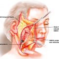
Vascular impairment is an infrequent, but dreaded, complication of facial filler injections. An understanding of the vascular anatomy and physiology is essential when injecting in the facial region ( Fig. 36.1 ). The face is perfused with vessels form both the internal carotid artery and the external carotid artery. The ophthalmic artery arises from the internal carotid artery and leads to the dorsal nasal, supraorbital, and supratrochlear arteries. The facial artery arises from the external carotid artery and gives rise to the facial artery, which is the source of the labial, angular, and lateral nasal arteries. Direct anastomoses between the anterior facial vessels enable emboli to traverse between the internal and external carotid systems. It is these anastomoses that have led to emboli that cause blindness.
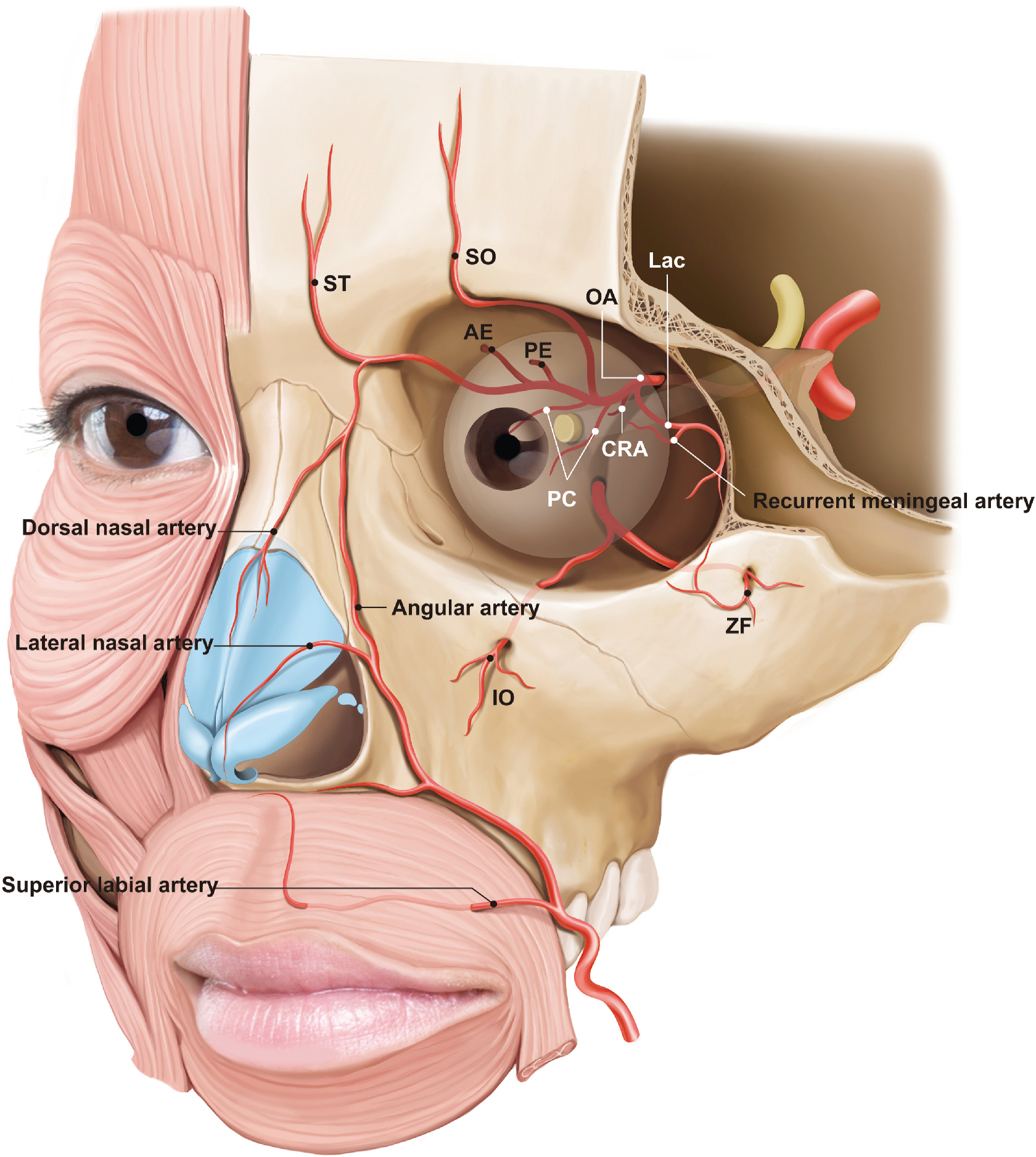
Ashton et al. studied the functional angiosomes of the face and their association with tissue necrosis. It has been reported that injection-mediated tissue necrosis frequently occurs at a site remote from the injection site, and Ashton et al. found that all complications, regardless of injection site, occur in only five facial regions. Choke vessels have the ability to control or “choke” the flow between angiosomes. Studies have shown that intravascular HA is highly inflammatory and thus can induce choke vessel spasm and reduce perfusion leading to necrosis. Alternatively, if adjacent angiosomes are connected by an anastomosis rather than a choke vessel, the HA can continue through the anastomoses until it hits a choke vessel in a distant angiosome causing remote necrosis. The choke vessel physiology has implications in the application of nitroglycerin paste after suspected filler-induced ischemia. If nitroglycerin paste causes vasodilation opening the choke vessels that are containing the HA, the HA can proceed to distant angiosomes exacerbating the extent of tissue loss. This theory is corroborated by some case reports of worsening necrosis after application of nitroglycerin paste.
History
The application of dermal fillers for facial reshaping differs from that for simple wrinkle and fold correction. Patients who present for facial reshaping may desire correction of asymmetry, change in facial shape, isolated alteration of specific anatomic features (e.g., nose, chin), or gender-related changes. It is important to obtain a detailed history of desires, expectations, and limitations of the result. For instance, chin augmentation with fillers has limitations because the chin pad has an amorphous shape and texture and does not exhibit the strong skeletal nature of a normal chin; similar limitations exist for filler rhinoplasty. A previous history of surgery or radiation is also important. In these patients, autologous fat grafting may be a better option, given the potential for stem cell regeneration. Finally, medications and medical conditions (e.g., bleeding disorders) should be noted and the associated risks should be discussed.
Physical examination
Facial reshaping with dermal fillers focuses on certain regional areas where the illusion of skeletal change can be created through the use of fillers.
Temporal region: The patient is examined to assess the degree of temporal hollowing and to also note any asymmetry.
Forehead: The forehead should exhibit a smooth, symmetrical, and slightly convex contour.
Glabella: The glabella should have a convex shape in the profile view. This contributes to the aesthetic quality of facial divergence, as discussed in Chapter 16 .
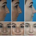
Nose: The nose should be evaluated as described in Chapters 28 and 29 . Specific anomalies amenable to placement of fillers are low dorsum, low radix, decreased tip projection, and asymmetry.
Midface region: The cheeks should be assessed for projection and symmetry. Bizygomatic width is also noted; this dimension has a profound influence on the perception of facial shape. Asian patients typically exhibit wide bizygomatic width and less projection in the medial malar region. Placing fillers in this region can create the illusion of a narrower face. In contrast, Western women desire augmentation of the lateral zygoma to restore cheek fullness. The effects of structural fillers in both sets of patients create the ideal oval face.
Mandible: The entire mandible is assessed for symmetry, width, and contour. Analyses of the mandible and the chin should be performed separately because each influences the perceived shape and position of the other. Masseter muscle bulk is assessed in addition to the bone dimensions of the gonial region.
Chin: The symmetry, projection, and shape of the chin are noted. The acuity of the labiomental crease and the soft tissue support of the underlying mentalis are also noted.
Imaging
For injection of dermal fillers, skeletal imaging is not routinely obtained. Two-dimensional (2D) and 3D images are part of the record and are useful in assessing the results of volume augmentation. It is important to carefully review the images because subtle asymmetries and morphologic abnormalities frequently are more obvious on a review of images than on physical examination.
Indications and contraindications
Contraindications to the use of fillers include active infection near the site of injection, known allergy/hypersensitivity to the material or to the lidocaine mixed in the syringe with the filler, and therapeutic anticoagulation. No causal relationship has been established between the use of fillers and autoimmune diseases, such as dermatomyositis/polymyositis, lupus erythematosus, rheumatoid arthritis, or scleroderma; thus the use of fillers is not contraindicated in these patients. Additionally, immunosuppression has not been found to increase the risk of complications linked to the use of fillers.
Preoperative planning
Based on the patient’s concerns, the physical examination, and photographic analysis, a treatment plan is developed. A wax makeup pencil serves as a useful tool to mark the patient with reference lines such as a midline. Hinderer’s lines can be drawn on the cheeks to aid in location of malar injection sites. Areas of vascular risk can be denoted to remind the surgeon to inject in low-risk areas.
Primary operative approach
Facial reshaping in the Asian patient
When evaluating the face, the aesthetic standards of the East and the West are not exactly the same due to ethnic and regional variations as well as anthropologic differences. Western faces demonstrate clear facial outlines, narrow cheekbones, well-defined 3D structure, obvious light-and-shadow effects, rough skin texture, skin relaxation, and lack of elasticity. The Asian face is full, the malar complex is large, the facial form lacks clear outlines, and the contrast between highlights and shadows is not clear. Asian skin is delicate and elastic. For these reasons, rejuvenation of Western patients focuses on skin resection and some volume restoration. The Western patient is typically a candidate for lifting and filling, whereas rejuvenation of the Asian patient focuses on filling, altering facial divergence, and tapering the face to the ideal oval shape.
However, whether it is the East or the West, regardless of ethnic and cultural differences, the classic canons of aesthetic facial form are similar: symmetry, equilibrium, proportion. The highest realm of facial filler injection should be regarded as artistic creation applied within professional medical guidelines. It is imperative that the surgeon has complete comprehension of the principles of facial aesthetics to achieve an artistic result. It is obvious that achieving beauty and facial enhancement with injection of fillers is not a simple procedure. The surgeon using fillers is similar to a sculptor applying clay—the needle is a tool to create a beautiful face as a work of art, and it is not easy. When applying fillers to the face to create artistic structural and dimensional beauty, the clinician with a background in skeletal surgery has an advantage over a nonsurgical injector. A surgeon has an expert understanding of surgical anatomy and thus has the ability to visualize the three dimensions of the recipient’s facial anatomy; this gives the surgeon an understanding of how the amount, viscosity, and depth of the injection will affect the facial shape. Additionally, a thorough knowledge of facial anatomy, both superficial and deep, enables the surgeon to minimize the risk of inappropriate filler injection, which could result in tissue necrosis or skin slough.
In 2007, Dr Cui introduced the concept of overall design and creation of an aesthetic image of the human body. This concept includes systematic overall design and aesthetic evaluation before surgery, psychologic counseling during perioperative treatment, and the comprehensive use of multiple treatment methods. After the surgery, makeup, clothing, modeling, etiquette training, and appropriate intervention in the social survival state are necessary complementary strategies to create a beautiful image full of charm and vitality.
If we regard facial injection as an artistic endeavor within the parameters of safe and ethical medical guidelines, then the creation of beauty cannot be simply achieved by using a single technology or treatment method; rather, it should be based on a systematic and holistic design. We have done a retrospective survey, according to the visual analogue scale (VAS) evaluation method, and found that satisfaction from simply receiving a treatment is far less than that from the results of a systematic overall design.
In the past, the medical model was a simple biomedicine model. Now it is a biopsychosocial medicine model—that is, even if an operation is performed well, it only accounts for about 30% of the result. The model also needs to take into account the influence of the psychologic and social factors related to the surgeons. This is particularly relevant in the area of aesthetic surgery.
Beauty enhanced through the injection of dermal fillers should also follow the principle of a systematic overall design. We have always emphasized that technology can provide basic and necessary tools that can only solve the most basic problems. What is more important is the surgeon’s understanding of the aesthetic facial form and what is beautiful.
Some cosmetic patients are not satisfied after an isolated, localized area of injection. For example, to improve the nasolabial folds, injections are commonly limited to the region of the nasolabial crease. After the injection, the acuity of the nasolabial fold may be attenuated, but with injection of a larger volume of the filler, the face would look fatter, compromising patient satisfaction. What causes the perception of a fatter face? This problem arises when the surgeon fails to visualize the face as a whole and only addresses parts of the whole. Therefore it is particularly important to consider the artistic aspect of making sculptural changes to the face through aesthetic surgery. As Canadian Dr. Trudeau says, surgery is done “sometimes to heal, often to help, always to comfort.”
Before performing a dermal filler injection, careful attention must be paid to the overall design and construction of the aesthetic image of the human body. Artistic success in this endeavor brings better physical appearance, psychologic satisfaction, and social recognition to the patient. The modern aesthetic approach emphasizes the role of social and psychologic factors in the treatment.
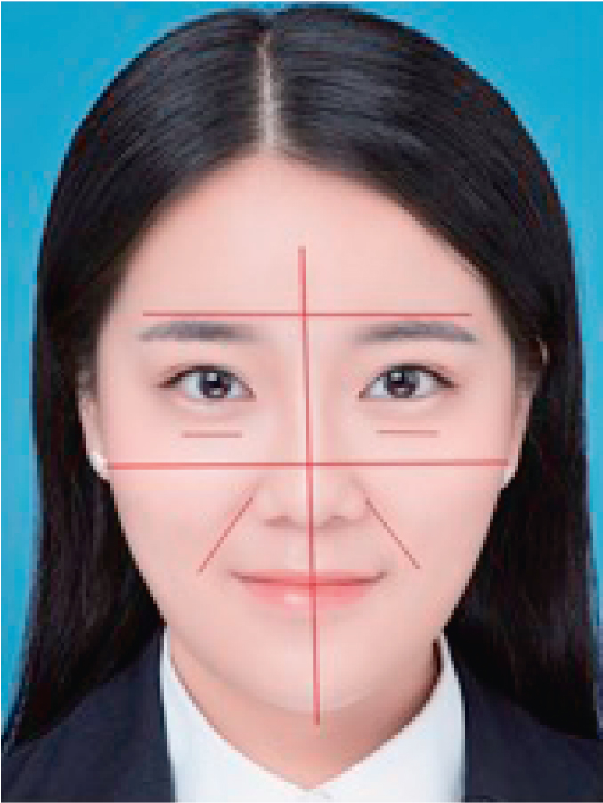
When we approach the concept of overall design creation of the aesthetic image of the human body, how do we find a simple and effective way to express the concept? In our approach, while planning treatment, we determine the locations for filler placement by using several anatomic landmarks. The first horizontal line passes the arch of the brow and the temporal region. The second horizontal line passes through the malar cheek pad.
The midline is marked with a line passing through the forehead, glabella, nose, lip, and chin. Two additional lines are marked obliquely parallel to the nasolabial folds, and a final line is marked at the tear trough ( Fig. 36.2 ).
Related posts:
Stay updated, free articles. Join our Telegram channel

Full access? Get Clinical Tree


