Fig. 11.1
Schematic representation of the penetration pathways of topically applied substances
While the intercellular penetration process has been recognized for decades, the existence of follicular penetration could be demonstrated not before a few years ago. Follicular penetration experiments require measurements at high spatial resolution (Jung et al. 2006). Only in recent years, suitable techniques for such measurements could be provided. These systems are based mainly on optical methods specifically developed or optimized for that purpose (Tfayli et al. 2012; Lademann et al. 2012; Mura et al. 2012; Forster et al. 2011; Haag et al. 2011). A further challenge is that while intercellular penetration can be usually investigated in vitro or ex vivo, follicular penetration must be analyzed in vivo or ex vitro on porcine ear model skin (Patzelt et al. 2008). This is due to the fact that skin contracts immediately after excision during surgical intervention. Although the interfollicular fibers can be expanded by stretching the excised skin to its original size for the measurements, the very dense network of fibers surrounding the hair follicles remains contracted, which is an almost irreversible process (Patzelt et al. 2008). The effect of contraction of the hair follicles in excised human skin was initially demonstrated by Patzelt et al. (2008), who could moreover show that the follicular penetration of a fluorescent dye-containing formulation was reduced by 90 % in the case of ex vitro investigations on excised skin in comparison to in vivo investigations, whereby methods and skin models were identical.
As a consequence, the follicular orifices are not accessible for the penetration process in ex vivo models, which has to be considered when certain methods are employed such as the diffusion cell experiment. Diffusion cell experiments (Okuda et al. 2011; Baert et al. 2010) are widely used for penetration investigations on split or full-thickness skin. However, the hair follicles reach deeply into the subcutaneous tissue, which is mostly removed before the skin probes are clamped onto the diffusion cell meaning that the hair follicles are somewhere cut in their middle. This would imply, if the hair follicles were accessible and not plugged by the contraction process, that the topically applied substances would pass directly through the hair follicle stump into the receptor medium. Since, however, the hair follicles are plugged due to contraction, intercellular penetration can be investigated more or less properly by this method whereas follicular penetration is not measurable. Thus, follicular penetration must be investigated either in vivo or ex vivo on porcine ear model skin, which strongly resembles human skin in terms of composition and structure. The advantage of the porcine ear skin model is that the skin remains tightly attached to the cartilage even after truncation of the ear and does not contract. However, it has to be taken into consideration that the porcine hair follicles extend deeper into the skin and are larger in diameter than human hair follicles (Lademann et al. 2010; Hutton et al. 1978).
The present chapter will summarize the easy and wide application possibilities of the different stripping methods, which can be applied for investigating intercellular and follicular penetration pathways.
11.2 Tape Stripping
One of the oldest methods used in penetration studies is the tape stripping method. Briefly, tape stripping comprises the successive removal of adhesive films pressed onto the skin after the topical application and penetration of the substance under investigation. With each adhesive tape, a certain amount of corneocytes is removed including the topically applied substances contained within this layer. The tape stripping method is illustrated in Fig. 11.2 in detail. After application with a syringe Fig. 11.2a and homogeneous distribution by means of a saturated rubber glove finger Fig. 11.2b, the applied formulation is then allowed to penetrate for a determined time. Thereafter, the individual adhesive strips are pressed one by one onto the skin by a roller Fig. 11.2c or stamp leading to a smoothing of the skin surface and are then removed quickly Fig. 11.2d. The smoothing of the skin surface represents an important step in the tape stripping protocol as by pressing the adhesive film onto the skin, the influence of furrows and wrinkles can be avoided or at least minimized (Lademann et al. 2005). In Fig. 11.2c, a roller is used for smoothing (Mohammed et al. 2012; Bettoni et al. 2012; Lademann et al. 2006, 2009) the skin surface. The principle of smoothing the skin surface by a roller is represented in Fig. 11.3 using optical coherence tomography (OCT). After rolling the skin, the previously structured skin surface (Fig. 11.3a) has been converted into a flat surface (Fig. 11.3b). During the rolling process, the adhesive film is permitted to contact the surface of the respective skin area completely. The adhesive films pressed onto the skin are subsequently removed and analyzed for the amount of stratum corneum and the amount of the topically applied substance. In Fig. 11.4, the image of a typical tape strip after removal from the human skin is shown. The corneocyte cover is clearly recognizable on the adhesive film.


Fig. 11.2
Images presenting the tape stripping method. (a) Application of the formulation with a syringe. (b) Distribution of the formulation using a saturated rubber glove finger.(c) Adhesive tapes are pressed onto the skin by using a roller. (d) Adhesive tapes are rapidly removed from the skin
Fig. 11.3
Influence of the roller smoothing the skin surface. (a) Skin furrows before rolling. (b) Smoothed skin surface after rolling (Lademann et al. 2005)
Fig. 11.4
Microscopic image of a tape strip removed from the skin
Thus, tape stripping is a well-established method to investigate the penetration of topically applied substances into the complete stratum corneum; however, it is not suited to remove cells from the viable epidermis located beneath the stratum corneum. In Fig. 11.5, a biopsied human skin sample prior to (Fig. 11.5a) and after tape stripping (Fig. 11.5b) is shown, demonstrating that this procedure is capable of removing the stratum corneum completely. Prior to tape stripping, the wrinkles and furrows in the skin surface structure are clearly recognizable, yet, whereas a flat skin surface is visible after tape stripping. This is due to the tape stripping procedure causing a light swelling of the skin surface.

Fig. 11.5
Histological sections from biopsies removed prior to (a) and after (b) tape stripping, demonstrating that the stratum corneum can be removed completely (Lademann et al. 2005)
When the tape stripping method was introduced, the number of the individual tapes had initially been considered as a measure for the stratum corneum depth at which the tape strip was removed. Applying the tape stripping method with growing expertise and experience, it became evident that the tape number is not a reproducible value characterizing the depth of the stratum corneum at which the tape strip is removed. This is attributable to the fact that different formulations influence the amount of stratum corneum adhering to the tape strip differently. Whereas greasy formulations may reduce the adhesive strength of a tape strip, meaning that less stratum corneum is removed per tape strip, an ethanolic formulation may increase the amount of stratum corneum removed. In addition, also the pressure used for pressing the adhesive film onto the skin and the type of adhesive film decisively influences the amount of stratum corneum removed by each tape strip. The literature reports of a variety of methods to quantify the amount of stratum corneum on the respective tape strips which further allow the determination of the depth at which the tape strips were removed. This can be performed, for instance, by weighing the tape strips prior to and after their application to the skin (Weigmann et al. 2005). A decisive shortcoming of this method is, however, that the weight of the first tape strips is not only determined by the corneocytes, but also by the topically applied substances penetrated into the uppermost layers of the corneocytes. Moreover, in deeper layers of the stratum corneum interstitial fluid may escape, which could also increase the weight. As an alternative method, Lindemann et al. (2003) proposed to use the pseudoabsorption at 430 nm as a measure for the amount of stratum corneum on the tape strips. The pseudoabsorption characterizes the attenuation of the light penetrating through the tape strip. It is determined by the absorption, reflectance, and scattering of the corneocytes. These signals are known to depend on the wavelength, and the shorter the wavelength, the stronger the signal, indicating that the scattering effect is more pronounced in shorter wavelength. However, wavelengths below 400 nm are unsuitable for these investigations as a variety of formulations, including sunscreens, have absorption bands in this spectral range. A third method for the determination of the amount of stratum corneum on the tape strips, which is meanwhile commercially available, was suggested by Voegeli et al. (2007, 2009). This method uses the infrared absorption of the corneocytes for determining the amount of stratum corneum. The measuring device is based on an infrared densitometer permitting indirect measurements of the stratum corneum protein content on the removed tape strip by means of absorption. This measuring method is a nondestructive technique so that the tape strips can subsequently be used for other analyses. It provides a clear advantage as it is not influenced by the absorption of applied formulations in the ultraviolet (UV) spectral range, and it is not influenced by nanoparticles. A further method suggested by Dreher et al. is based on staining the corneocytes after removal so that the amount of stratum corneum on the tape strips can be determined by absorbance measurements using a UV/VIS spectrometer (Dreher et al. 2005).
Taking into consideration the above-mentioned facts, it became established as a standard that instead of the number of tape strips, the amount of stratum corneum is now used for the calculation of penetration profiles of drugs into the stratum corneum. A comparison of the traditional and the extended method is schematically shown in Fig. 11.6, highlighting the advantages of the extended method. The traditional tape stripping procedure only considers the amount of substance in relation to the tape strip number, whereas the information about the amount of stratum corneum is neglected in contrast to the extended method where this essential part of information is likewise considered. In this way, it can be clearly seen that although the amount of corneocytes may vary between the individual tape strips, on average it is declining with the increasing tape strip number. Therefore, based on the investigations of the amount of removed stratum corneum, the tape strips were used to calculate the profile of the horny layer. Here, the space between two horizontal lines corresponds to the amount of stratum corneum removed with the specific tape strip. In the deeper layers of the stratum corneum the distances between the horizontal lines are getting smaller. The top line of the horny layer profile shown in this figure represents the surface of the stratum corneum, while the bottom horizontal line represents the boundary to the living cells.

Fig. 11.6
Principle of the calculation of the penetration profile. The amount of corneocytes on each tape strip is determined by UV/VIS spectroscopy. The concentration of the applied substance on each tape strip is determined by traditional methods such as UV/VIS spectroscopy, X-ray fluorescence (XRF), atomic absorption spectroscopy (AAS), high-performance liquid chromatography (HPLC), or gas chromatography in combination with mass spectroscopy (GC/MS). The combination of both results allows the calculation of a penetration profile
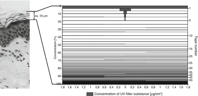
The amount of topically applied substance removed with a single tape strip can be determined by classical analytical methods like high-performance liquid chromatography (HPLC) and mass spectroscopy. If the information on the amount of topically applied substance detected on the respective tape strips is added to the horny layer profile, a penetration profile is obtained. This is illustrated in Fig. 11.7 showing part of a histological section. The adjacent penetration profile of a UV filter exhibits the distribution of this substance in the stratum corneum.
 Retardation Strategies for Sunscreen Agents
 Confocal Raman Spectroscopy as a Tool to Investigate the Action of Penetration Enhancers Inside the Skin
 Human Native and Reconstructed Skin Preparations for In Vitro Penetration and Permeation Studies
 Confocal Microscopy for Visualization of Skin Penetration
 Finite and Infinite Dosing
 Basic Mathematics in Skin Absorption


Related posts:
Retardation Strategies for Sunscreen Agents
Confocal Raman Spectroscopy as a Tool to Investigate the Action of Penetration Enhancers Inside the Skin
Human Native and Reconstructed Skin Preparations for In Vitro Penetration and Permeation Studies
Confocal Microscopy for Visualization of Skin Penetration
Finite and Infinite Dosing
Stay updated, free articles. Join our Telegram channel
Full access? Get Clinical Tree
