Nasal hump excision is common during septorhinoplasty. Without appropriate restoration of the middle nasal vault, cosmetic and functional problems may ensue. Recently, spreader flaps have become an established alternative to traditional spreader grafts. Typical indications include primary rhinoplasty patients with hump noses, hump/tension noses, and moderately hooked or crooked noses. When suitable patients are selected, spreader flaps and their modifications represent a reliable alternative to the standard spreader graft, and when all of the necessary prerequisites are met, this technique obviates the need for additional cartilage grafting in most cases.
Key points
- •
Reconstruction of the middle nasal vault after nasal hump removal is almost always necessary to prevent postoperative functional and cosmetic imperfections including the inverted V deformity.
- •
Spreader grafts are the gold standard for restoring the stability and contour of the middle vault after hump reduction; recently, spreader flaps have become reliable treatment alternative in select cases.
- •
Improvements and modifications of the basic spreader flap technique allow precise adjustments to middle vault contour to further expand the utility of spread flap reconstruction; however, appropriate patient selection is crucial to a satisfactory surgical outcome.
Introduction and treatment goals
A considerable number of patients who express the desire for cosmetic rhinoplasty require contouring and subsequent stabilization of the middle nasal vault. Some of these patients may present with an overly narrow humped middle vault and a (natural) visible delineation between the nasal bones and the upper lateral cartilages (ULC). Endonasal examination in these patients often reveals pinching of the internal nasal valve with an accompanying reduction in valve patency and premature collapse of the ULCs upon inspiration. This phenomenon is particularly common in patients who present with a high, peaked nasal dorsum, as seen in the so-called tension nose deformity. However, patients with short nasal bones, long and weak ULCs, and thin nasal skin are also at increased risk for middle vault distortion and collapse after nasal hump reduction. In fact, surgical detachment of the ULC from the dorsal septum can incite pinching, malposition, and/or concave collapse of the ULC in virtually any nose, and when predisposing factors are not properly recognized and treated, unsightly cosmetic aftereffects of nasal surgery frequently occur. Typically, this manifests as inward collapse of the lateral nasal sidewalls (often accompanied by a slight middle vault saddle deformity) and symptomatic nasal airway obstruction. When the middle vault narrowing is severe in comparison to upper vault width, a stigmatic upside-down V-shaped shadow becomes visible at the bony–cartilaginous junction, an unsightly contour abnormality known as the “inverted V” deformity. In addition to the V-shaped shadow, the dorsal aesthetic lines are often disrupted or “washed out,” particularly after over-resection of the dorsal hump.
The importance of maintaining a functional internal nasal valve and reconstructing the middle nasal vault immediately after hump reduction is now widely recognized among rhinoplasty experts. The groundwork was laid by Sheen, who first advocated spreader grafts for middle vault stabilization and contour enhancement. Today, spreader grafts have become the gold standard for preserving or restoring contour and structural integrity of the middle vault. However, the more recent advent of spreader flaps has added a second option for middle vault reconstruction after hump reduction in select patients. Instead of trimming the ULCs to match the newly established dorsal profile, the excess vertical height is used to create bilateral inwardly folded cartilage flaps that are sutured to the upper margin of the dorsal septum to strengthen and stabilize the surgically weakened middle vault. In principle, the in-folded ULCs behave similar to traditional spreader grafts by maintaining width at the apex of the nasal valve and thereby increasing the threshold for inspiratory nasal valve collapse. This benefit is derived almost entirely by the springlike effect of the partially folded ULCs, which mimic the natural anatomic configuration of a well-functioning ULC–septal junction. However, only patients with adequate ULC length and reasonably firm cartilage are satisfactory candidates for spreader flap fabrication.
Oneal and Berkowitz first described the use of in-folded ULC flaps in 1998 and coined the term “spreader flaps.” Later, further modifications were made, especially by Rohrich and colleagues, Byrd and colleagues, Gruber and colleagues, Ozmen and colleagues, and Neu. However, in our opinion, these modifications failed to fully optimize nasal valve function and airway patency. Furthermore, the modifications offered only limited opportunities for customization of the middle valve width according to the individual functional and cosmetic goals. The modifications we describe herein are further refinements of the spreader flap technique and serve to address these shortcomings.
Preoperative planning and preparation
Preoperative findings and planned surgical objectives are discussed in detail with the patient, and objectives vary according to individual patient preferences. Patients are also fully informed about the risks and benefits of the planned procedure. Morphing software may be of value in this context, but the patient must be told that the images generated from such software are only approximations and by no means a guarantee of a particular postoperative result. Patients are also advised not take any nonsteroidal anti-inflammatory drugs or anticoagulants for 10 to 14 days before surgery.
Immediate preoperative preparation includes cutting the nasal vibrisse and disinfecting the nasal vestibule. All incision and osteotomy lines are infiltrated with an injection solution containing 2% lidocaine and adrenaline 1:200,000 to minimize intraoperative bleeding.
Surgical technique
Basic Spreader Flaps
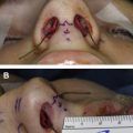
For surgical exposure, we routinely use the external rhinoplasty approach. This begins with degloving the entire skeletal framework in a sub-superficial musculoaponeurotic system (SMAS) dissection plane via a transcolumellar incision. Dissections should be carried out in a supraperichondrial and subperiosteal planes, respectively. Starting from the anterior septal angle, bilateral submucosal tunnels are then elevated on the undersurface of the ULC–septal cartilage junction and extended cranially beneath the bony vault. A “component” cartilaginous hump reduction is then begun by sharply dividing the ULC from the dorsal septum while preserving the underlying mucosa. In this manner, the overprojected ULC are not trimmed and can be used for spreader flap creation. Next, sharp reduction of the cartilaginous dorsal septum is performed to establish the new middle vault profile line. After resecting the cartilaginous septal hump, fibrous attachments of the ULC to the undersurface of the nasal bones are released in the midline using blunt dissection over an approximately 0.5-cm wide strip on both sides. Because the ULC release is confined to the area of planned bony hump resection, the detached cephalic ends of the ULC (which may extend as far as 10–12 mm beneath the rhinion) are protected during hump reduction, whereas the more lateral bony attachments of the ULC to the nasal bones remain intact. By preserving the uppermost extensions of the ULC, minor postoperative contour irregularities of the open roof can sometimes be prevented. After separation of the ULC and elevation of their perichondrium in 0.5- to 1-cm wide strip, both medial edges can be invaginated medially as turn-in flaps and temporarily positioned alongside the dorsal septum for suture fixation. Our method for flap fixation differs from previously described techniques. First, the distal rolled ends of the ULC are grasped and pulled caudally while they are sutured to the upper (caudal) border of dorsal septum ( Figs. 1 and 2 ). As a rule, we find that 1 internal fixation suture is adequate for secure fixation. However, in cases of skeletal instability at the keystone area, a second suture can be added cranially for additional stabilization. We prefer a 4-0 Polydioxanone suture for flap fixation. The knot is buried between the ULC and septum to prevent visible contour irregularities. Moreover, hiding the knot keeps a possible foreign body reaction hidden beneath the skeletal framework. Care is taken to match spreader flap height to the existing height of the dorsal septum to create a smooth and straight dorsal profile. In virtually every case of spreader flap placement, in-folding and fixation of the ULC introduces modest laterally directed tension across the ULC. This beneficial tension tightens the ULC to minimize inward collapse and thereby helps to maintain and/or improve internal valve patency. Moreover, depending on the natural rigidity of the ULC, a springlike effect is often generated at the ULC fold that further contributes to valve patency and stabilization against sidewall collapse. In this way, spreader flaps can be used to reconstruct the contours and the functional stability of the middle vault after hump reduction. However, modifications of the basic technique can also be used for further contour refinements of the middle vault according to individual patient requirements.