Vertical arch division is a mainstay of tip surgery, and its applications are expanding. It allows deprojection of the overprojected tip, and modifies rotation, length, and lobule definition. These parameters can be altered in a controlled, predictable fashion when the alar cartilage is preserved and overlapped, maintaining its strength. Cartilage overlay techniques aim to preserve normal anatomy and establish support for the nasal framework. We discuss the uses of vertical arch division when applied to the M-arch model, an expansion of the nasal tip tripod concept, which provides for a utilitarian approach to surgical techniques for the nasal tip.
Key points
- •
The M-arch model is a contemporary expansion of the nasal tip tripod theory, which helps to provide a conceptual framework for understanding the anatomy and dynamics of the alar cartilages.
- •
Shortening the M-arch, effected by vertical arch division at a carefully chosen point along the contour of the alar cartilages, can help to achieve substantial, controlled, and predictable adjustments in projection and rotation, producing refinement of the tip when indicated.
- •
After vertical arch division, reconstitution and overlap of the split ends of the alar cartilage maintains normal anatomy and strength, limits postoperative shifting, and allows the tip to remain stable in its optimal position.
- •
Depending on the distance of the vertical arch division from the tip defining point, the relative effects on deprojection, rotation, and tip refinement can be determined.
- •
Divisions closer to the lobule have a greater effect on narrowing the lobule, and to a lesser degree on rotation, whereas the reverse principal applies when divisions are placed further from the tip.
Introduction
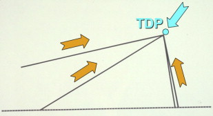
The nasal tip is widely appreciated by rhinoplasty surgeons as the most conceptually and technically challenging aspect of the nose to master. The vast number of tip-altering techniques used to modify and establish the 3 principal parameters of nasal aesthetics—length, projection, and rotation—is a testament to the complexity and level of difficulty involved in surgically controlling the lower third of the nose. As is frequently the case when applying challenging and intricate surgical principles in practice, simplification is often a reliable path to understanding. It is for this reason that rudimentary concepts tend to withstand the test of time, amid a myriad of more complicated paradigms. One seemingly eternal simplification of tip dynamics, Jack R. Anderson’s nasal tip tripod concept (1969), has greatly advanced the universal understanding of the tip among rhinoplasty surgeons and has facilitated the development of various techniques. This tripod concept defines the conjoined medial crura (MC) as the central leg of a tripod and the lower lateral crura (LC) as the 2 side legs. The position of the tip-defining point (TDP) can be manipulated by altering the length(s) of these legs ( Fig. 1 ). Shortening the legs decreases projection (retrodisplacement) and, depending on the integrated degree of shortening of the MC and LC, there will be a variable effect on rotation and, subsequently, on nasal length.
The M-arch model, first described by the senior author in 2006, expands on the concept of the nasal tip tripod by conceptualizing the tip as continuous paired arches. This model is more accurate, both anatomically and dynamically, when considering the actual architecture of the tip, and provides for a more detailed and utilitarian approach to the application of surgical techniques. The essence of the model is that these paired arches have a specific length. Furthermore, each of the components of the M-arch—the MC, intermediate crura (IC), and LC—has a length. These component lengths, and their combined overall length, establish tip projection and rotation, and indirectly nasal length. Shortening the length of the M-arch, implemented by vertical division at a carefully chosen point along its contour, can help to achieve substantial, controlled, and predictable adjustments in projection and rotation, while also producing refinement of the tip when indicated. The senior author has gradually evolved toward the application of crural cartilage overlap (overlay) techniques, in lieu of division or excision, with or without end-to-end reconstitution of the cartilage, as was typically performed in the past. Overlap of the alar cartilage helps to strengthen and spatially fixate the alar cartilages, and ultimately the tip complex, producing more reliable positioning of the TDP. In cases where the arch length needs to be increased, a variety of cartilage grafts and steal techniques are applied to increase the relative lengths of the M-arch components. Herein, we have addressed the use of crural overlay techniques to decrease the length of the arch, thereby achieving our desired projection, rotation, nasal length, and lobule definition. The M-arch model does not directly address changes in tip projection and rotation effected by maneuvers that weaken the support structures of the tip, such as transfixion incision or weakening of ligaments in the intercrural or scroll regions or excision of premaxillary spine. However, if these maneuvers or others are performed, their effects can be integrated easily into the M-arch model by invoking how they individually affect the length of the specific M-arch components.
Introduction
The nasal tip is widely appreciated by rhinoplasty surgeons as the most conceptually and technically challenging aspect of the nose to master. The vast number of tip-altering techniques used to modify and establish the 3 principal parameters of nasal aesthetics—length, projection, and rotation—is a testament to the complexity and level of difficulty involved in surgically controlling the lower third of the nose. As is frequently the case when applying challenging and intricate surgical principles in practice, simplification is often a reliable path to understanding. It is for this reason that rudimentary concepts tend to withstand the test of time, amid a myriad of more complicated paradigms. One seemingly eternal simplification of tip dynamics, Jack R. Anderson’s nasal tip tripod concept (1969), has greatly advanced the universal understanding of the tip among rhinoplasty surgeons and has facilitated the development of various techniques. This tripod concept defines the conjoined medial crura (MC) as the central leg of a tripod and the lower lateral crura (LC) as the 2 side legs. The position of the tip-defining point (TDP) can be manipulated by altering the length(s) of these legs ( Fig. 1 ). Shortening the legs decreases projection (retrodisplacement) and, depending on the integrated degree of shortening of the MC and LC, there will be a variable effect on rotation and, subsequently, on nasal length.

The M-arch model, first described by the senior author in 2006, expands on the concept of the nasal tip tripod by conceptualizing the tip as continuous paired arches. This model is more accurate, both anatomically and dynamically, when considering the actual architecture of the tip, and provides for a more detailed and utilitarian approach to the application of surgical techniques. The essence of the model is that these paired arches have a specific length. Furthermore, each of the components of the M-arch—the MC, intermediate crura (IC), and LC—has a length. These component lengths, and their combined overall length, establish tip projection and rotation, and indirectly nasal length. Shortening the length of the M-arch, implemented by vertical division at a carefully chosen point along its contour, can help to achieve substantial, controlled, and predictable adjustments in projection and rotation, while also producing refinement of the tip when indicated. The senior author has gradually evolved toward the application of crural cartilage overlap (overlay) techniques, in lieu of division or excision, with or without end-to-end reconstitution of the cartilage, as was typically performed in the past. Overlap of the alar cartilage helps to strengthen and spatially fixate the alar cartilages, and ultimately the tip complex, producing more reliable positioning of the TDP. In cases where the arch length needs to be increased, a variety of cartilage grafts and steal techniques are applied to increase the relative lengths of the M-arch components. Herein, we have addressed the use of crural overlay techniques to decrease the length of the arch, thereby achieving our desired projection, rotation, nasal length, and lobule definition. The M-arch model does not directly address changes in tip projection and rotation effected by maneuvers that weaken the support structures of the tip, such as transfixion incision or weakening of ligaments in the intercrural or scroll regions or excision of premaxillary spine. However, if these maneuvers or others are performed, their effects can be integrated easily into the M-arch model by invoking how they individually affect the length of the specific M-arch components.
Nasal tip anatomy and definitions
The nasal tip is composed of the conjoined MC, IC, and diverging lower LC ( Fig. 2 ). The TDPs, which are highlighted by an external light reflex, are formed by the apices, or most anterior projections, of the domal arches, which have varying degrees of acuity related to the underlying shape and angulation of the alar cartilages.
The nasal lobule includes the soft tissue anterior to the nostril apex, caudal to the supratip break of the dorsum, and anterior to the lateral alar side walls. The lobule can be subdivided into the supratip, infratip, and paired lateral supratips, each of which neighbors the TDPs. The nasal base includes the infratip lobule, columella, alar side walls, and nasal sill.
Beyond the cartilages, the thickness or thinness of the skin–soft tissue envelope (S-STE) has a significant impact on the degree of tip definition. Thin skin reveals the details of cartilage anatomy, whereas thick skin blunts the appearance of well-defined domes, both for the better or worse.
The cephalic border of the nasal tip is the transition into the scroll region, or junction of the LC with the caudal upper lateral cartilage, via fibrous attachments. The caudal edge of the tip consists of the soft tissue triangles of the alar rims, which extend below the caudal margin of the alar cartilages. The lateral edge of the tip is the transition into the alar–facial groove and bony pyriform fossa.
With respect to additional attachments, the MC cartilages are fixed onto the posterior septal angle, and extend anteriorly to the apex of the nostrils, which correspond to the medial crural angle (junction of the MC and IC). The medial crural angle follows a gentle curvature to transition seamlessly with the IC, which is defined as the segment of the tip cartilage that extends from the medial crural angle to the TDP. The lateral crus extends from the TDP to the hinge area, or junction of the LC with the pyriform fossa (see Fig. 2 ). Sesamoid cartilage may be present at this transition point. This area is also called the foot of the LC, analogous to the foot of the MC, where the MC embraces the posterior septal angle.
Although neighboring anatomic structures transmit their intrinsic forces to the tip by way of ligamentous and fascial attachments, influencing its overall appearance, the underlying structure of the alar cartilages constitutes the foundation and primary determinant of nasal tip anatomy and its dynamics. Its construct can be altered by surgical refinements guided by the tripod concept and the M-arch model. The M-arch anatomy can be best described as a pair of adjoining arches in much the same way as the “golden arches” of the McDonald’s Restaurant Corporation (Oak Brook, IL). This symbol is widely recognized and thus serves as a useful tool to represent both the anatomic construct of the alar cartilages as well as the impact of surgical modifications. An important feature of the M-arch is that the lateral legs of the alar cartilages are not in the same horizontal plane as the medial legs, but rather oriented in a posterosuperior position. As described in the senior author’s original article, the golden arches facing a headwind would best illustrate the anatomic configuration of these cartilages.
From an anatomic perspective, one of the major advantages of the M-arch model, with respect to the more classic tripod concept, includes its consideration of the tip as a continuous arch, as opposed to a 3 straight-legged structure. It also recognizes the importance of the medial, intermediate, and lateral crural arches in continuity, instead of the MC and LC being attached at a hinge. The M-arch model defines yet another arch, the domal arch. This is essentially an arch within an arch, and consists of the intermediate crus (ie, the IC) and anterior component of the LC. The M-arch concept’s inclusion of the intermediate crus, and subsequently its consideration of how alterations in the arch can affect lobule definition, is an additional advantage. The tripod concept predates the labeling of the intermediate crus and simply does not address the lobule.
Nasal tip dynamics
In essence, the goal of rhinoplasty is to alter the architecture of the bony and cartilaginous structure of the nose to create an underlying foundation for the S-STE to redrape. The resultant aesthetic of the nose is largely contingent on appropriately addressing the 3 fundamental parameters—length, projection, and rotation—which are determined by the position of the TDPs, and the balance of these parameters with tip refinement, as well as the construct of the nasal dorsum and nasal base.
The tip receives its structural support from a multitude of sources, none greater than the size, shape, strength, and resiliency of the alar cartilages themselves. The curvature of the conjoined medial and LC, which include the IC, creates an inborn tension in the structure, somewhat like a “sprung horseshoe.” The LC provides anterior tension to the tip, thrusting the complex anteriorly and inferiorly. This vector is counteracted and stabilized by the anterior and superior thrusting force of the MC. The ultimate position of the TDP is primarily determined by the resultant vector of these 2 forces, modified by the intrinsic strength of the cartilage ( Fig. 3 ). Additional sources of support include the attachment of the feet of the MC with the posterior septal angle, the scroll region, the hinge area of the LC, and the interdomal ligaments.