2 Skull: External and Internal Views
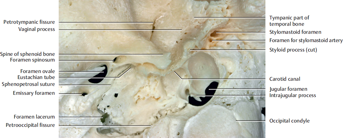
Fig. 2.1. The external surface of the skull base. Close-up view of the stylomastoid foramen.
The facial nerve exits from the stylomastoid foramen and is supplied by the stylomastoid artery, which usually originates from the posterior auricular artery. The stylomastoid artery enters into the skull from the foramen adjacent to the stylomastoid foramen.
The foramen ovale allows passage between the middle cranial fossa and the infratemporal fossa of the mandibular division of the trigeminal nerve, the lesser petrosal branch of the glossopharyngeal nerve, the accessory meningeal branch of the maxillary artery, and some emissary veins. Behind the foramen ovale lies the foramen spinosum, which transmits the middle meningeal vessels and meningeal branch of the mandibular division of the trigeminal nerve.
The jugular foramen is located between the temporal bone and the occipital bone. The structures that traverse the jugular foramen are the sigmoid sinus and jugular bulb, the inferior petrosal sinus, the meningeal branches of the ascending pharyngeal and occipital arteries, the glossopharyngeal, vagus, and accessory nerves with their ganglia, the tympanic branch of the glossopharyngeal nerve (Jacobson’s nerve), the auricular branch (also known as the mastoid branch) of the vagus nerve (Arnold’s nerve), and the cochlear aqueduct. The intrajugular process is a small, curved process which partially or completely divides the jugular foramen into lateral and medial parts.
Behind the foramen spinosum, the bone is raised to form the spine of the sphenoid to which the sphenomandibular ligament is attached. The posterior margin here is grooved and is related to the cartilaginous component of the Eustachian tube (see Fig. 10.18).
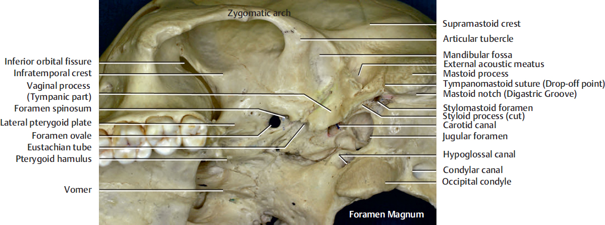
The digastric muscle is attached to the mastoid notch (digastric groove), the anterior end of which indicates the stylomastoid foramen. The tympanomastoid suture is a landmark of facial nerve trunk. The facial nerve (stylomastoid foramen) can be identified at 6 to 8 mm medial to the inferior “drop-off “point of the tympanomastoid suture.
Jacobson’s nerve arises from the petrous ganglion of the glossopharyngeal nerve. It enters the tympanic cavity via the inferior tympanic canaliculus and contributes to the tympanic plexus. It contains both sensory and parasympathetic fibers. The sensory fibers supply the middle ear. The parasympathetic fibers leave the plexus as the lesser petrosal nerve and enter the otic ganglion.
Related posts:


Stay updated, free articles. Join our Telegram channel

Full access? Get Clinical Tree


