Skin coloration is highly diverse, partly due to the presence of pigmentation. Color variation is related to the extent of ultraviolet radiation exposure, as well as other factors. Inherent skin coloration arises from differences in basal epidermal melanin amount and type. Skin color is influenced by both the quantity and distribution of melanocytes. The effectiveness of inherent pigmentation for protecting living cells also varies. This article discusses skin color, pigmentation, and ethnicity in relation to clinical practice. Color perception, skin typing/classification, and quantitation of pigmentation are reviewed in relation to ethnicity, environmental stresses/irritants, and potential treatment effects.
Key points
- •
Skin color classification is a cornerstone of clinical practice. Quantitation of pigmentation in the context of response to stress is essential for determining cutaneous treatments.
- •
The quantity and distribution of melanin-producing melanocytes varies with inherent skin color. Both individually distributed and clustered melanosomes are significantly larger in African versus white skin, and of intermediate size in Asians.
- •
The greatest increase in melanosome protein levels after ultraviolet exposure occurred in black people. The melanin distribution varied with skin color, moving from the lower to middle layers and to a greater extent in black skin.
- •
Genetic studies across pigmentation types have identified at least 120 genes associated with skin pigmentation and the melanocortin 1 receptor gene seems to predominate.
- •
The relationship between ancestry/ethnicity and skin pigmentation shows a significant but modest correlation and high variability.
Introduction
Human skin color uniformity and distribution communicate physiologic health status. Facial images with uniform skin coloration are judged as younger versus images with highly variable color. For example, individuals were asked to adjust the red color component of facial images until it represented a healthy appearance. All observers increased red color but dark-skinned observers increased the redness of dark skin images more than for lighter skin images. Japanese women with darker and more yellow skin color were classified as older. This article discusses skin color, pigmentation, and ethnicity as they relate to clinical practice. Color perception, skin typing/classification, and quantitation are reviewed in relation to ethnicity, environmental stresses/irritants, and potential treatment effects.
Color perception
Humans detect color when retinal cone cells receive and mediate light via peak absorption at 564 to 580 nm (red), 534 to 545 nm (green), and 420 to 440 nm (blue) wavelengths of the color spectrum. Visible light interacts with skin components; namely, melanins (yellow to black), oxygenated hemoglobin (red), deoxyhemoglobin (blue-purple), bilirubin (yellow), and carotene (yellow). Approximately 5% is reflected back and the remainder is transmitted, absorbed, or scattered. The stratum corneum transmits light. The epidermis and dermis absorb light because of melanin and hemoglobin. Melanin is synthesized by the melanocytes and transferred to keratinocytes throughout the epidermis. Oxygenated blood (dermal capillaries, vascular plexus) and deoxygenated blood (dermal venules) contribute to skin color. Epidermal carotene and bilirubin contribute to yellow coloration. Bilirubin is deposited in phospholipid membranes and leaked into extravascular regions. Light is scattered within the epidermis, dermis, and subcutaneous fat.
Two kinds of skin pigmentation are considered here. Constitutive pigmentation is the inherent type; that is, the color of sun-protected areas such as the upper inner arm ( Table 1 ). Facultative pigmentation is produced as a result of ultraviolet (UV) exposure, either chronically or in response to a specific dose of radiation. Facultative pigmentation varies for an individual, reflecting changes in UV intensity with changes in season. Minimal erythema dose (MED) is the minimum amount of ultraviolet radiation required to produce minimum erythema (see Table 1 ).
| Classification | Melanin Content (μg/mg) |
|---|---|
| Ethnic Group | |
| Asian | 3.9 |
| Black/African American | 15 |
| Hispanic/Latin American | 8 |
| Native Hawaiian/other Pacific Islander | 11 |
| White | 4.5 |
| Fitzpatrick Skin Phototype | |
| 1 and 2 | 4 |
| 2.5 | 5 |
| 3 and 3.5 | 5.7 |
| 4 | 11.5 |
| 4.6–6 | 13 |
| MED Range (J/m 2 ) | |
| ≤225 | 3.5 |
| 226–300 | 4 |
| 301–400 | 7.3 |
| 401–600 | 9.5 |
| 601–800 | 12.3 |
| ≥801 | 14.3 |
Skin pigmentation protects living cells from the injurious effects of environmental UV radiation and repairs the DNA damage that occurs. Epidermal melanin diminishes UV penetration and removes reactive oxygen species generated during UV exposure. UV radiation can suppress the immune system’s ability to prevent actions of tumor antigens both systemically and locally, as shown by changes in and reduction of the levels of Langerhans cells. Constitutive pigmentation varies in the effectiveness of the protection it affords living cells.
Skin color classification
Skin color classification is a cornerstone of clinical practice and is used to decide on the treatment of cutaneous conditions. The Fitzpatrick skin type system originated in 1975 from a need to better predict patient response to the UVA radiation used in psoriasis treatment. Patients of similar visual skin color reacted differently to the same UVA dose, with some developing severe burns. The classification was broadened to include response to environmental stress, namely UV radiation. Therefore, the UV response serves as a surrogate for predicting the reactivity to other stresses, such as laser energy. Inherent skin coloration (constitutive pigmentation) and reaction to UV radiation (facultative pigmentation) gave rise to 6 types. Fitzpatrick phototype I always burns, never tans; phototype II usually burns, sometimes tans; phototype III may burn, usually tans; phototype IV rarely burns, always tans; phototype V has moderate constitutional pigmentation; and phototype VI has marked constitutional pigmentation. Total UV exposure is associated with the incidence of squamous cell carcinoma, which is higher in light skin and high reactivity to UV radiation. The lightest skin types experience the highest incidence of melanoma.
Limitations of the Fitzpatrick system have been noted, because of difficulties with self-reports of the effects of UV exposure. The relationship between self-assessed Fitzpatrick type and skin color at the wrist (sun protected) was determined among 3386 ethnically diverse subjects. The darkest Fitzpatrick types (V and VI) were self-assigned in 17.7% of participants with the lightest skin. Statistical modeling showed the selected Fitzpatrick type to be 1 classification off, on average. To address these limitations, a color-based method for skin type self-assessment was developed from evaluations of 120 white, Hispanic, and African people. They selected their skin coloration from a color figure (available at www.jaad.org ) and questions about effects of UV exposure. Melanin index was measured spectroscopically. Efforts to develop more objective, quantitative skin color measurements to assist clinicians in predicting response to skin treatments are ongoing.
Quantitation of Skin Color
Expert visual assessment of skin color is routine in clinical practice but is limited by high variation and low reliability. Digital imaging (standardized lighting, focal length, positioning, color correction) and spectroscopic methods have been used to improve objectivity and to quantify features (eg, dyspigmentation, erythema). Digital photographs (collected as RGB, ie, red, green, blue colors), are typically converted to L*a*b* color (L*, lightness-darkness; a*, red-green; and b*, yellow-blue) because it better replicates human color perception.
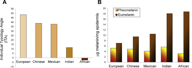
The impact of melanin on lightness (L*), red-green (a*), and blue-yellow (b*) as a function of ethnicity (European, Chinese, Mexican, Indian, African) was investigated in the experiments by Alaluf and colleagues. For sun-protected skin sites, lightness was highest for Europeans and lowest for Africans. Indian skin was significantly darker than European, Chinese, and Mexican skin. Red color (a*) was significantly higher for Indian and African versus the other groups. Yellow color (b*) was significantly higher for Indian and African versus all others and for Chinese and Mexican versus European. L* was significantly and inversely correlated with levels of eumelanin and total melanin overall and for all ethnic groups (correlation coefficients, −0.77 to 0.89). Although significant, L* was inversely correlated with pheomelanin amounts with lower correlations (−0.23; range, −0.53 to 0.83).
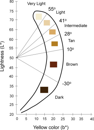
To quantify pigmentation, the individual typology angle (ITA) was determined from L* and b* values using this equation:
The values constitute 6 groups of skin pigmentation: ITA greater than 55° (very light), 41° to 55° (light), 28° to 41° (intermediate), 10° to 28° (tan), −30° to 10° (brown), and less than−30° (dark). The ITA values and categories, shown in Fig. 1 , were reported for 3500 subjects from white, African, Asian, Hispanic, and Brazilian ethnic groups. ITA values correlated with skin pigmentation. Skin erythema, with confounders from perfusion, is typically quantified by a* color value. Fig. 2 shows the ITA values (see Fig. 2 A) for each ethnicity, along with eumelanin and pheomelanin levels (see Fig. 2 B) from Alaluf and colleagues (described earlier). The associations among skin classifications are shown in Fig. 3 along with corresponding melanin index ranges (spectroscopic assessment), minimum erythema dose ranges, and colors typified by each classification.
Related posts:

Stay updated, free articles. Join our Telegram channel

Full access? Get Clinical Tree


