Secondary breast augmentation

3 Secondary breast augmentation


Introduction


Breast augmentation has been performed using breast implants since the early 1960s. Since then, millions of women worldwide have undergone breast augmentation. As the technique, surgical approach and implants have improved, results have become more predictable and stable. Given the sheer numbers of women who have breast implants in place, the number of women presenting for reoperative breast surgery continues to increase. In fact, many plastic surgeons who focus on breast aesthetics are seeing their practices evolve into specialty centers for revision breast surgery.


A consistent theme in this chapter will be prevention. The need to perform revisionary surgery has several consequences. It places the patient at an unnecessary risk. The best way to avoid risk is to avoid surgery. Revision surgery is costly to the patient. It is also costly to the surgeon and to the surgical practice. Most importantly, it is never as easy to get as good and as predictable a result as it is the very first time that the breast is operated on.


Revision breast augmentation can be a very difficult and challenging undertaking. The normal anatomy that exists in a primary procedure is no longer present. The surgeon must deal with a variety of factors, some predictable and some not predictable. These include: external and internal scarring, previously dissected tissue planes, alteration to the deep and superficial blood supply to the breast, alteration to the pectoralis major muscle (PMM), thinning and stretching of the overlying soft tissues and secondary effects related to the presence of a capsule around the pre-existing implant.


There are many reasons why a woman presents for secondary implant surgery, however the most common reason is for the management of capsular contracture.1 Implant malposition is another common presentation and can be categorized into malpositions that are superior, inferior, lateral or medial. Other common indications include the management of rippling or implant edge palpability, size change, device failure and finally the correction of soft tissue changes secondary to hematoma, infection, radiation or previous surgery. Regardless of the presentation, the management of the secondary implant patient requires a systematic approach to identify the underlying problems, recognize the limitations, and develop a plan that is safe and maximizes the chances for a predictable outcome.


Diagnosis/patient presentation


The first step in evaluating the revision patient is to develop a clear understanding of their concerns and expectations. This may be easier to explain for some patients than others. Often, patients are abstract in the discussion of their concerns. Phrases such as, “this isn’t what I wanted”; “I am very unhappy with my breasts”; “My friends tell me this can’t be right, it should be fixed”, demonstrate displeasure with the outcome but are hardly specific. To correct a problem, one must understand what the problem is, why it occurred and have a reasonable plan as to how to fix it. There is not a solution for every concern, nor is it reasonable to try to correct every outcome that is felt to be less than perfect.


A detailed history should include patient goals and expectations, general health and an assessment of risk factors for compromised healing including smoking history, systemic illness or previous radiation to the breast. The patient’s breast health should be reviewed including the presence of past breast disease, family history of breast cancer and the timing of previous breast imaging. All prior breast surgeries should be well documented and operative notes obtained, if available. It is impossible to plan for a successful revision procedure without having a clear understanding as to what was done in the past. Table 3.1 lists the key points of information that should be known about previous breast procedures.


Table 3.1 Previous breast surgery


Physical examination should focus on three components: the implant, the soft tissues, and the musculoskeletal framework. These three variables will completely define the dynamics of the presenting problem. The implant must be examined for position under the breast tissue and stability within the implant pocket. Malposition should be classified into medial (symmastia), lateral, inferior (double bubble) or superior. Implant stability within the pocket should be assessed with the patient in multiple positions including standing with arms at side, arms over head, lying supine and leaning forward. The presence of capsular contracture should be noted along with any areas of rippling or palpable implant edges.


Examination of the soft tissues is very important not only to determine a surgical plan but to inform the patient what is realistically achievable with surgical correction. Any asymmetries should be recorded and discussed. Measurements include sternal notch to nipple distance, nipple to IMF distance and breast width. An attempt should be made to compare the width of the breast to the dimensions of the underlying implant. Nipple position on the breast mound should be noted, along with degree of ptosis or glandular ptosis. The quality and quantity of breast tissue in all four quadrants should be assessed to determine adequacy of soft tissue coverage. This may be done by subjective assessment or by more objective measures such as the pinch test. The breast and axilla is examined for abnormal masses and finally all scars are recorded for both location and quality.


As breast implants require a stable foundation to sit on, examination of the musculoskeletal anatomy is important. Pre-existing scoliosis or chest wall asymmetries should be recorded. Abnormal contour of the sternum either concave or convex will affect medial positioning of implants. Similarly, patients with asymmetric contour of the rib cage either superiorly or inferiorly will affect the upper pole fullness or the stability of the implant at the level of the IMF. The pectoralis muscle should be examined as it may have been damaged or excessively released with previous surgeries. In cases of Poland syndrome, the muscle may be absent or rudimentary.


Investigations should be patient specific. Implants can be imaged with a combination of ultrasound, mammography or MRI. The soft tissues should be imaged as necessary for abnormal masses or calcifications. In general, ultrasound and mammography are used to image the breast tissue and assess for actual breast pathology. The author routinely requires a screening mammogram within one year prior to any elective breast surgery for all women over the age of 40. MRI is indicated for assessment of integrity of the breast implant and specifics of soft tissue or skeletal asymmetries. It is also useful in identifying the exact plane of the implant and its relation to the pectoral muscle. In patients who have undergone multiple procedures, the exact implant location and the status of the pectoral muscle can often be difficult to determine. The value of any radiologic investigation should be weighed against the cost including actual financial cost of the test, radiation exposure and patient time. Concerns within the breast tissue or axilla may require a referral and opinion from a breast oncologic surgeon.


Classification


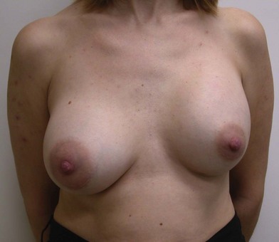
Revision breast augmentation surgery can be classified into three categories: related to the previous surgery, related to the soft tissues, related to the implant. Problems related to the previous surgery are most common. There are many decisions that go into planning any breast surgery. Improper pocket selection, improper implant selection, over or under dissection of the pocket, infection and postoperative bleeding can all affect the final outcome. Patients who would benefit from a skin tightening procedure but instead undergo the insertion of a large implant will likely have a short term gain with unacceptable results in the longer term. Figure 3.1 shows a woman who underwent bilateral subpectoral augmentation and presented with concerns of breast asymmetry. Upon surgical exploration, it was determined that the right pectoral muscle had been released along the IMF to the level of the sternum, but the left muscle had not been released at all, resulting in a unilateral high riding implant.


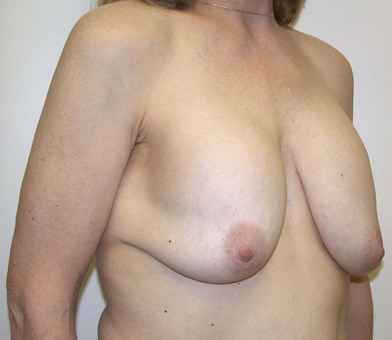
The second category includes issues related to the soft tissues. Breast tissue will undergo normal physiologic changes throughout a woman’s lifetime. These changes may include atrophy, hypertrophy, thinning or stretching. Changes are exaggerated by events such as pregnancy, breast-feeding or fluctuations in weight. The presence of an implant may hasten some of these changes, especially thinning of the skin, the development of striae and stretch marks or the development of ptosis and pseudoptosis. Even if the implant itself remains in a normal position, without complications, the development of soft tissue changes on their own can be enough to drive the need for revision surgery. Figure 3.2 shows a woman with normal, soft implants and overlying breast ptosis. This “waterfall” deformity requires a mastopexy to bring the breast tissue back up to where the implant sits.


The final category includes issues related to the implant itself. This would include capsular contracture, implant malposition, implant deflation, rupture or implant palpability and rippling. The remainder of this chapter will focus on how to prevent and treat these causes of revision breast surgery.


Patient selection/indications


The decision to proceed with a revision breast augmentation should only be made after careful consideration of the patient’s concerns and a thorough discussion regarding goals and expectations. As the primary surgeon, it is possible to decrease revision rates by committing to basic core principles that include; patient education, informed consent, preoperative planning, proper implant selection using tissue based planning, precise surgical technique and structured postoperative care. Each indication for a revision can find its roots embedded in one or more of these principles. For example, a patient whose implant was improperly matched to their soft tissue envelope will likely develop implant malposition, rotation or rippling. A patient who is concerned about an ongoing asymmetry but did not understand that this asymmetry existed preoperatively will not be prepared to accept the continued difference in her two breasts. Surgery that uses imprecise technique increases the likelihood of infection, contracture, hematoma and implant malposition. A device placed through too small an incision will be more likely to fail.


The best way to manage a revision is to have avoided it in the first place. Having said that, errors do occur, soft tissues do not always behave as planned, well intentioned decisions are not always born out to be correct, patient’s bodies continue to change and the principles of gravity continue to exist. For these reasons, the need for revision augmentation surgery will persist.


The surgeon and patient should spend time performing a risk/benefit analysis. On one side of the equation are the reasons why the patient wishes a revision. The patient may have symptoms such as pain from a capsular contracture or a tight, poorly healed scar. She may have restriction of movement that affects her ability to do common activities. Other concerns may relate to the effect on quality of life. Asymmetry, improper sizing, malposition or contracture may limit the wearing of certain clothing styles and this may impact on the patients self image, self-esteem and confidence, all reasons for having the augmentation done in the first place. These concerns must be balanced against the inherent risks and complications of the revision as well as the likelihood for being able to meet the patients stated goals. The commonly used phrase; “the enemy of good is better” should be carefully considered and discussed with the patient. There is nothing worse than performing surgery to correct a relatively minor problem only to be left with a major one.


Not all revisions of previous breast augmentations require the use of an implant. Patients who have had multiple previous procedures, painful contractures, implant ruptures or complex malposition problems may be best served by implant removal with or without capsulectomy. Often, breast reshaping in the form of a mastopexy can be performed simultaneously or at a later date. For patients with saline implants, consideration can be given to preoperative deflation of the implant. This can be done in the office using a sterile prep-tray, needle and suction. Patients must sign a consent committing to the plan for definitive surgery. This short procedure allows both the patient and surgeon to assess the quantity and quality of breast tissue as well as visualize any existing asymmetries. On several occasions, the author has had patients decide to accept the breast as is (other than removal of the deflated device) or to perform a lift only.


As with any procedure, patients undergoing revision breast augmentation must have realistic expectations. There is not a solution for every problem and in some circumstances, it is best not to offer a surgical option.


Any implant can be used in reoperative breast surgery, however there are certain circumstances where a particular implant may offer a unique benefit. The surface of an implant may affect issues of rippling, palpability and capsule formation. Patients with rippling may gain benefit from the use of a smooth device whereas patients with repeated contractures may have less chance of a recurrence with a textured implant. In situations of implant malposition and capsular contracture, the author prefers using textured surface devices. This decision is not absolute and requires a new pocket for the implant and adequate soft tissue cover to minimize the risk of traction rippling. In most cases of secondary implant surgery, it is the author’s view that everything possible should be done to attempt to correct the presenting problem. This usually requires the selection of a new device. The cost of new implants as well as surgical costs, are the responsibility of the patient. In primary implant surgery, the author discusses financial implications of subsequent surgery as part of the consent process. Patients are made aware that they may be responsible for the costs of any revision procedure and for the cost of new implants, if indicated. The author reserves the right to determine his surgical fee based upon the reason for revision and time since primary surgery. In Canada, the treatment of conditions such as symptomatic capsular contracture, infection or hematoma are considered insured services.


In situations where an implant is too wide for the breast, higher profile devices can be selected and in cases of breast asymmetry, the surgeon may select implants of different projection. Round implants produce a shape that is very different from anatomic devices and do not carry the added risk of implant rotation. Shaped implants however, have the potential to correct various asymmetries, produce a natural upper breast contour and have a low rate of capsular contracture.24


Specific contraindications exist when using shaped gel implants in revision surgery. Of course, a patient must be willing to accept the use of a gel device. Outside of North America, this is rarely an issue, but there are a number of women within Canada and the United States who continue to have a greater comfort level with saline implants. Understanding a woman’s goals is key to selecting the best implant. Patients who would like a full looking upper pole are not good candidates for a shaped implant. The form stable nature of the gel results in a breast that is slightly firmer than what is seen with more responsive gel devices. Patients who are unwilling to accept this will do better with a softer round gel or saline implant.


The location and length of the surgical scar is an important consideration for most implant patients. In revision surgery, there will be a pre-existing scar. Surgery should always be performed through an incision that allows for direct access and precise correction of the underlying problem. If that cannot be done through the previous scar, then a new incision should be selected. When using a gel implant, it is critical to have a long enough incision to allow for implant insertion without damaging the device. This is generally in the range of 4 cm for a round implant and 5 cm for a form stable shaped device. This incision length limits the choice for scar location. Most commonly, the incision will be placed in the inframammary fold or through a large periareolar incision. If an axillary approach is selected, it is imperative that the incision be of adequate length. Patients who are not willing to accept the required scar length and location are not candidates for gel implants.


Treatment/surgical technique


Careful preoperative planning is a cornerstone of any successful breast augmentation. This statement is especially true in revision surgery. It is absolutely imperative to have a detailed understanding of all previous breast surgeries. Whenever possible, surgical records should be obtained and reviewed. Often, women presenting with breast implant problems have had multiple procedures. These may have included mastopexies, reductions, biopsies or lumpectomies. Each of these surgeries will have left scars and have caused an alteration to the normal blood flow within the breast and to the nipple areola complex. For each previous breast implant procedure, it is important to know the implant fill, surface, size, pocket and incision used. Any complications such as infection, contracture or malposition should be recorded, including the methods by which these complications were treated.


The exact surgical plan will be determined by the specifics of the patient. Revision procedures may involve implant removal with or without a mastopexy. When the plan calls for insertion of a new implant, there are several fundamental decisions that need to be made. Pocket selection is a very important consideration. Options for the implant pocket are summarized in Table 3.2.


Table 3.2 Options for implant pocket


When the capsule around the implant is normal, there is adequate soft tissue coverage and the implant/soft tissue relationships are acceptable then the same pocket can be utilized. This is most common with revisions for size change or minor malpositions. In the case of minor contractures, an open capsulotomy or partial capsulectomy may be indicated. With Baker III/IV contractures, implant malposition, rippling or edge palpability a site change is usually indicated. When revision surgery calls for the use of a shaped gel implant, a pocket change is mandatory to allow for a precise pocket to implant fit. Site change can take the form of a capsulectomy, subglandular to subpectoral conversion, subpectoral to subglandular conversion or the creation of a new subpectoral pocket on top of a previous subpectoral capsule.5 A variation on site change is the conversion to a dual plane pocket as described by Spear.6 This approach is helpful in many cases of contracture or superior implant malposition.


A second critical consideration is the implant type, shape and size. There is no single formula that can be applied to all cases of revision augmentation but certain principles can be followed. In cases of contracture, consider the use of a textured surface implant. Several studies suggest that there is a lower capsule contracture rate with textured implants, especially when the implant is placed in the subglandular space.7,8,9 Consider the use of smaller implant sizes. Many problems following breast augmentation are as a result of soft tissue changes secondary to a large implant. It is usually better to use a smaller implant size with a skin tightening procedure rather than filling a skin envelope with larger volume devices. Shaped gel implants have a central role to play in implant selection for revision augmentation. Although they usually require a new pocket for insertion, they have many benefits including a lower contracture rate, less rippling, and shape stability. The variety of sizes allows for correction of asymmetries and the form stable nature of the gel allows the implant to shape the breast rather than the breast shaping the implant. Selection of the implant size must be based on defined measurements of implant height, width and projection to insure stability of the implant under the soft tissues.2,10


The final preoperative decision relates to the soft tissues. In cases of ptosis, glandular ptosis, skin stretch, asymmetry or nipple malposition, an alteration to the soft tissue envelope may be necessary (Fig. 3.3). Combining a mastopexy with revision of an augmentation is a complex procedure with many variables, some unpredictable. It should be undertaken with caution and only in patients with few risk factors for delayed wound healing. The soft tissues should be handled with extreme care and all skin undermining should be kept to a minimum to decrease the likelihood of skin flap or nipple loss.


The technical steps required to perform a successful breast augmentation are beyond the scope of this chapter, however many of the principles are shared when performing revision breast augmentation. Preoperative markings are done with the patient in a standing or sitting position. When using shaped implants, these markings are critical, as the implant pocket must be dissected just large enough to allow the implant to be inserted. The surgical steps will vary depending upon the individual case but usually follow a similar pattern; removal of the old implant; management of the existing capsule; creation or preparation of a pocket for the new implant; insertion of the new implant, and then contouring of the soft tissues. It is always advisable to use precise dissection techniques under direct vision. In primary augmentation, the use of sizers is rarely indicated but in revision cases, sizers may be used more frequently. When using a shaped implant, it is important to use a sizer slightly smaller then the anticipated implant size. Too large a sizer, will result in over-dissection of the pocket and increase the likelihood of rotational problems with the final implant.


Implant insertion is performed using a no touch or minimal touch technique. This is done to decrease the risk of implant contamination and eventual biofilm formation or infection. Minimal digital palpation is used to insure that the implant is sitting flush with the chest wall and in the desired location. Closed suction drains are usually necessary in revision procedures, especially when performing a capsulectomy or pocket change. In thin patients with minimal breast tissue, it is important to place the drain in such a way that there is a lengthy subcutaneous tunnel. This will minimize the likelihood of bacterial tracking especially following drain removal.


Contracture


Capsular contracture is classified according to the classification proposed by Baker. The Baker Classification is still used today and is very simple. The original Baker classification defines four classes11,12:


Capsular contracture represents the most common reason for secondary breast implant surgery.1 The treatment of capsular contracture begins with prevention. Multiple factors have been implicated in the development of contracted capsules and they are listed in Table 3.3.


Table 3.3 Etiology of capsular contracture


Many of these factors can be controlled through precise surgical technique, proper patient and implant selection, patient education and defined post surgical protocols. Maintaining a clean, non contaminated pocket is critical in preventing contractures. The author places an OpSite shield over the nipple at the start of surgery to minimize contamination from the lactiferous ducts. After creation of the pocket, irrigation is performed with an antiseptic solution. Many recommendations have been made as to the ideal solution for irrigation and include betadine, bacitracin and antibiotic solutions. Adams has published extensively on this area and recommends a triple antibiotic mixture that includes 50 mL of povidone-iodine, 1 g of cefazolin and 80 mg of gentamicin in 500 mL normal saline.13 This author has used a 50/50 mixture of povidone-iodine and normal saline for 15 years with good success. Following preparation of the pocket, the surgical gloves are changed prior to opening the implants. The implants are bathed in the irrigation solution and then inserted. Various techniques have been reported for implant insertion. These range from cleaning the skin around the incision to a complete no touch technique.14 Other options include using an insertion sleeve or a lubricated funnel. Prophylactic antibiotics are given prior to surgery and for several days after surgery.


Our present understanding of the entire etiology of capsular contracture is lacking. It is necessary for us to develop a better understanding of the physiologic and biochemical processes responsible for capsular contracture if we expect to completely eradicate this problem. Until then, a certain percentage of implant patients will continue to present for treatment.


The decision to treat a contracture is usually based upon the aesthetic consequences of the contracture and the presence of patient symptoms. For minor degrees of contracture (Baker class II), it may be best to take an expectant approach. Given that there is no treatment that can guarantee success, the risks of treating low grade contractures often outweigh the potential benefits.


There are very few nonsurgical options for the treatment of established capsular contractures. In 2002, Schlesinger et al. reported on the use of Zafirlukast (Accolate) for the nonsurgical treatment of capsules.15

Only gold members can continue reading. Log In or Register to continue

Stay updated, free articles. Join our Telegram channel

Feb 21, 2016 | Posted by in General Surgery | Comments Off on Secondary breast augmentation

Full access? Get Clinical Tree

Get Clinical Tree app for offline access