Fig. 7.1
Typical appearance of infected, surgically repaired distal tibia. (a) Exposed hardware, screws backing out. (b) Removal of hardware reveals nonviable bone requiring debridement
An infection occurring in a surgically treated fracture of the distal tibial plafond may prove impossible to eradicate (Bourne et al. 1983; Childress 1965; Coonrad 1970; Cox 1965; Gay and Evrard 1963; Green et al. 1996; Green and Roesler 1987; Ruedi and Ailgower 1979; Ruedi and Allgower 1969; Scheck 1965). The presence of nonviable bone fragments and hardware within the septic focus, combined with a thin or deficient soft tissue envelope, makes reconstructive surgery extremely challenging (Leach 1984; Ovadia and Beals 1986; Pierce and Heinrich 1979).
Rarely, the treating physician may elect to leave the initial internal fixation in place, even though the hardware is exposed in an open wound. In our series, we selected this option for two patients; both had excellent stable internal fixation with only exposed screw heads and radiographic evidence of progressive osseous healing. Generally, the infection in such cases starts out as localized skin slough over a screw head or other prominent piece of hardware several weeks or months after the original injury. Inflammatory changes must be limited to the skin immediately surrounding the slough. By leaving the hardware in place, the surgeon may aid fracture healing, but only if fixation is secure (Burwell and Charnley 1985).
When considering whether to “wait out” the local skin slough (leaving the hardware in place), the physician should avoid wishful thinking, hoping that the infection does not involve deeper osseous tissues. The compression-type pilon fracture often creates nonviable bone fragments that can become the focus of persistent sepsis if microorganisms enter through an area of skin breakdown. Serial radiographs should show disuse osteopenia of all bone fragments; nonviable bone will remain relatively dense on X-ray films as time passes.
In some cases, early healing of a fibular fracture, combined with evidence of progressive healing of the tibial fracture, may suggest that a septic medial (or anterior) tibial buttress plate (if present) can be removed to eliminate the infection. (Fractures originally requiring such a plate, however, have the potential for angulating, especially if there is substantial supramalleolar comminution. In general, these fractures will collapse into varus or apex posterior angulation if not united. Furthermore, motion at such a nonunion site helps perpetuate the infection, leading to an unstable, infected nonunion at the level of the leg where there is only limited soft tissue cover. For this reason, removal of hardware before union of the distal tibial fracture should be accompanied by a strategy (either external skeletal fixation or a non-weight-bearing cast) to prevent fracture angulation.
7.2.2 Classification System
Kallem and Waddell’s classification of distal tibial fractures(Kallem and Waddell l979) recognizes two basic categories based on the mechanism of injury: rotation fractures and compression-type fractures. With rotation fractures, the distal tibial fractures into two or more large fragments (with minimal or no anterior tibial comminution), combined with a transverse or short oblique fracture of the distal fibula. The compression-type fracture has marked anterior tibial comminution, multiple distal tibial fragments, and superior migration of the talus. (The distal fibula may or may not be fractured in the compression-type injury.)
7.2.3 Series Report
At the Problem Fracture Service at Rancho Los Amigos Medical Center, the author treated 13 infected fractures involving the distal tibial metaphysis and plafond.
7.2.3.1 Demographics
The demographic details of this series of patients is of interest. The average age of the patients was 43 years. Ten patients had no associated injuries, while three patients suffered a variety of injuries, including a peroneal palsy, a severe head trauma, and polytrauma with multiple long-bone fractures.
Ten of the patients in this series had compression-type fractures, and three had rotation fractures. Eight patients had open injuries, and five sustained closed fractures.
7.2.3.2 Mechanism
The mechanism of injury was a fall in seven patients, a motorcycle accident in three patients, a twisting injury in one, an assault in one, and unknown in one patient. Seven of the 13 patients were initially treated with open reduction and internal fixation. Interestingly, all but one of the seven patients with internally stabilized fractures experienced a loss of fixation that would have required hardware removal and reoperation even if an infection had not developed.
7.2.3.3 Cultures
All patients had culture-positive drainage at the time of the initial evaluation at our clinic. Staphylococcus aureus was the most common organism encountered, followed by Enterobacteriaceae species and Pseudomonas aeruginosa. Ten patients had positive cultures for more than one organism. Since these injuries became infected from without inward, polymicrobial contamination is not surprising.
7.2.3.4 Pre-Ilizarov Protocol
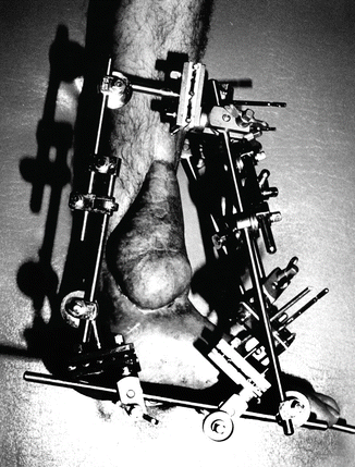
Our protocol for managing the infected pilon fracture employs the basic principles of septic fracture care (Cave 1965; Green 1982, 1983; Leach 1984; Muller 1982). Debridement of all nonviable bone is essential, along with removal of all hardware that may serve as a nidus of infection. Following debridement, the distal tibia must be stabilized to prevent angulation and shortening (Weller 1982). Of the 13 patients in our series, 11 were placed in an external fixator following hardware removal. Since the fixator must span the ankle joint, full pins are needed distally, almost always into the calcaneus (Fig. 7.2).


Fig. 7.2
Stable external fixation with Hoffmann apparatus. Note that the frame spans the area of surgery with pins in the calcaneus and forefoot. A microvascular flap covers the area of resection
Rigid external fixation may have to remain in place 8–12 months during reconstruction. Eventually, the calcaneal transfixion pins may become loose or septic, necessitating their premature removal. Placing one or more pins or wires pin across the midmetatarsal arch or forefoot when the frame is initially applied allows the construction of a triangular frame connecting the single metatarsal full pin to the pin cluster in the calcaneus and another pin cluster in the mid-tibia. Extraordinary stability and fixator longevity can be achieved in this manner, which creates a triangle between the tibia approximately and the heel and forefoot distally.
7.2.4 Mounting Principles
External skeletal fixation of the calcaneus is technically demanding. Numerous fixator configurations are possible for this type of reconstruction. All of them, however, should, at the very least, completely surround the limb, with either a quadrilateral system or a circular system. In this manner, transfixion implants in the foot are easily secured to the frame.
7.2.4.1 Quadrilateral Configurations
With quadrilateral external fixation, multiple pins within the same pin gripper placed into the tuber of the calcaneus should be in an oblique row, slanting downward and posteriorly, following the angle of the bone. Fluoroscopy aids pin placement with such devices.
7.2.4.2 Monolateral Configurations
Attempting to obtain consolidation after resection of the distal tibia and upper talus with a monolateral fixator is difficult and probably unwise. To do so, one would have to use threaded half pins, most likely inserted from the lateral side of the limb. Unfortunately, the cancellous nature of the calcaneus will not hold half pins for the many months required for the kind of surgery contemplated here. Instead, pin loosening becomes a distressing problem. The calcaneus is a small bone to start with and can quickly turn to Swiss cheese in appearance if implants work loose.
7.2.4.3 Ring Configurations
Certain fixators with ring configurations allow tensioned smooth or beaded wires to crisscross the calcaneus, thereby stabilizing it. The addition of one or more wires in the forefoot completes the distal configuration.
As for the proximal tibial mounting, any stable configuration that uses either wires alone, half pins alone, or combinations of pins and wires (including centrally threaded or fully threaded transfixion pins that attach to the frame on both sides of the limb) will do.
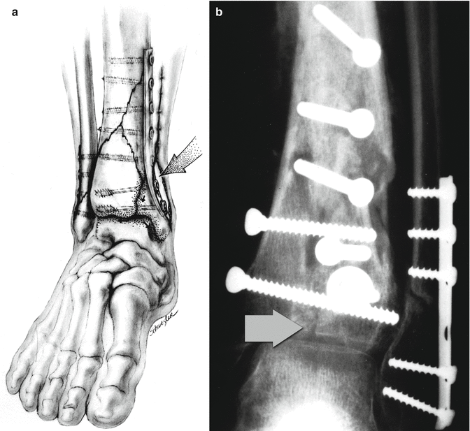
In two of our patients, and several others we have seen in consultation, thorough debridement of the infection, including removal of hardware and excision of nonviable bone from the distal tibia, failed to cure the infection. In these cases, we also observed progressive radiographic narrowing of the ankle joint caused by a subclinical pyarthrosis of the ankle. When wound breakdown over hardware or elsewhere occurs, it is best to assume that the joint infection tracks down a fracture line from the open wound into the ankle joint itself. Once there, the microbes establish a chronic infection that drains to the wound surface via a persistent fracture defect in the tibial plafond (Fig. 7.3a, b). For this reason, the usual clinical signs of joint infection—swelling, fever, and intense pain—may be absent. Instead, there is a slow, progressive bacterial degradation of articular cartilage manifested by joint-space narrowing and, possibly, juxta-articular bone erosion.


Fig. 7.3
Entry of microbe into the ankle joint space after wound breakdown. (a) Diagram showing passageway of bacteria along plate and screws into open fracture lines into the ankle joint. (b) Arrow points to open fracture line allowing entry of bacteria into the ankle joint from infected exposed hardware
7.2.5 Resection Principles
A chronic pyarthrosis of any joint is often difficult to eradicate under the best of circumstances and frequently necessitates arthrodesis (Weise and Weller 1982). When a chronic pyarthrosis of the ankle is combined with a chronic osteomyelitis of the distal tibial metaphysis, a classic joint-fusion procedure is impossible because debridement of both the distal tibia [metaphysis and the tibia] plafond leaves a large cavitary bone defect that opens anteriorly or anteromedially. With debridement of the upper talar articular surface as well, the defect extends down into the body of the talus.
7.2.5.1 Papineau Procedure
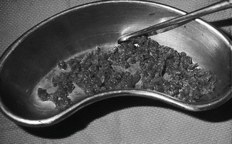
An open cancellous bone graft—the so-called Papineau procedure(Green 1994; Papineau 1973)—has been proven to be a reliable technique for filling in the defect created by extensive debridement of osseous tissue (Fig. 7.4).


Fig. 7.4
Fresh autogenous bone graft from the iliac crest, the “gold standard” for bone graft material
7.2.5.2 Microvascular Reconstruction
Free, microvascular, composite-tissue transfers—utilizing either the ipsilateral iliac crest or a rib—might also be useful for salvage. Union to the body of the talus, however, would be difficult to achieve. Alternately, coverage can be obtained with a soft tissue free flap, followed by closed bone grafting. (We do not use the contralateral fibula for reconstruction of an infected pilon fracture. In the event the reconstruction fails and the septic limb has to be amputated; we prefer to leave the good leg undamaged.)
7.2.5.3 Structural Considerations
When the reconstructive plan requires extensive bone grafting to fill a cavitary defect across the ankle joint, prolonged immobilization in an external fixator frame is required until the graft matures. Unfortunately, full corticalization of a cancellous bone graft takes 2–5 years, and prolonged bracing is necessary thereafter.
At the ankle, the foot projects forward at a right ankle to the tibia, creating a substantial bending movement in the “ankle” fusion site. A nonunion of the ankle graft mass—actually a motion-induced pseudoarthrosis—occurred in some patients in our series. One way to prevent this problem is to “corticalize” the graft by incorporating the distal fibula. For this reason, we do not remove the distal fibula when performing an arthrodesis of the ankle in infected pilon fractures. (By the time the patient has reached the point where secondary bone grafting of an osseous-articular defect is needed, any fibular fracture has probably already healed, permitting placement of a graft between the distal fibula and lateral side of the talus.)
The bone graft should be extended proximally between the distal tibia and fibula in the region normally occupied by the interosseous tibiofibular ligament. The goal of surgery is to create a solid mass of bone connecting the lower leg to the talus by whatever means possible.
In several cases, we noted that the infection of the distal tibial metaphysis, while perhaps requiring extensive debridement of the anterior cortex, generally spares the posterior and posterolateral distal tibia. When attempting fusion of the ankle for an infected pilon fracture, the surgeon might be able to abut the posterior cortex of the distal tibia to the posterior edge of the remaining upper talus, thereby enhancing the stability of the construct. This measure alone will do much to speed consolidation in cases managed with a bone graft in the ankle defect..
In spite of our best efforts, we had a substantial number of patients with unfavorable outcomes. Eight of the 13 patients had their fractures unite, but 2 of these 8 had persistent sepsis that we could not eradicate. There were two nonunions, and one patient required a below-knee amputation. Four patients required an ankle fusion to control chronic ankle pyarthrosis. The six patients whose fractures united and were free of infection (less than 50 % of the series) all had stiff ankles and scarred, dystrophic-looking skin around the distal tibia.
7.3 The Masquelet Technique
In virtually all circumstances, it is preferable to insert a bone graft into a closed rather than an open space. Likewise, an ideal situation is to have a cavity receptive to the bone graft already prepared and sterilized. During the last decade or so, the Masquelet technique has become popular among reconstructive surgeons (Donegan et al. 2011; Giannoudis et al. 2011; Karger et al. 2012).
A skeletal defect is filled with antibiotic impregnated bone cement, left in place long enough for a membrane to form around the cement mass. It has been shown that this membrane secretes growth factors, including BMPs, transforming growth factor-beta, and VEGF. When the membrane becomes mature, the cement spacer is carefully removed, leaving the surrounding membrane in place to serve as a bed for the bone graft.
7.4 Reamer Irrigator Aspirator (RIA)
Reports of donor site morbidity associated with the process of obtaining iliac crest bone graft has led surgeons to alternative sources. A popular method of attaining bone involves the use of a device, the Reamer Irrigator Aspirator (RIA), that extracts bone from the marrow cavity of long bones, especially the femur (Cuttica et al. 2010; Stafford and Norris 2010


Stay updated, free articles. Join our Telegram channel

Full access? Get Clinical Tree


