9 Revision surgery following breast reduction and mastopexy
Synopsis
 The plastic surgeon should know the pedicle created in the first operation and reuse it.
 “Tailor-tacking” (or skin staple tacking) is a powerful tool for simulating breast shape changes which are best evaluated with the patient sitting at 90° on the operating room table.
 Wide de-epithelialization of the peri-areola tissues with preservation of the subdermal plexus and the use of the interlocking Gore-Tex® suture technique is helpful where additional movement of the nipple is necessary in revision cases.
 Be extremely cautious in performing a mastopexy following a previous subglandular breast augmentation.
Introduction
Revision surgery following a breast reduction has been relatively uncommon in the author’s practice experience over the past 25 years – probably in the range of 2–3%. The main reasons patients seek or have revision surgery at various time points in the wound healing continuum are listed under the headings of sub-acute and long-term complications of breast reduction, listed in Table 9.1.
Table 9.1 Complications of breast reduction
| Acute complications |
| Sub-acute complications |
| Long-term complications |
Patient history
It is essential that the surgeon planning a reoperative procedure following breast reduction have as precise an understanding as possible about the blood supply to the nipple areola complex, as a revision surgery procedure may entail moving the NAC on a pedicle or altering breast shape adjacent to it.1 This is also true for revision of mastopexy and augmentation-mastopexy procedures. This information is most accurately obtained from a review of the previous operative record(s). The greatest margin of safety for ensuring nipple areola complex viability is obtained by using the previously developed pedicle. In patients you are evaluating after surgery performed elsewhere, these records can be obtained by asking the patient to request them in writing from the previous surgeon. In most US states, the law requires that permission must be given by the patient before you can speak with the previous surgeon.
Surgical re-intervention for acute problems
Hematoma
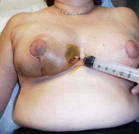
Reoperation in this setting is relatively rare. It mainly entails drainage of hematomas when they occur. The hematoma rate is ≤1–2%. It is marked by swelling, ecchymosis, and pain. It can occur at any location in the breast. For localized, nonexpanding and liquefied hematoma problems, a percutaneous aspiration may suffice (Fig. 9.1). It is essential to aspirate or drain hematomas since they may threaten the viability of the overlying skin3 and/or result in long-term palpable abnormalities and asymmetries in the breast(s). Any existing coagulopathies that may not have been identified preoperatively (e.g., von Willebrand’s disease4) must be sought and corrected.
Skin flap necrosis
Skin flap ischemia is rarely noted at surgery but rather it appears in the immediate postoperative period. In my experience, it is much more common in patients who smoke as is delayed healing of open wounds, wider scars, and fat necrosis. The author believes that this must be mentioned to all patients preoperatively, and a strong plea made to smoking patients to completely stop for 4 weeks prior to surgery.3 The incidence of complications following breast reduction has been linked to elevated BMI (>30), hypertension, previous breast incisions, and the amount of tissue resected.3
Nipple areola ischemia
Ischemia of the NAC is a dreaded but fortunately rare potential complication of every pedicled breast reduction procedure (probably in the range of 1%). It is most often related to arterial insufficiency, and it usually occurs in the setting of a large breast reduction (>1000 g resection), where a long pedicle is created to carry the circulation to the NAC, often with associated co-morbidities such as obesity and diabetes mellitus.2 Folding such a pedicle during closure additionally stresses the circulation. Therefore, in cases where NAC is suspected, the incision should be opened and the pedicle examined. If it is too bulky, it should be reduced in volume and the circulation re-evaluated. If this improves the situation, then re-closure of the wounds is attempted. If re-closure again causes a decrease in circulation, the pedicle might be trimmed further, the skin flaps might be made thinner, or some of the incisions may be left open.
If this still does not adequately address the problem, then consideration is given to removing the nipple from its position on the pedicle, resecting the distal pedicle, and applying the nipple as a full thickness skin graft more proximally on the pedicle itself (Fig. 9.2C,D)
Stay updated, free articles. Join our Telegram channel

Full access? Get Clinical Tree


