Introduction
The best way to treat a complication, is to avoid it. Unknown expert surgeon
Breast augmentation is a common operation, and it has been estimated that there are currently 5–10 million women with breast implants in the United States alone, based on different surveys. It is a requirement of proper informed consent that we inform our patients of the short- and long-term potential risks and possible complications of the procedure, not the least of which is that patients should expect to undergo additional revision procedures in their lifetime to maintain their outcome.
The patient undergoing breast revision will present to the plastic surgeon in a variety of ways. Many return to their original surgeon years later with still an acceptable outcome, yet want newer implants. Others may return after breastfeeding and desire a more youthful appearance of their breasts. Other patients will present after their 10th revision procedure elsewhere with a combination of tissue paper–thin tissues, striae/vertical skin pleating, oversized subglandular implant, and ptotic breasts. Many have unrealistic expectations of wanting to use the same periareolar approach and not wanting a breast lift. Every one of the patients seeking revision has a unique presentation, and it is our challenge to try to return their breasts to an aesthetic shape and softness.
This chapter will focus on evaluation and treatment of two of the more common reasons women seek breast revision: implant malposition and rippling. I will review the evaluation of implant pocket malposition and rippling and how to develop a surgical approach using straightforward techniques and recommended follow-up and postoperative care.
Preoperative Evaluation and Special Considerations
The patient interview is important for you to get to know the patient’s breast history and thorough medical and surgical history. You should know all you can about a patient, especially anything that may affect intraoperative and postoperative care. The more information you have about previous breast procedures, including incision(s), dissection technique, vascular pedicles, and implant characteristics, the more successfully you can plan your revision procedure. Try to obtain the medical records from her earlier breast surgeries, if possible.
The examination consists of standard breast measurements and evaluation of overlying soft tissue characteristics followed by a complete set of breast photographs to include a supine view, an animated view, and provocative views such as those producing rippling, wrinkling, and thinness of soft tissue coverage of the implant. Saline-filled implants are more likely to produce both malposition and rippling/wrinkling. Also take an image from behind to look for shoulder height asymmetry or scoliosis. Diagnostic ultrasound examination should be part of the examination, because it is useful to determine implant integrity, extracapsular gel, calcification, possible implant flip-over, double capsules, and periprosthetic fluid.
The physical examination of the patient undergoing breast revision is vital, but one will not fully appreciate the extent of implant malposition until in the operating room with the implant removed to inspect the pocket and chest wall directly. The intraoperative evaluation helps verify your exact plan of repair. I have often found at surgery that the implant pocket may be more spacious than expected. The implant capsule may be of benefit to your repair (“internal bra cup”) or may require partial or total removal if pathologic (i.e., calcified).
An effort should be made to determine as much as possible about previous surgical procedures such as scars, implant placement, and the size, style and type of implant. I have found cases in which operative notes informed me that previous submuscular implants were changed to a subglandular position, which changes my operative plan or at the least makes it more difficult. I choose to examine patients undergoing revision with them in the sitting upright position and then in the recumbent position. Measurements are taken and transcribed by staff, allowing the patient to demonstrate the aspects of their breasts that concern them with provocative maneuvers such as animation, bending over, or lying down. It is far better to see these issues before the procedure than to see them for the first time in the operating room.
If unable to determine the size, style, or type of implants currently implanted, it will be necessary to plan accordingly by the biodimensional process and bring along implants that can be chosen from to suit the patient’s desires. It is very helpful in revision breast surgery to acquire and review previous operative notes if they are available. Properly chosen implant sizers allow the surgery to be conducted and implants chosen until almost time to close incisions.
It is common to use a different implant with a different gel formulation at the time of the revision, which may further add to the complexity of revision, especially if the base width of the new implant is narrower than the existing implant. I have found that the newer generation, highly filled gel implants are less prone to wrinkling and rippling compared to earlier generation gel implants or saline-filled implants.
Type of Malposition and Its Management
Inferior Malposition
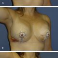
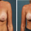
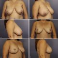
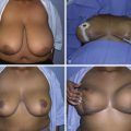
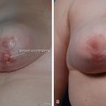
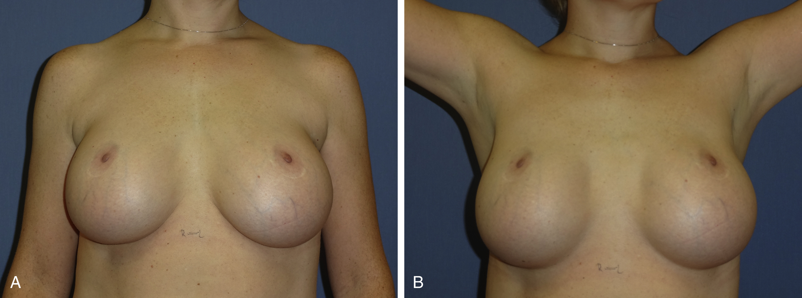
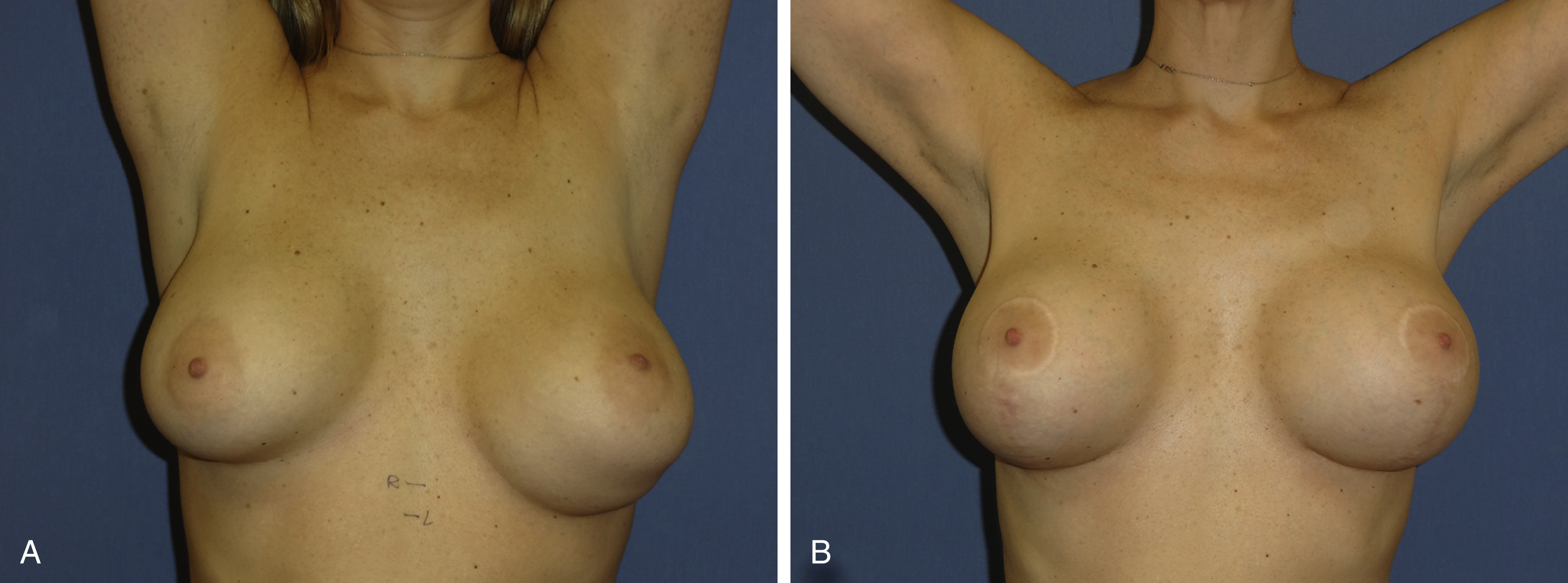
Inferior malposition occurs when the implant sits lower than the desired inframammary fold (IMF) typically allowing the fold incision to ride up on the breast mound if this approach was used. This can be seen with any incisional approach and typically leads to a thinning of the lower breast tissue that can cause a “star-gazing” deformity ( Fig. 10.1A, B ), asymmetry, and increased implant visibility and palpability because of the thin nature of the stretched breast and upper abdominal wall tissue ( Fig. 10.2A, B ). A variant of inferior malposition is the double bubble deformity ( Fig. 10.3A, B ). This can occur when an existing IMF is lowered to try to centralize the nipple–areolar complex on the augmented breast or in the incomplete release in a tuberous or constricted breast.
The repair of a double bubble deformity typically involves re-establishing the native IMF or softening a tight fold in a constricted breast with radial scoring of the parenchyma. A capsulotomy in the upper pocket may be needed to achieve upward position of the implant that occurs with raising of the IMF. Although a double bubble is more commonly seen in a submuscular augmentation, I do not personally recommend a site change to subglandular placement as a primary treatment.
There also should be a distinction made between lower pole stretch and inferior malposition. Lower pole stretch occurs when the IMF is in the proper position, but the lower pole soft tissue is attenuated, leading to an increased nipple-to-IMF position. Repair of lower pole stretch will involve removal of breast skin in the IMF to shorten the nipple-to-IMF distance. It is possible that these can exist together but the approach to repair is different.
Repair of inferior malposition involves a repositioning of the IMF to a higher position with the goal being to centralize the nipple on the breast mound. Unless the transaxillary or transumbilical approach was used to place the implants initially, the original surgical approach can be used. My preference is through the IMF, which allows a clear view into the implant pocket for capsulorrhaphy with or without the need for additional support of a synthetic mesh or acellular dermal matrix (ADM) for support. If a mastopexy is planned, the vertical limb provides excellent visualization for repair.
Preoperatively the surgeon must decide on the location to which the IMF should be elevated. If the implant characteristics are not going to change, you can estimate this position by pushing up on the lower implant to a point where the nipple–areolar complex is essentially centered on the breast mound. The new IMF is marked and then measured from the nipple. If the patient desires a smaller volume implant, the preoperative measurements remain important but the final design of the new IMF can best be planned with an implant sizer in place intraoperatively. For an IMF approach, the incision is placed on this new IMF line just lateral to the breast midline.
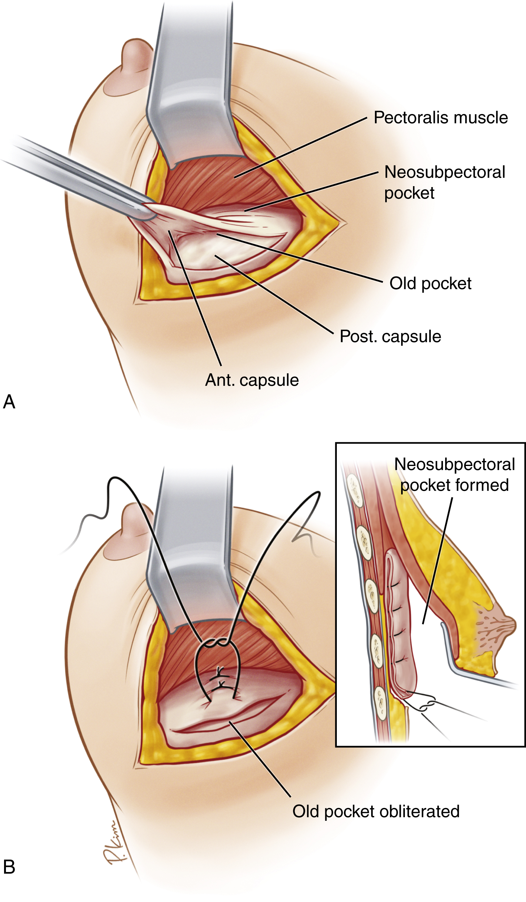
There are basically four described techniques to elevate the IMF: (1) capsulorrhaphy; (2) neosubpectoral pocket, as described by Maxwell and Gabriel ( Fig. 10.4A, B ); (3) capsular flap (see Fig. 10.7 ); and (4) resection of a strip of capsule with suture approximation of the raw edges. The latter is discouraged because of an already weak characteristic of the capsule and is mentioned only for completeness.
If the malposition is purely inferior, with the implant removed, tack the posterior capsule to chest wall with three interrupted 3-0 Ethibond sutures at the 6-o’clock position. It is important to inspect the capsular surface to be closed and either gently abrade with a lap sponge or treat with intermittent cautery to encourage adhesion of the two surfaces. This technique is described with a ball-tipped cautery in an effort the decrease the surface area of the capsule to in effect tighten the pocket. Next, the sizer is placed at this point to determine if you are pleased with the elevation of the fold and evaluate if the superior pocket needs to be opened to ensure there is no undue tension on the inferior repair. The sizer is then removed, and under direct lighted vision, a running permanent suture of the plastic surgeon’s preference is placed, grasping the capsule anteriorly and posterior capsular tissue posteriorly to include a purchase of perichondrium for substantial support when possible. I prefer a softer, braided suture such as 3-0 Ethibond. The anterior breast tissue is classically thin and vigilance is necessary to avoid dimpling or puckering. Once the capsulorrhaphy has been tapered to its endpoint the suture is then run back on itself for strength. The implant sizer is again placed to determine the position and smoothness of the repair.
By reducing the volume of the three-dimensional capsular space, you will be able to determine by bimanual palpation how much capsulotomy will be required anteriorly to allow some degree of implant mobility and breast softness. I commonly place a third and subsequent fourth row of capsulorrhaphy sutures for support. Evaluation of your repair should be accomplished by bringing the patient to an upright position with the arm boards brought down to the patient’s sides. This view will allow you to see if there is flattening or overcorrection of the IMF. Once the fold repair is determined to be satisfactory, the pocket is irrigated, the surgeon’s gloves are changed, and the new implant is placed with minimal skin contact, preferably with an insertion sleeve. Closure of the incision is in three layers with absorbable monofilament sutures.
The barbed suture is another option for fold repair. Reported advantages of this technique over traditional suturing techniques is said to include speed in execution and better tissue control with even distribution along the deep suture line and tension-free epidermal closure. In a study of wound complications with barbed sutures it was reported the barbed sutures were associated with significantly higher rates of minor wound complications, specifically if a two-layer closure was used. Readers are encouraged to become familiar with any new technique they adopt to include the risks, benefits, and potential complications.
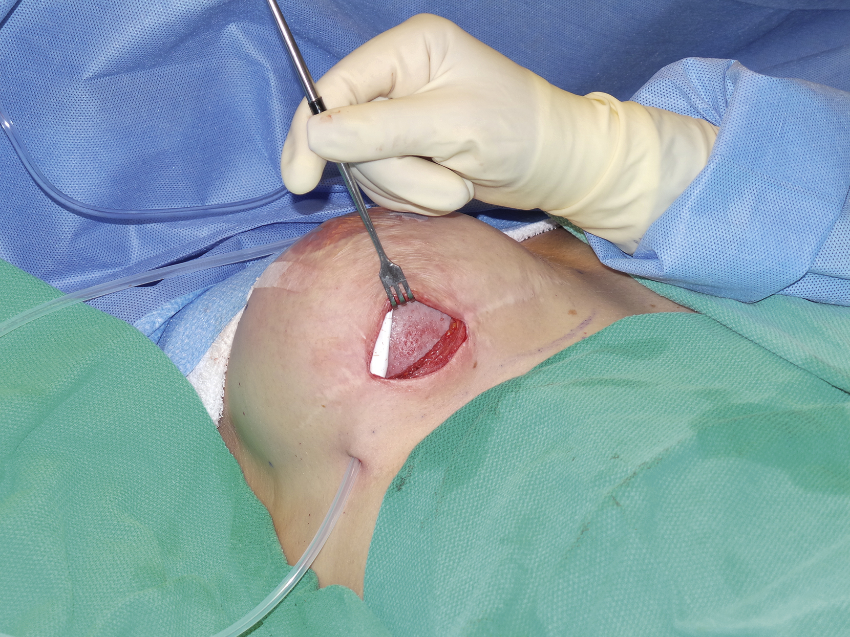
In some cases of weak or thinned tissue in the face of previous failures of repair, you may choose to support your repair with an ADM or a synthetic absorbable mesh (Gala-FLEX). Regardless of what you choose to use, it is your responsibility to know the product, the technique, and how to care for it postoperatively. Placing these materials directly against the capsule internally is possible, but you are more likely to have integration if a new raw surface is provided after partial or total capsulectomy ( Fig. 10.5 ).
External support of the internal repair is mandatory, and patients are instructed to wear a supportive postoperative bra at all times for the first 6 weeks. Patients should not immerse their incisions for 4 weeks, and physical activity is restricted per her surgeon’s recommendations. These patients undergoing revision should be seen frequently through their early recovery so that you can guide their recovery massage and garment wear and encourage activity restrictions. My choice for revision surgery uses smooth, round implants exclusively and recommends implant displacement exercises starting 2 days after the drains have been removed. It is critical to modify these exercises to protect any dimensions of repair.
Lateral Malposition
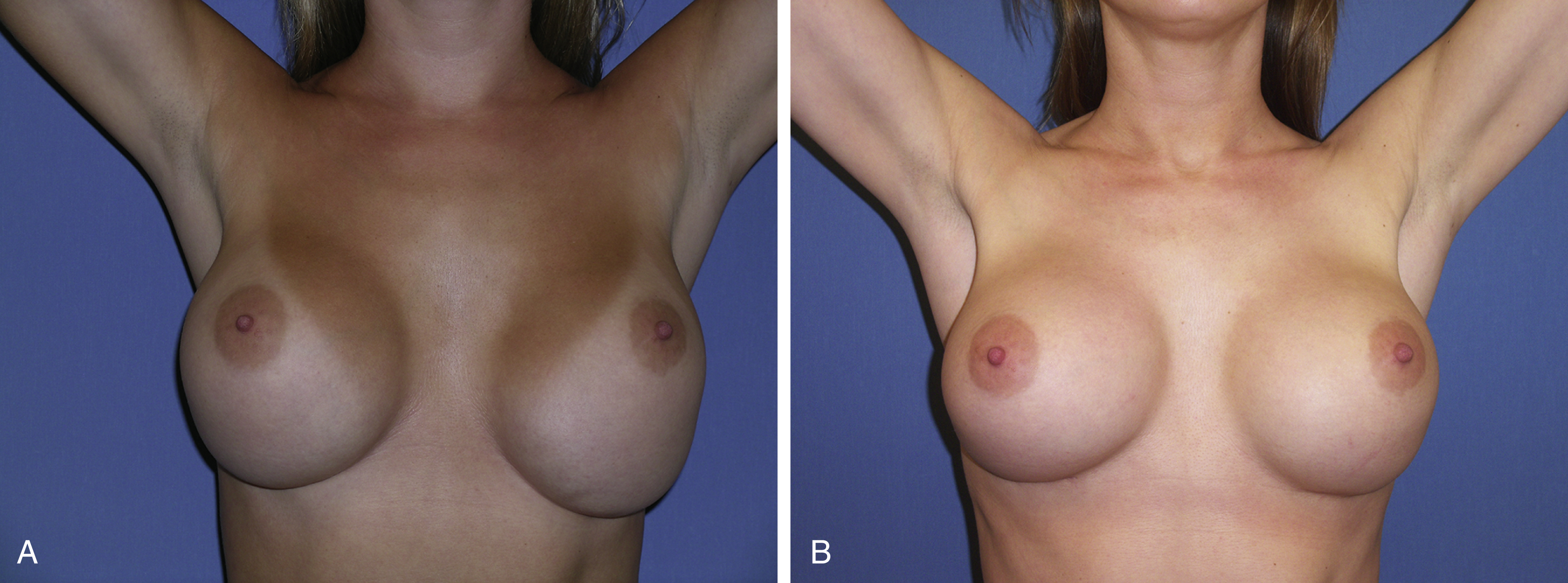
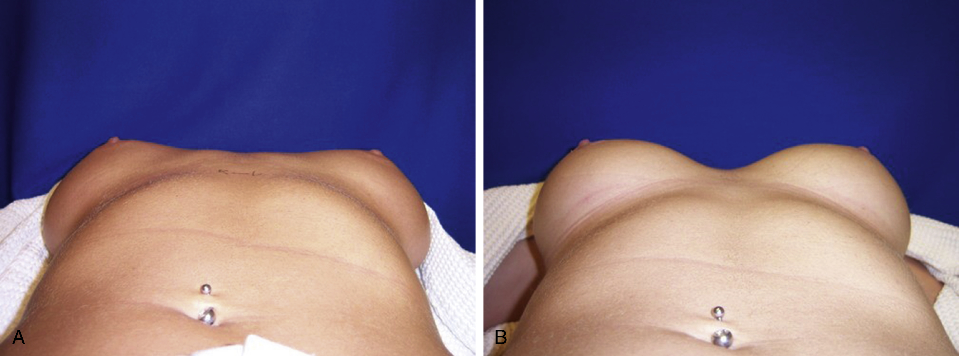
It is important to lay the patient undergoing breast revision supine in the examination room to check for unrecognized lateral displacement that would otherwise be an unexpected finding in the operating room ( Fig. 10.6A, B ). The primary cause of lateral malposition is overdissection laterally at the time of initial augmentation. Poor implant selection in dimensions and volume can also lead to overexpansion laterally. At the initial augmentation, we must be conservative in the lateral dissection and rarely does it need to extend beyond the anterior axillary line. With a sizer or permanent implant in place, this lateral pocket can be modified by cautious cautery or gentle digital dissection. The lateral pocket will suffer the most when an oversized or too-wide implant is placed.
Although there are talented plastic surgeons achieving satisfactory results with the transaxillary route, I commonly find lateral malposition after a primary transaxillary augmentation in revision cases. The takeaway is that if you perform this technique, view your postoperative patients in a supine position after 6 months to assess your incidence of lateral malposition.
The patient seeking breast revision will many times present with a combination of lateral displacement in addition to inferior malposition. The choice of the new implant is critical in the repair because you do not want a wider implant pressing against the repair.
As with inferior malposition the use of capsulorrhaphy is a powerful tool, whether the malposition is purely lateral or a combination of inferolateral in dimensions. I find it helpful to use the lateral border of the pectoralis minor as a medial landmark to follow when securing the lateral capsule to the chest wall. If the tissues are thin or attenuated, you may want to support your repair with ADM or synthetic mesh.
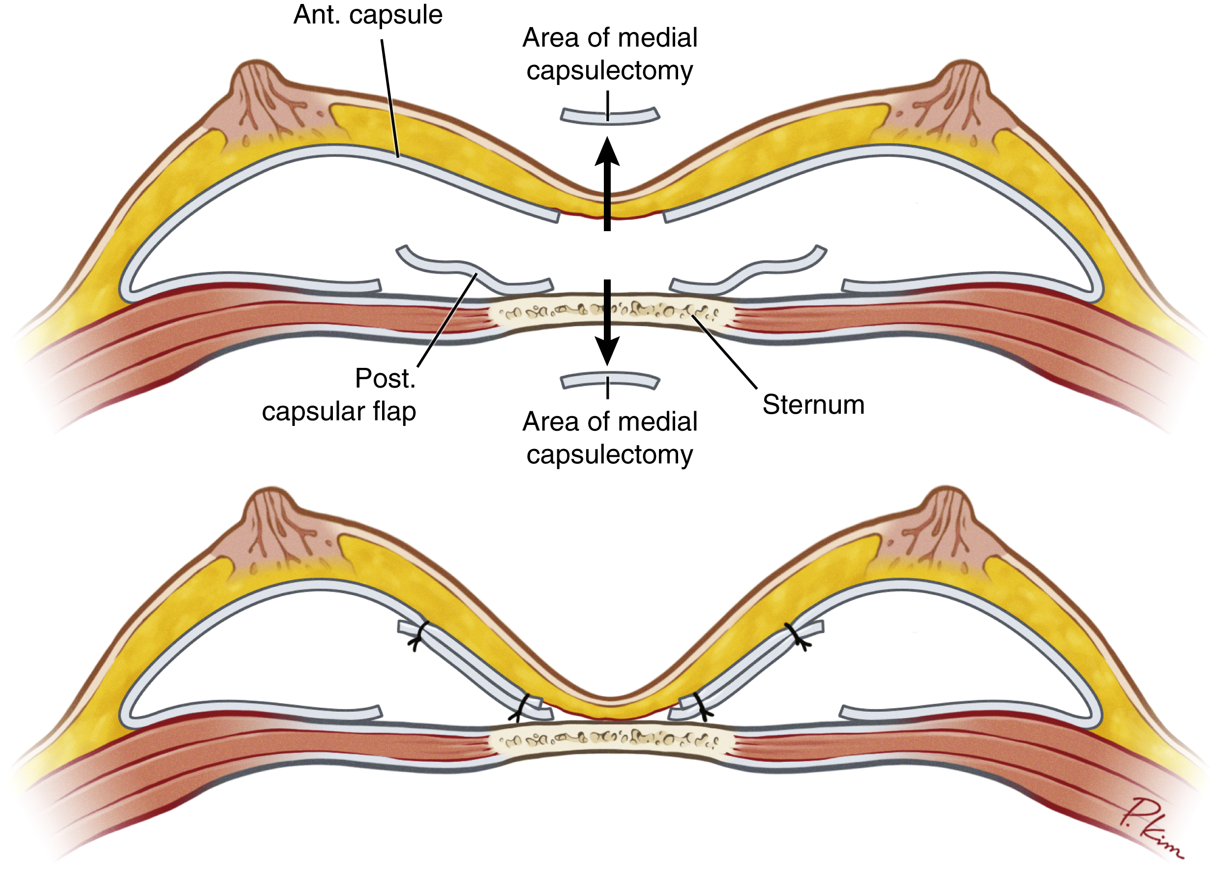
The single or double capsular flap is a technique initially described by Parsa et al. ( Fig. 10.7 ) can be employed in medial or lateral malposition cases; however, the existing capsule must be supple yet substantial enough to support the repair. This procedure is described as marking the area to define the new limit of implant excursion and then elevating the proximal capsule to just short of that mark, knowing there will be additional stretch depending on the quality of the capsule itself. To not stress your repair, it is wise to consider downsizing the volume of the implant for this procedure.