Chapter 20 Respiratory care
 Access the complete reference list online at http://www.expertconsult.com
 IN THIS CHAPTER
  PowerPoint Presentation Online
Introduction
The multitude of respiratory complications caused by smoke inhalation, thermal burns, and their treatment epitomize the clinical challenges which confront respiratory care practitioners. Smoke inhalation injury and its sequelae impose demands upon the respiratory care practitioners who play a central role in its clinical management. These demands may range from intubation and resuscitation of victims in the emergency room to assistance with diagnostic bronchoscopies, to performance of pulmonary function studies, monitoring of arterial blood gases, airway maintenance, chest physiotherapy, and mechanical ventilator management.1 Additional demands are placed upon the respiratory care practitioner in the rehabilitation phase in determining disability or limitations diagnosed by pulmonary function studies or cardiopulmonary stress testing. In some countries outside the United States, the duties of the respiratory care practitioner are augmented by a combination of physicians, nurses, and physiotherapists. It is imperative that a well-organized, protocol-driven approach to respiratory care of the burn patient be utilized so that improvements can be made, and the morbidity and mortality associated with inhalation injury can be reduced (Box 20.1). This chapter provides an overview of the common hands-on approaches to the treatment of inhalation injury, with emphasis on mucociliary clearance techniques, pharmacologic adjuncts, mechanical ventilation, infection control, and the late complications associated with inhalation injury.
Box 20.1 Inhalation injury treatment protocol
• Titrate humidified oxygen to maintain SaO2s >90%
• Cough, deep breath exercises every 2 h
• Turn patient side to side every 2 h
• Chest physiotherapy every 4 h
• Aerosolize 3 cc of 20% N-acetylcysteine every 4 h with a bronchodilator
• Alternate aerosolizing 5000 units of heparin with 3 cc of normal saline every 4 h
• Nasotracheal suctioning as needed
• Early ambulation on postoperative day 5
• Sputum cultures for intubated patients every M-W-F
• Pulmonary function studies prior to discharge and at outpatient visits
Bronchial hygiene therapy
Therapeutic coughing
Series of three coughs
The patient is asked to start a small breath and small cough, then a bigger breath and harder cough, and finally a really deep breath and hard cough. This technique is especially effective for postoperative patients who tend to splint from pain.2
Tracheal tickle
The therapist places the index and middle finger flat in the sternal notch and gently massages inward in a circular fashion over the trachea. This is most effective in obtunded patients or in patients coming out of anesthesia.2
Cough stimulation
Patients with artificial airways cannot cough normally since a tube is either between the vocal cords (endotracheal) or below the cords (tracheostomy). Adequate pressure cannot be built up without approximation of the cords. These patients may have a cough stimulated by inflating the cuff on the tube, giving a large, rapid inspiration by a manual resuscitation bag, holding the breath for 1–2 s, and rapidly allowing the bag to release and exhalation to ensue. This technique is normally performed by two persons and is made more effective by one therapist performing vibration and chest compressions from the time of the inspiratory hold, all during exhalation.2 Cough and deep breathing exercises are encouraged every 2 h to aid in removing retained secretions.
Chest physiotherapy
Chest physiotherapy has come to mean gravity-assisted bronchial drainage with chest percussion and vibrations. Studies have shown that a combination of techniques are effective in secretion removal.3–6
Bronchial drainage/positioning
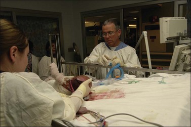
Bronchial drainage/positioning is a therapeutic modality that uses gravity-assisted positioning designed to improve pulmonary hygiene in patients with inhalation injury or retained secretions. There are 12 basic positions in which patients can be placed for postural drainage. Due to skin grafts, donor sites, and the use of air fluid beds, clinical judgment dictates that most of these positions are not practical. In fact, positioning in the Trendelenburg and various other positions may acutely worsen hypoxemia. Evidence has shown that a patient’s arterial oxygenation may fall during positioning.7 To accomplish the same goal it is common practice, in intensive care units, to turn patients side to side every 2 h so as to aid in mobilizing secretions (Fig. 20.1).
Percussion
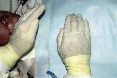
Percussion aids in the removal of secretions from the tracheal bronchial tree. Percussion is done by cupping the hand so as to allow a cushion of air to come between the percussor’s hand and the patient. If this is done properly, a popping sound will be heard when the patient is percussed. There should be a towel between the patient and the percussor’s hand in order to prevent irritation of the skin.8 Percussion is applied over the surface landmarks of the bronchial segments that are being drained. The hands rhythmically and alternately strike the chest wall. Incisions, skin grafts, and bony prominences should be avoided during percussion (Fig. 20.2).
Vibration/shaking
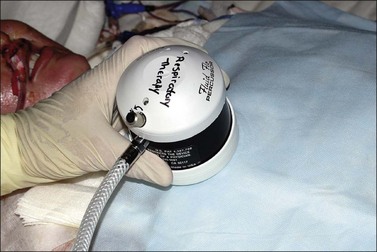
Vibration/shaking is a shaking movement used to move loosened secretions to larger airways so that they can be coughed up or removed by suctioning. Vibration involves rapid shaking of the chest wall during exhalation. The percussor vibrates the thoracic cage by placing both hands over the percussed areas and vibrating into the patient, isometrically contracting or tensing the muscles of their arms and shoulders. Mechanical vibrations have been reported to produce good clinical results. Gentle mechanical vibration may be indicated for patients who cannot tolerate manual percussion (Fig. 20.3). Chest physiotherapy techniques should be used every 2–4 h for patients with retained secretions. Therapy should continue until breath sounds improve.
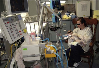
Early ambulation
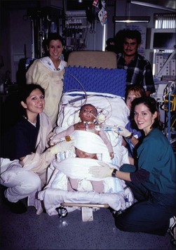
Early ambulation is another effective means of preventing respiratory complications. Patients routinely should be helped out of bed on postoperative day 5 and encouraged to ambulate and sit in a chair. With appropriate use of analgesics, even patients on continuous ventilatory support can be helped out of bed and into a chair (Fig. 20.4). The rocking chair (Fig. 20.5) has several beneficial effects:
• the patient can breathe with regions of the lungs which are normally hyperventilated;
• muscular strength and tone are preserved;
• contractions are prevented and exercise tolerance is maintained.9
Airway suctioning
Airway suctioning is another method of clearing an airway. Normal bronchial hygiene is usually accomplished by the mucociliary escalator process. When these methods are not effective in maintaining a clear airway, tracheobronchial suctioning is indicated.10–13 Nasotracheal suctioning is intended to remove from the trachea accumulated secretions and other foreign material which cannot be removed by the patient’s spontaneous cough or less-invasive procedures. Nasotracheal suctioning has been used to avoid intubation which was solely intended for the removal of secretions. Nasotracheal suctioning refers to the insertion of a suction catheter through the nasal passages and pharynx into the trachea in order to aspirate secretions or foreign material. The first step in this process is to hyperoxygenate the patient with 100% oxygen. The patient should be positioned in the Fowler’s position and the catheter slowly advanced through the nares to a point just above the larynx. The therapist or nurse then listens for air sounds at the proximal end of the catheter. When airflow is felt to be strongest and respiratory sounds are loudest, the tip of the catheter is immediately above the epiglottis. On inspiration, the catheter is advanced into the trachea. After the vocal cords have been passed, a few deep breaths are allowed and the patient is reoxygenated. Suction is begun while the catheter is slowly withdrawn from the trachea. The patient should not be suctioned for more than 15 s without being reoxygenated. Suctioning is not without potential hazards.14,15 Complications include irritation of the nasotracheal mucosa with bleeding, abrupt drops in PO2, vagal stimulation, and bradycardia. Preoxygenating and limiting suction time have been shown to decrease or eliminate the fall in the PO2.12–14 Sputum cultures should be performed for microbiological identification when they are clinically indicated. Patients on mechanical ventilation should have sputum cultures performed once per week.
Pharmacologic adjuncts
Bronchodilators
Bronchodilators can be helpful in some cases. Inhalation injury to the lower airways results in a chemical tracheobronchitis which can produce wheezing and bronchospasms. This is especially true for patients with preexisting reactive airway diseases. Most drugs used in the management of bronchospasms are believed to act on the biochemical mechanism which controls bronchial muscle tone. Aerosolized sympathomimetics are effective in two ways: they result in bronchial muscle relaxation and they stimulate mucociliary clearance. The newer bronchodilators are more effective and have fewer side-effects than the older generation drugs. Some of the newer compounds used in the United States are worthy of note. First, metaproterenol is available in cartridge inhalers, as a liquid to be aerosolized, as an oral medication in tablets, or as a syrup. The recommended oral dose is 10–20 mg every 6–8 h; as an inhaled bronchodilator, 1–2 puffs every 3–4 h. Its duration of action as an inhaled bronchodilator is 1–5 h.16
Albuterol can also be administered orally, parenterally, and by aerosol. Albuterol is available in a metered cartridge inhaler and its standard dose is 1–2 puffs three to four times daily. Aerosolized albuterol has a duration of action of approximately 4–6 h.17
Racemic epinephrine is used as an aerosolized topical vasoconstrictor, bronchodilator, and secretion bond breaker. The vasoconstrictive action of racemic epinephrine is useful in reducing mucosal and submucosal edema within the walls of the pulmonary airways. A secondary bronchodilator action serves to reduce potential spasm of the smooth muscles of the terminal bronchioles. Water, employed as a diluent for racemic epinephrine, serves to lower both adhesive and cohesive forces of the retained endobronchial secretions, thus serving as a bond-breaking vehicle. Racemic epinephrine has also been used for the treatment of post-extubation stridor.18 Its mode of action is thought to be related to the vasoconstrictive activity, with the resultant decrease in mucosal edema. Aerosolized treatments may be given every 2 h as long as the heart rate is not increased.
Hypertonic saline offers a theoretically more effective form of mucokinetic therapy. The deposition of hypertonic droplets on the respiratory mucosa results in the osmotic attraction of fluids from the mucosal blood vessels and tissues into the airway. Thus a ‘bronchorrhea’ is induced, and the watery solution helps to dilute down the respiratory tract secretions and to increase their bulk, thereby augmenting expectoration. Furthermore, there is evidence that hypertonic saline has a direct effect on the mucoprotein DNA complexes, and by reducing the cohesive intramolecular forces, the salt helps reduce the viscous properties of the mucoid fluid.19 Excessive use of hypertonic saline is not recommended because irritation can occur in the respiratory tract, absorption can occur, and burn patients who cannot tolerate the sodium load may develop edema.
Finally, aerosolized acetylcysteine is a powerful mucolytic agent in use in respiratory care. Acetylcysteine contains a thiol group; the free sulfhydryl radical of this group is a strong reducing agent which ruptures the disulfide bonds that serve to give stability to the mucoprotein network of molecules in mucus. Agents that break down these disulfide bonds produce the most effective mucolysis.20 Acetylcysteine is an irritant to the respiratory tract. It can cause mucosal changes, and it may induce bronchospasm. For this reason, patients are evaluated for signs of bronchospasm, and a bronchodilator may be added if necessary. Acetylcysteine has proven to be effective in combination with aerosolized heparin for the treatment of inhalation injury in animal studies.21
Heparin/acetylcysteine combinations have been used as scavengers for the oxygen free radicals produced when alveolar macrophages are activated either directly by chemicals in smoke or by one or more of the compounds in the arachidonic cascade.22 Animal studies have shown an increased P/F ratio, decreased peak inspiratory pressures, and a decreased amount of fibrin cast formation with heparin/acetylcysteine combinations.23 In a retrospective review, Desai et al. have shown that the use of heparin/N-acetylcysteine is effective in pediatric patients with inhalation injury.24 Results indicate a significant decrease in the reintubation rates, incidence of atelectasis, and improved mortality for patients treated with heparin/N-acetylcysteine therapy. Therefore a standard treatment for patients with inhalation injury might include 5000–10 000 units of heparin and 3 mL normal saline nebulized every 4 h, alternating with 3–5 mL of 20% acetylcysteine for 7 days. This insures that the patient receives an aerosolized treatment every 2 h. Baseline and daily clotting studies are recommended for the entire length of the aerosolized treatments.
Mechanical ventilation
Over the past 30 years, and especially over the past decade, there has been an increase in new ventilatory techniques which present alternatives for the treatment of patients with smoke inhalation. Unfortunately, although the number of options available to the clinician has appeared to increase exponentially, well-controlled clinical trials defining the specific role for each of the modes of ventilation and comparing them to other modes of ventilation have not been forthcoming. Based upon current available data, the recommendations from the American College of Chest Physicians consensus conference on mechanical ventilation generally serve as guidelines.25 The general consensus concludes:
• The clinician should choose a ventilator mode that has been shown to be capable of supporting oxygenation and ventilation and that the clinician has experience in using.
• An acceptable oxygen saturation should be targeted.
• Based primarily on animal data, a plateau pressure of greater than 35 cmH2O is cause for concern. With clinical conditions that are associated with a decreased chest wall compliance, plateau pressures greater than 35 cmH2O may be acceptable.
• To accomplish the goal of limiting plateau pressures, PCO2 values should be permitted to rise (permissive hypercapnia) unless other contraindications exist that demand a more normal PCO2 or pH.
• Positive end-expiratory pressure (PEEP) is useful in supporting oxygenation. An appropriate level of PEEP may be helpful in preventing lung damage. The level of PEEP required should be established by empirical trials and reevaluated on a regular basis.
• Large tidal volumes (8–10 mL/kg) with PEEP may be needed to improve oxygenation if the use of protective ventilatory strategies becomes ineffective. Peak flow rates should be adjusted as needed to satisfy patient inspiratory needs.25 Care must be taken to avoid the consequences of utilizing high ventilator pressures if large tidal volumes are required.
Animal studies performed showed a reduction in ventilator-induced injury with a reduction in plateau pressures to 35 cm of water by increasing positive end-expiratory pressure (PEEP) and decreasing tidal volume.26 A number of clinical trials have been conducted to support this treatment strategy.27–29 A meta-analysis of these trials was conducted by the Cochrane Anesthesia Review Group in 2007.30 It showed a reduction in mortality and duration of mechanical ventilation with the use of plateau pressure at <31 cm water and tidal volume at <7 mL/kg body weight. In light of this evidence, the tidal volumes used when initiating mechanical ventilation should be 6–8 mL/kg of predicted body weight. If the patient becomes obstructed with fibrin cast and presents with an acute increase in PCO2 and decrease in PaO2, the clinician should first provide aggressive pulmonary toilet, then consider changing over to volume ventilation with higher tidal volumes. If ventilation continues to worsen, tidal volumes of 8–10 mL/kg may be needed to provide adequate mechanical ventilation.
Modes of ventilation
Assist-control mode
In the assist-control mode of ventilation, in which every breath is supported by the ventilator, a back-up control rate is set; however, the patient may choose any rate above the set rate. Using this mode of ventilation, the tidal volume, inspiratory flow rate, flow waveform, sensitivity, and control rate are set.31–33 Advantages are that assist-control ventilation combines the security of controlled ventilation with the possibility of synchronizing the breathing pattern of the patient and ventilator, and it ensures ventilatory support during each breath. Disadvantages are as follows:
• Excessive patient work occurs in case of inadequate peak flow or sensitivity settings, especially if the ventilator drive of the patient is increased.31–33
• It is sometimes poorly tolerated in awake, nonsedated subjects and can require sedation to insure synchrony of patient and machine.
• It can cause respiratory alkalosis.
• It may worsen air trapping with patients with chronic obstructed lung disease.24
Synchronized intermittent mandatory ventilation
Related posts:
Stay updated, free articles. Join our Telegram channel

Full access? Get Clinical Tree


