Conjunctival prolapse
|
| History of prior congenital ptosis repair or craniofacial surgery |
| Adequacy of superior conjunctival fornix |
| Presence of symblepharon |
| Corneal examination |
| Upper eyelid position and contour |
| Presence of lagophthalmos |
Introduction
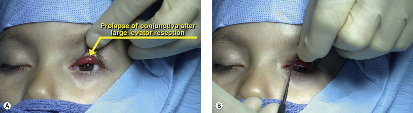
Prolapse of the superior conjunctiva is an uncommon complication that may occur after levator resection surgery for congenital ptosis. This condition is more likely to occur with large and supermaximal resections (22 mm or more) of the levator ( Chapter 14 ). Craniofacial surgery involving the superior orbit with cerebrospinal fluid leak has also been reported to cause superior conjunctival prolapse even in the absence of levator surgery.
The superior cul-de-sac is formed by the suspensory ligament of the fornix which contains fine, sheath-like fibers between Müller’s muscle and the conjunctiva. With large levator dissections, these fascial connections may be severed. This results in a disparity in length between the redundant conjunctiva and the now relatively short levator. Fluid and hemorrhage may then fill this potential space, causing the conjunctiva to prolapse inferiorly.
Clinically, prolapse of the superior fornix can result in conjunctival hyperemia, keratinization, and corneal irritation as well as a displeasing appearance. Furthermore, the prolapsed tissue can obstruct the visual axial and induce astigmatism that may promote amblyopia.
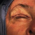
If prolapse of the conjunctiva is noted during levator resection, prophylactic fornix-deepening sutures should be placed at the time of ptosis surgery. Spontaneous resolution is not expected with this complication. An alternative treatment for conjunctival prolapse is excision of the prolapsed tissue; however, this may result in significant forniceal shortening, symblepharon, loss of ocular surface stem cells and restrictive strabismus. Additionally, resection of conjunctiva may also include Müller’s muscle, which could lead to postoperative eyelid retraction. Our preferred treatment is placement of fornix-deepening sutures, which encourages adhesions between the levator and the conjunctiva.
Surgical Technique