Reconstruction of Penile Urethral Strictures: Staged Procedures

div class=”ChapterContextInformation”>


© Springer Nature Switzerland AG 2020
F. E. Martins et al. (eds.)Textbook of Male Genitourethral Reconstructionhttps://doi.org/10.1007/978-3-030-21447-0_11


11. Surgical Reconstruction of Penile Urethral Strictures: Staged Procedures


Paurush Babbar1, Hadley M. Wood1 and Kenneth W. Angermeier1  


(1)
Center for Genitourinary Reconstruction, Glickman Urological and Kidney Institute, Cleveland Clinic, Cleveland, OH, USA


Kenneth W. Angermeier


Keywords

Penile urethral strictureStaged proceduresUrethroplastyBuccal mucosa graftReconstructive urology


11.1 Introduction


Staged urethroplasty involves utilizing more than one surgical intervention in the treatment of urethral stricture, diverticulum or fistula. While single stage procedures are favored for obvious reasons, there is a well-established role for staged procedures. Indications include: strictures related to lichen sclerosis (LS), prior reconstructive surgery for hypospadias or urethral stricture, poor tissue quality or a paucity of viable genital skin for flap reconstruction, and an absent lumen precluding substitution urethroplasty. Current staged procedures involve tissue transfer, most commonly using buccal mucosa in the modern era [1, 3]. This chapter will outline the relevant preoperative evaluation in patients with penile urethral stricture or fistula, and describe the surgical technique and reported outcomes of staged urethroplasty for penile urethral reconstruction.


11.2 Preoperative Evaluation


The preoperative evaluation in patients being considered for urethral reconstruction is critical for adequate preoperative counsel and surgical planning. Evaluation should include a detailed history with specific attention to pertinent pediatric urological history, prior surgical interventions, previous history of genitourinary trauma or infection, recent urethral instrumentation, current voiding pattern, and a history of urinary retention especially if associated with renal insufficiency. Additional important patient factors that should be documented include presence of urinary incontinence, erectile dysfunction or chordee, history of malignancy and relevant treatment modalities used (especially radiation), and oral pathology or tobacco use. The aforementioned list is by no means exhaustive, but rather a starting point to frame the subsequent discussion for treatment of urethral stricture. Physical examination is also key when deciding on surgical approach, especially for a penile urethral stricture. Assessment should include documentation of quality and availability of penile skin and soft tissue, circumcision status, location and severity of genital scars, and the presence of LS.


The next step is radiographic evaluation and typically includes a retrograde urethrogram (RUG) with or without voiding cystourethrogram (VCUG) to understand stricture length, location and other associated urethral or bladder abnormalities (Fig. 11.1). In an optimal situation, we prefer to wait at least 6–12 weeks following last urethral instrumentation to proceed with imaging to allow stricture maturation. If a VCUG is needed to fully evaluate proximal urethral anatomy, the bladder may be filled with a Glidewire and open end catheter, or a suprapubic catheter placed to aid the procedure. Retrograde cystoscopy using a small caliber cystoscope or antegrade flexible cystoscopy are helpful confirmatory measures in select cases.

../images/468369_1_En_11_Chapter/468369_1_En_11_Fig1_HTML.png

Fig. 11.1

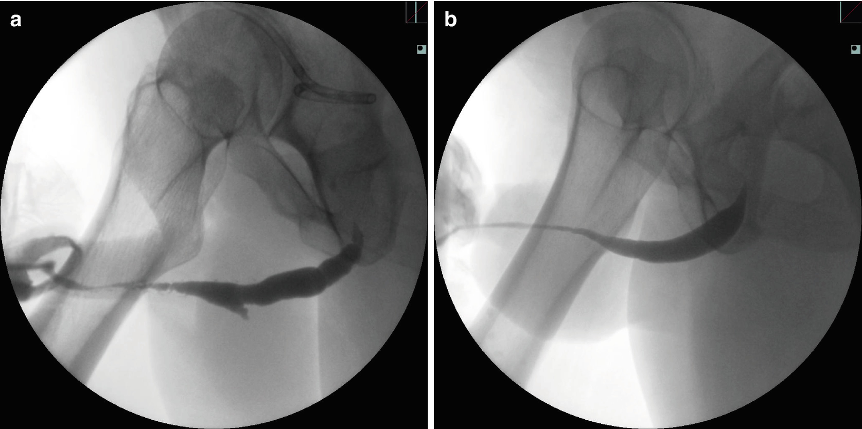
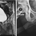
Penile urethral stricture related to: (a) multiple prior surgical procedures for penoscrotal hypospadias and subsequent complications, (b) lichen sclerosis (LS)


Managing expectations and helping the patient understand the timeline for urethral reconstruction is integral to the success of the operation. The period of urethral rest prior to the first stage procedure must be discussed, as well as a possible need for a suprapubic catheter to allow for this if the patient is requiring frequent dilation or performing self-catheterization. Between the first and second stage, a period of 4–6 months is typical for graft take and stabilization, and the patient needs to understand the process of sitting to void through a penile or upper scrotal urethrostomy during this time. Sexual intercourse is allowed approximately 6–8 weeks after the first procedure, and the patient is made aware that semen will also emanate from the urethrostomy. Following the second stage procedure, a catheter or urethral stent is left in the urethra for a period of 18–21 days to permit urinary diversion and urethral healing. Intercourse is again allowed after 6–8 weeks . From a surgical standpoint, patients must also understand the longer term risks of stricture recurrence, urethral fistula or diverticulum, spraying of the urinary stream, and potential need for additional procedures.


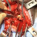
11.3 Surgical Technique: First Stage for Penile Stricture


The patient is placed in the supine position for penile urethral reconstruction, but in dorsal lithotomy if there is to be concurrent work on the bulbar urethra or for a panurethral stricture. If there is an area of the reconstruction where penile skin may need to be incorporated, attention should be paid to hair pattern to ensure use of non-hair bearing tissue in the final repair. After the patient is prepped and draped, a glans traction suture is placed to assist with surgical exposure. The incision is then marked out along the ventral penis in the midline, incorporating the urethral meatus and glans as needed, and may be extended proximally to the upper scrotum along the midline raphe depending on stricture length. To confirm the distal limit of the stricture, a 20-22F bougie is placed in the urethral meatus and advanced to the point of resistance. An incision is made through the skin and dartos and then into the urethral lumen. The penile incision and the urethrotomy are extended through the entire length of the stricture until healthy wide caliber urethra is encountered. This maneuver is aided by passage of a Glidewire or open end catheter in some cases. Once healthy normal caliber urethra is reached, urethrostomy is performed by suturing adjacent genital skin to the spatulated urethral opening full-thickness using 4-0 or 5-0 Vicryl (Fig. 11.2). If the penobulbar junction has been reached within the upper scrotum and more proximal ongoing stricture within the bulbar urethra is identified, this can usually be reconstructed using a one-stage type repair using a ventral or dorsal onlay of oral mucosa with the urethrostomy created at the distal end of the bulbar repair (Fig. 11.3). The legs are raised within adjustable stirrups to allow a separate perineal incision for this part of the procedure. For any stricture, flexible cystourethroscopy is recommended to evaluate the proximal urethra and bladder once normal caliber urethra is reached.

../images/468369_1_En_11_Chapter/468369_1_En_11_Fig2_HTML.png

Fig. 11.2

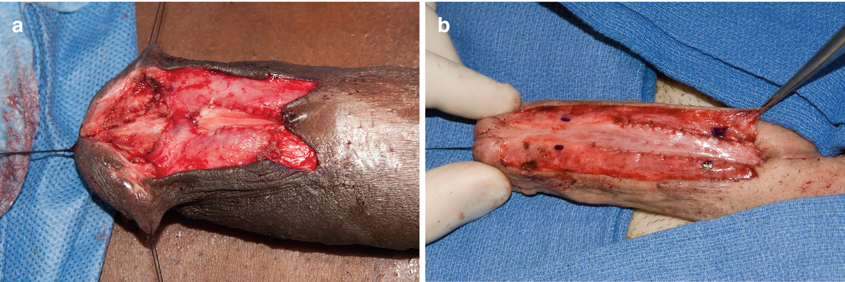
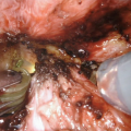
(a) Urethral excision and urethrostomy in patient following prior distal hypospadias repair; (b) Urethrotomy and penoscrotal urethrostomy in setting of LS


../images/468369_1_En_11_Chapter/468369_1_En_11_Fig3_HTML.png

Fig. 11.3

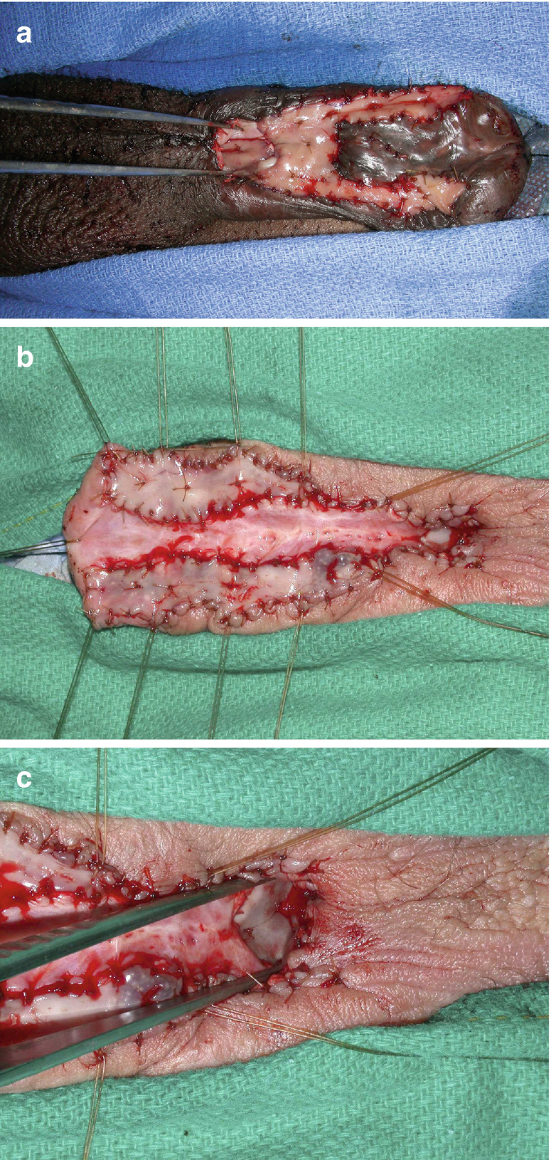
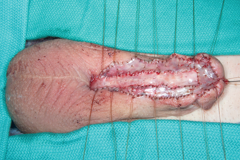
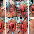
(a) Complex first stage hypospadias patient with proximal dorsal buccal mucosa graft seen between forceps at proximal urethrostomy; (b) Proximal dorsal graft at urethrostomy opening for extensive LS stricture; (c) Closer view of urethrostomy and graft


Once the above has been completed and urethrostomy established, the existing urethral plate is then examined. Portions of the urethral plate may require complete excision if severely diseased, obliterated or hair-bearing. However we have found that significant portions of the urethral plate, usually starting just proximal to the level of the corona, often appear viable and may be preserved and greatly aid second stage tubularization even in the setting of LS (Fig. 11.4). Prior to grafting, a 5-0 running absorbable suture may be used to close the edges of the corpus spongiosum and urethral mucosa on each side to aid hemostasis. In some cases it is necessary to mobilize and rotate some of the adjacent dartos and tack it medially to the urethral plate to optimize the graft bed, and aid subsequent graft mobility and tension-free closure. The length of graft needed for penile urethral reconstruction is then measured. In a patient with a normally virilized phallus, we try to obtain a final urethral plate of approximately 3 cm width for optimal tubularization , although this is not always quite possible within the glans. A prepubertal phallus may accommodate a smaller caliber urethra, particularly if the glans is diminutive, and adjustments are made accordingly. Oral mucosal is then harvested in the usual fashion, either from one inner cheek or both depending on the amount needed and oral anatomy. Once the buccal graft(s) are harvested and appropriately thinned, the mucosa is spread and fixed onto the opened glans and adjacent to the urethral plate using 4-0 or 5-0 absorbable sutures peripherally, following by multiple quilting sutures to aid apposition and take. The graft(s) can be configured along either side of the urethral plate to fill in areas necessary to achieve the desired width symmetrically throughout the repair (Fig. 11.5). We have not routinely perforated our grafts. When the stricture involves the distal urethra into the glans, it is critical to ensure that the glans cleft is deep enough and glans flaps mobilized to provide a graft width adequate for closure at the second stage. In some forms of hypospadias or LS, the glans may be insufficient for adequate grafting, and in this instance a coronal or subcoronal meatus may be more feasible after discussion with the patient. In the setting of a panurethral stricture, there may be a segment of urethral plate between the proximal one-stage repair and distal first-stage repair where there is no further buccal mucosa available. In this setting, we have preferred to suture adjacent proximal penile skin to the urethral plate to complete the first stage repair as opposed to harvesting additional tissue (Figs. 11.3b and 11.4). This segment can then be tubularized using lingual mucosa at the time of second stage closure, or by using the adjacent penile skin if no evidence of LS. Urethral catheter is inserted at this time, or in some cases a urethral stent if the repair is distal and a suprapubic catheter already present.

../images/468369_1_En_11_Chapter/468369_1_En_11_Fig4_HTML.png

Only gold members can continue reading. Log In or Register to continue

Stay updated, free articles. Join our Telegram channel

Aug 4, 2021 | Posted by in General Surgery | Comments Off on Reconstruction of Penile Urethral Strictures: Staged Procedures

Full access? Get Clinical Tree

Get Clinical Tree app for offline access