Research model
Radiation modality
Chamber model (Narayan and Cliff [1])
19 × 5 mm rectangular ear chamber
31, 5 cGy/min
Total dose: 75, 6 Gy
Skin graft (Tadjalli et al. [9])
4 × 3 cm rectangular skin graft
Total dose: 15, 20, 25 or 30 Gy
Skin flap (Virolainen [3])
2 cm diameter abdominal skin flap
16 MeV electrons at a source-to-skin distance of 100 mm
2.5 Gy/min
Total dose: 20 Gy
Cremaster muscle (Siemionow et al. [4])
4 × 4 cm field over the groin and scrotum
300 kV, 12 mA, Thoraeus filter, exposure time of 11.24 min (5.62 min for each side)
Source to target distance of 51 cm
Total dose: 8 Gy irradiation was delivered half from the front and other half from the back
Hindlimb-cremaster muscle composite tissue (Ayhan et al. [5])
Total-body irradiation
MV photon beam at a dose rate of approximately 340 cGy/min
Total dose: 2 Gy
TRAM (Lin et al. [6])
3 × 9 cm rectangle over the abdominal wall
6 MV electron energy with 5 × 25 cm cutout
Total dose: 50 Gy in 25 fractions (200 cGy/fraction)
5 days a week for 5 weeks
Rabbit Ear Chamber Model
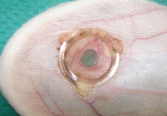
One of the oldest techniques for in vivo microscopy is the chamber model. These tissue window models are very useful for the studies of wound healing process and visualisation of circulation. Skin chambers, ear windows, cranial and spinal windows, bone window, lung window are the examples of the chamber models. The dorsal skin chamber in mice, hamster, rats and rabbits are widely used in skin microcirculation, ischemia reperfusion, wound healing and tumor growth studies. Especially rabbit ear chamber model has the advantage of clear most view of the blood vessels. Dimitrievich et al. used this model to show radiation damage to microvasculature in 1977 [7]. Rabbit ear chamber was also used by Narayan and Cliff to Show the effects of 75 Gy radiation on the skin microcirculation [1]. In this model, a metal ear chamber is implanted to the rabbit ear. A fold of depilated dorsal skin of the animal is taken and cut out surgically a round area of skin layer completely (consisting of epidermis, dermis, muscle and fat). Then the skinfold is fixed like a sandwich between two frames of chamber and the window is closed with a sterile coverslip to avoid drying, infection or mechanical trauma of unprotected side of opposite skin. The coverslip can be removed and reclosed between treatments or evaluations. Disadvantage of this model is that only few capillaries can be seen in the model, and small changes in the number of capillaries may cause significant bias on results (Fig. 12.1).


Fig. 12.1
Rabbit-ear chamber. Arterioles and venules can be observed microscopically in the center window (Kotsu et al. [8])
Skin Graft Model
Effect of radiation on skin graft survival was evaluated by Tadjalli et al. in 1999 in rat skin graft model [9]. In this model, a rectangular dorsal skin was removed and reapplied as a full thickness skin graft after defatting and removal of the panniculus carnosus. The animals received a single dose 15, 20, 25 or 30 Gy external radiation after 4 weeks postoperatively. The skin grafts were evaluated before irradiation and weekly thereafter for 4 weeks. Areas of graft loss were measured by a software program. The animals were euthanized 4 weeks after irradiation. Histologic samples were obtained from intact and ulcerated graft sites and evaluated for epidermal thickness or ulceration, adnexial spacing, granulation tissue in areas of healing, vascular changes, inflammatory infiltrates and fibrosis. In graft survival analysis, it was observed that the average decrease graft survival was higher at high doses of radiation (25 Gy, 30 Gy). In histopathological evaluation, vascular changes, deep ulcerations, edema were more frequently seen in high dose radiation groups. In conclusion, skin graft loss was significantly increased when single doses of 25 Gy or more are delivered 4 weeks after grafting procedure.
Skin Flap Model
Angelos et al. have developed a free-flap animal model to study the effects of irradiation on flap revascularization [2]. Rat ventral faciacutaneous flap model, based on the inferior epigastric artery and vein, was used in the study. Rats were subjected to either 23 or 40 Gy irradiation to their ventral abdominal wall. Rats were placed in the supine position on the irradiation table and a lead shield was placed to isolate the templated flap region. For animals receiving 23 Gy, irradiation was administered in three divided doses of 766 cGy over 5 days. For animals receiving 40 Gy, irradiation was administered in five divided doses of 800 cGy over 10 days.
After a recovery period of 28 days post irradiation, the animals underwent a ventral fasciocutaneous flap procedure. Following elevation of the flap, a vascular clip was applied to the vascular pedicle for 2 h to simulate average ischemic time during free-tissue transfer. At the end of the 2-h period of ischemia the occluding clip removed and the wound closed. After this procedure, pedicle ligation was performed 10 days postoperatively. And also some animals were subjected to 40 Gy of irradiation and underwent pedicle ligation at 8 or 14 days to determine if time to pedicle ligation affects percentage of flap viability. Percentage of flap viability was evaluated on postligation procedure day 5 as a marker for flap revascularization. Evaluation for viable and non-viable flap areas was performed on standardized digital photographs. Then cutouts of viable tissue from the template were weighed to express a percentage of the total template weight, effectively giving the area percentage of viable flap tissue. Consequently, rats receiving a 40-Gy total dose of irradiation had a significantly lower average flap viability than animals receiving 23 Gy when pedicle ligation was performed 10 days postoperatively and longer duration between pedicle ligation and 40 Gy total dose of irradiation led to significant increases in flap viability.
Virolainen and Aitasalo introduced another animal model for investigate the effect of postoperative irradiation on free skin flaps [3]. This model was designed to assess differences in wound healing between free skin flaps with and without 20 Gy single dose postoperative radiation. The abdominal skin flap, 2 cm in diameter and based on the epigastric vascular pedicle (epigastric island flap) was dissected from the left groin region. The vessels were divided just proximal to the bifurcation of the epigastric vessels. Then end-to-end microvascular anastamoses were performed. The skin of right groin was exposed to radiation 1 week postoperatively. A single fraction of 20 Gy was delivered at a mean dose of 2.5 Gy/min at the surface level. The field was treated with 16 MeV electrons at a source-to-skin distance of 100 mm. Four weeks later tensile strength of flaps, total nitrogen and hydroxyproline contents and histological changes were evaluated. In the results of the study they have seen the single postoperative fraction of 20 Gy did not affect the tensile strength of the skin. Histologically the flaps healed in a similar fashion with non-irradiated group. Differences in hydroxyproline and total nitrogen contents between postoperatively irradiated and non-irradiated animals were also insignicant. In conclusion, according to this study postoperative irradiation with a single dose 20 Gy did not affect the survival of free skin flaps in rats.
Cremaster Muscle Model
The first attempt to quantify microvascular changes in muscle circulation was performed by Siemionow et al. in 1999 upon description of the cremasteric muscle island muscle flap [4, 10


Stay updated, free articles. Join our Telegram channel

Full access? Get Clinical Tree


