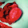
Fig. 3.1
(a) Rat femoral lymphatic vessel divided and anastomosed. Please note that there is no suitable venous structure near to perform lymphaticovenous anastomosis. (b) Femoral lymphatic vessels cannulated by 8.0 Ethilon as an aid for anastomosis. (c) 11.0 Ethilon sutures placed and anastomosis accomplished. (d) Aproximator removed
Based on our experiences we have published a technical paper on lymphaticovenous anastomosis model [7]. In this study we have dissected the thoracic duct of laboratory rat (Wistar), anastomosed to anterior facial vein (end-to-end) and in some cases to external jugular vein (end-to-side). The thoracic duct was found via an incision to the left side of the neck. After incision left submandibular gland, sternocleidomastoid muscle, omohyoid muscle and left clavicle was excised with attention. Next, left external jugular vein was cleared off from surrounding soft tissues using angled vannas microscissors. Then medially the junction between subclavian vein and thoracic duct was visualized. After excision of the omohyoid muscle, careful dissection of common carotid was performed. Between the juction and carotid sheath it is possible to see thoracic duct retrogradely from the junction to superior direction. Meticulous sharp dissection will provide better exposure of the thoracic duct. Incision starting from mandible to thorax on the left side of the neck. Physiologic reflux of the blood to the duct lumen during respiration and central venous pulsation can be observed from the duct wall. Under the fascia (deep neck fascia) where ductus lies whitish hue of the brachial plexus can also be observed (Fig. 3.2).
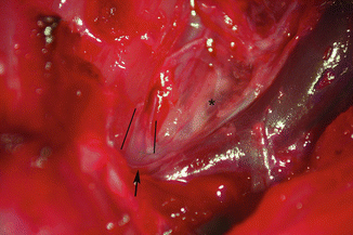
Fig. 3.2
Magnified photograph of the area after dissection in order to approach thoracic duct and veins. Two black lines demarcates the edges of the thoracic duct just above the junction. Arrow shows the direction of blood reflux which is possible to note real-time. Asterisk shows fibers of brachial plexus under the neck fascia
Following the dissection and exposure of the thoracic duct, dividing and anastomosis to a nearby venous structure was performed. Anterior facial vein seems to be the best option in order to mimic lymphaticovenous anastomosis scenario because of its calibre (0.5–0.8 mm) and end-to-end anastomosis geommetry (Fig. 3.3). External jugular vein is available for end-to-side practice (Fig. 3.4
 Microcirculation Model for Invasive Animal Monitoring
 Epineural Sleeve End-to-End Repair
 Somatosensory Evoked Potential Model for Assessment of Nerve Regeneration
 Peripheral Nerve Surgery Models Crush Injury and Epineural Patch
 Heterotopic Vascularized Ovarian Autotransplantation Model in the Sheep
 Defect Repairs After Resections of Laryngeal Cancer, Hypopharyngeal Cancer, and Cervical Esophageal Cancer


Related posts:
Microcirculation Model for Invasive Animal Monitoring
Epineural Sleeve End-to-End Repair
Somatosensory Evoked Potential Model for Assessment of Nerve Regeneration
Peripheral Nerve Surgery Models Crush Injury and Epineural Patch
Heterotopic Vascularized Ovarian Autotransplantation Model in the Sheep
Defect Repairs After Resections of Laryngeal Cancer, Hypopharyngeal Cancer, and Cervical Esophageal Cancer
Stay updated, free articles. Join our Telegram channel
Full access? Get Clinical Tree
