Key Terms
Acrodermatitis continua of Hallopeau
Impetigo herpetiformis
Pustulosis of the palms and soles
Pustules are defined as papular lesions filled with an exudate of acute inflammatory cells. In most cases, the acute inflammatory cells are neutrophils; however, less often the cells are eosinophils or an admixture of the two cell types. This chapter excludes pustular lesions that are follicular based (acute folliculitis), which is discussed in Chapter 23 . Not all pustular eruptions are discussed in this chapter (see box).
- •
Acute generalized exanthematous pustulosis
- •
Acropustulosis of infancy
- •
Candidiasis
- •
Intraepidermal immunoglobulin A (IgA) pemphigus
- •
Pustular psoriasis
- •
Reactive arthritis (Reiter disease)
- •
Pustular arthropod reactions
- •
Subcorneal pustular dermatosis
- •
Transient neonatal pustular melanosis
Important History Questions
How long have the pustules been present?
Some pustular reactions develop acutely, such as pustular drug eruptions or pustular arthropod reactions, whereas pustular psoriasis and reactive arthritis may be chronic or recurring.
Do you have a known personal or family history of psoriasis?
Pustular psoriasis can be tricky to diagnose when it initially presents. The presence of a personal or family history of psoriasis would strongly suggest the possibility of pustular psoriasis.
Do you have a past or current history of arthritis or painful joints?
This question is obviously looking for potential joint disease due to psoriasis or reactive arthritis.
Have you started any new medications?
This is a very important questions because pustular drug eruptions are always high in the differential diagnosis. One should be suspicious of any antibiotic, especially if it belongs to the penicillin class. This question is also important even for patients with known psoriasis because a number of drugs (e.g., oral or intramuscular corticosteroids, terbinafine) will precipitate a pustular reaction in an otherwise classic plaque type of psoriasis.
Had you spent any time outdoors before you developed the rash?
This question is directed at finding any potential source of pustular arthropod reactions.
Does anyone else in the family have a rash?
This question is directed at finding a potential common arthropod exposure, such as flea bites.
How are you treating this rash?
Many patients use home-based, over-the-counter (OTC), or prescription remedies that may alter a reaction. One of the most common is a topical corticosteroid, which may actually produce follicular pustules and confuse the clinical picture.
Important Physical Findings
How old is the patient?
Some pustular disorders occur in young children (e.g., acropustulosis of infancy), others are primarily in young adults (e.g., vaccinia infection), and others occur in any age group.
What are the distribution and arrangement of the pustules?
Some pustular disorders have a distinct distribution (e.g., acropustulosis of infancy, acrodermatitis continua). Some pustular disorders present as solitary lesions, whereas others may demonstrates annular or circinate arrangements (e.g., some cases of pustular psoriasis, intraepidermal IgA pemphigus).
Are the pustules based on hair follicles or non-follicular skin?
If the pustules are based on hair follicles, refer to Chapter 23 .
Acute Generalized Exanthematous Pustulosis
ICD10 code L27.0
DRUG ERUPTION
Analgesics
- •
Acetaminophen
Antimalarial Agents
- •
Hydroxychloroquine (Plaquenil)
- •
Quinidine
Antipsychotic Agents
- •
Olanzapine
Antibiotics
- •
Amoxicillin
- •
Ampicillin
- •
Cefazolin
- •
Cephradine
- •
Cephalexin
- •
Clindamycin
- •
Cotrimazole
- •
Metronidazole
- •
Penicillin
- •
Norfloxacin
- •
Vancomycin
Antiseizure Agents
- •
Carbamazepine
- •
Phenytoin
Calicum Channel Blockers
- •
Nifedipine
Miscellaneous
- •
Chromium picolinate
- •
Mercury
- •
Radiocontrast dye
Pathogenesis
Acute generalized exanthematous pustulosis (AGEP) is a reproducible pustular drug hypersensitivity produced by many drugs; the most common offenders are antibiotics (cephalosporins, cotrimoxazole are most frequently implicated), anticonvulsants, and hydroxychloroquine. This distinct drug eruption has been reproduced with topical patch testing; however, the exact immunologic basis for this reaction is not understood.
Clinical Features
- •
The patient has a history of a new medication within the past 1 to 5 days.
- •
There is an acute (often dramatic) onset of macular (typically confluent) erythema that can affect any site, although it usually affects large areas of the trunk and proximal extremities.
- •
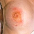
Erythema studded with varying numbers of 1- to 2-mm pustules ( Figs. 12.1–12.3 ) is found. Pustules may be present on some regions of the body and absent on others.
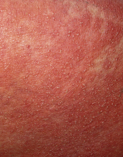
Fig. 12.1
Numerous small pustules on a background of erythema caused by amoxicillin.
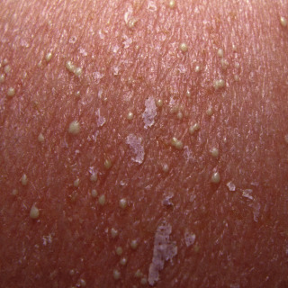
Fig. 12.2
Acute generalized exanthematous pustulosis due to ceftriaxone. This lesion demonstrates uniform pustules, background erythema, and early desquamation.
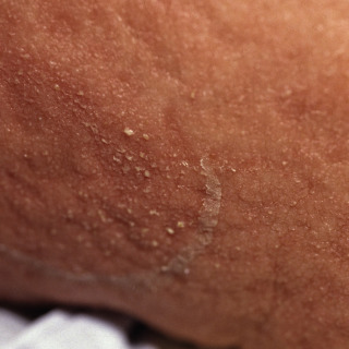
Fig. 12.3
Close-up of a pustular drug eruption due to hydroxychloroquine that demonstrates erythema studded with small pustules. This eruption is several days old, and there is already early desquamation of the top layer of skin.
- •
Less commonly, patients may demonstrate erythematous indurated papules or small plaques that may resemble erythema multiforme.
- •
Patients may complain of burning, itching, or both.
- •
A low-grade fever is common.
Diagnosis
- •
The history of a new drug (especially antibiotics), combined with toxic erythema and pustules, is very strongly suspicious of AGEP.
- •
Complete blood count (CBC). Leukocytosis is commonly present.
- •
Typically, a 3- or 4-mm punch biopsy will demonstrate a subcorneal or, less commonly, a follicular-based pustule containing numerous neutrophils, with variable numbers of eosinophils. The biopsy results, although not specific, can be strongly supportive of the diagnosis.
Treatment
- •
Withdraw the suspected offending drug(s).
- •
No treatment is necessary in most cases because this is a self-limited disease.
- •
Sedating antihistamines such as diphenhydramine can be used for patient comfort at night.
- •
Medium-potency corticosteroids (e.g., triamcinolone, fluocinonide) in a cream or ointment base can be used for for pruritus.
- •
Severe cases may require a short burst of oral prednisone (typically, 40 mg for 3 or 4 days).
- •
Patients need to be labeled as allergic to the implicated drug because these reactions will occur with subsequent challenge.
Clinical Course
The erythema typically fades over several days, followed by desquamation similar to that seen in sunburn.
Pustular Psoriasis
ICD 10 codes L40.1, L40.2, and L40.3
GENETIC DISORDER
Pathogenesis
The pathogenesis of pustular psoriasis is the same as for classic papulosquamous psoriasis in that it is a genetic disorder with a trigger, such as an infection. Patients may demonstrate features of both the papulosquamous and pustular types or move back and forth between the two phenotypic presentations. In some cases, pustular psoriasis may be precipitated by oral corticosteroids. The reason for this is not understood.
- •
Aceclofenac
- •
Antitumor necrosis factor therapy (e.g., adalimumab, etanercept, infliximab)
- •
Clopidogrel
- •
Fexofenadine
- •
Systemic corticosteroids
- •
Terbinafine
Clinical Features
- •
Patients may have a past, present, or family history of plaque-type or pustular psoriasis.
- •
Patients may demonstrate a localized (e.g., confined to palms and soles) or generalized distribution.
- •
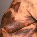
The primary lesion is diffuse erythema studded with pustules that are not infrequently distributed around the edges in an annular fashion ( Fig. 12.4 ).
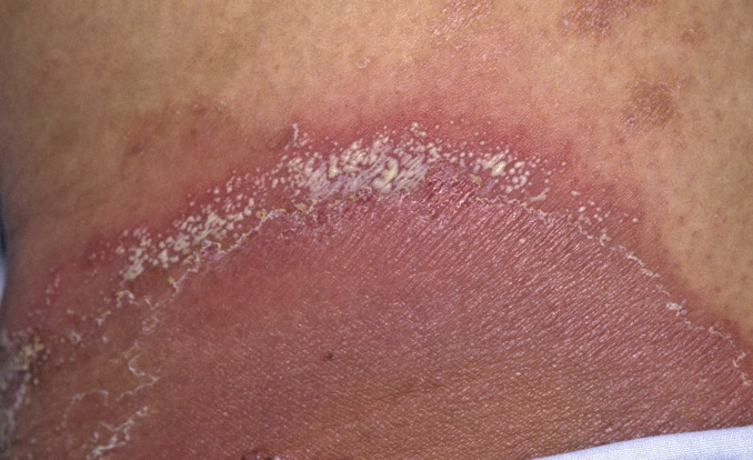
Fig. 12.4
Patient with generalized pustular psoriasis presenting as markedly erythematous plaques, with numerous small pustules arranged around the periphery of the lesions.
- •
Pustular psoriasis confined to the fingers is frequently associated with marked nail dystrophy and underlying arthritis.
- •
A patient experiencing acute flares may demonstrate associated fever and leukocytosis.
- •
Patients are more likely to have associated active psoriatic arthritis.
- •
Patients with pustular psoriasis are more likely to develop geographic tongue, scrotal tongue, and iritis.
- •
Rare cases may be life-threatening, with associated shock and/or renal failure.
- •
Impetigo herpetiformis is now considered to be acute pustular psoriasis associated with pregnancy.
- •
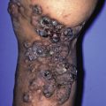
Pustulosis of the palms and soles (pustulosis palmaris et plantaris) is a variant that was formerly known as pustular bacterid (of Andrews) in older texts ( Fig. 12.5 ).
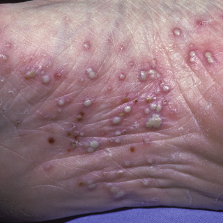
Fig. 12.5
Patient with pustular psoriasis of the palms and soles. This is a close-up of the instep of the foot.
(From the Fitzsimons Army Medical Center Collection, Aurora, CO.)
- •
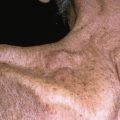
Acrodermatitis continua of Hallopeau is a rare variant of pustular psoriasis. It consists of pustules on one or more distal fingers or, less commonly, on the toes ( Fig. 12.6 ). This variant is often associated with nail dystrophy and distal psoriatic arthritis.
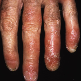
Fig. 12.6
Hand of a patient with acrodermatitis continua demonstrating marked erythema, pustules, crust, and destruction of the nail.
Diagnosis
- •
The diagnosis should be suspected in a patient with a personal or family history of psoriasis who has an acute or chronic pustular eruption.
- •
Peripheral leukocytosis is commonly present.
- •
A 3- or 4-mm punch biopsy of a pustular area will demonstrate a subcorneal pustule containing neutrophils. Although not specific, the histologic findings are often supportive.
- •
X-ray studies are indicated in the acrodermatitis continua of Hallopeau variant to exclude underlying psoriatic arthritis.
Treatment
- •
Withdraw oral corticosteroids or offending drugs, if possible.
- •
The recommended treatment is an oral retinoid, with etretinate being the treatment of choice. It can be combined with light therapy (narrow-band ultraviolet B [UVB], UVB, or psoralen and ultraviolet A [PUVA]).
- •
Treat with a biologic agent, such as adalimumab, etanercept, or infliximab. Paradoxically, some biologic agents have also been reported to precipitate pustular psoriasis.
- •
Oral cyclosporine, dapsone, and methotrexate have also been anecdotally reported to be useful.
- •
A medium-potency corticosteroid (e.g., triamcinolone, fluocinonide) in a cream or ointment base is helpful, although most cases only demonstrate partial improvement.
Acropustulosis of Infancy
ICD10 code L08.89
CAUSE UNKNOWN
Pathogenesis
Acropustulosis of infancy, also known as infantile acropustulosis, is, as the name implies, a pustular disorder that tends to affect the acral areas of infants. The pathogenesis is not understood, although there is a higher than expected incidence of atopic dermatitis. There also appears to be a higher than expected incidence in warmer climates, suggesting the possibility of an exaggerated arthropod reaction.
Clinical Features
- •
Typically, this first develops in infants between the ages of 2 and 6 months.
- •
This disorder may affect infants of any ethnic group but is more common in children with a darker skin color.
- •
Lesions classically recur in crops every 2 to 6 weeks.
- •
The primary lesion appears as markedly pruritic. 1- to 2-mm pustules on an erythematous base ( Fig. 12.7 ). In some cases, the primary lesions may have the appearance of cloudy vesicles.
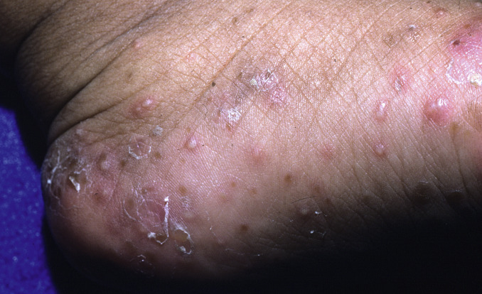
Fig. 12.7
Numerous small pustules and cloudy vesicles on the foot of a child. The lesions are in various stages of development, with some lesions demonstrating resolution and scale.
(From the William Weston Collection, Aurora, CO.)
- •
As the name implies, the distribution is primarily to the feet ( Fig. 12.8 ) and hands ( Fig. 12.9 ), although lesions may extend more proximally. In rare cases, lesions may affect the head and neck area or trunk.
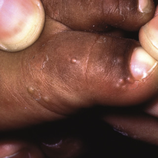
Fig. 12.8
Close-up of typical small pustules and cloudy vesicles on the toe of a young child.
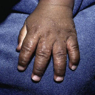
Fig. 12.9
Numerous pustules and cloudy vesicles on the hand of a young child.
(From the Fitzsimons Army Medical Center Collection, Aurora, CO.)
- •
Lesions typically resolve spontaneously over 1 to 3 weeks.
Diagnosis
- •
The clinical presentation of crops of small pruritic pustules on an erythematous base in an infant, particularly an infant with darker skin, is essentially diagnostic, although the differential diagnosis still includes scabies or another arthropod reaction.
- •
Important features include the absence of pruritic lesions in other members of the family. If other members of the family are affected, an arthropod reaction, especially scabies, needs to be excluded.
- •
In difficult cases, the lesions can be scraped for a mineral preparation to exclude scabies. The contents of the blister can also have Wright staining performed because the presence of numerous neutrophils favors acropustulosis of infancy, and the presence of numerous eosinophils would favor an arthropod reaction.
- •
A biopsy can be done in problematic cases and can be strongly supportive of the diagnosis; however, in most cases, it is not needed.
Treatment
- •
Topical corticosteroids are the mainstay of therapy. This disorder usually requires potent to superpotent topical corticosteroids.
- •
In severely pruritic patients, oral antihistamines can be added.
- •
In very severe cases, oral dapsone, 1 to 2 mg/kg per day, can be added. However, given the side effects associated with oral dapsone in infants, the risk-benefit ratio needs to be carefully considered.
- •
The patient’s parent(s) need to understand that none of these therapies alters the course of the disease.
Clinical Course
This is usually a chronic disorder, with children continuing to get new crops of lesions up to the age of 3 years, although the succeeding attacks tend to diminish in intensity.
Related posts:

Stay updated, free articles. Join our Telegram channel

Full access? Get Clinical Tree


