| Functional ptosis affecting vision |
| Cosmetically displeasing ptosis |
| Ptosis developing after cataract, refractive surgery, or prolonged contact lens use |
| Ptosis repair under general anesthesia or when patient cooperation is not necessary |
| Prior facial surgery or trauma |
| Dry eye symptoms/lagophthalmos present |
| Quality of Bell’s phenomenon |
| Prior refractive surgery |
| Co-existent dermatochalasis and/or eyebrow ptosis |
| Amount of levator function and response to topical phenylephrine |
| No history of glaucoma, strabismus, cicatricial conjunctival disease, cicatricial entropion or fornix shortening or congenital ptosis |
| Degree of ptosis present |
| Rule out myasthenia gravis |
| Presence of Hering’s reflex |
| Desire for upper eyelid crease (particularly with Asian patients) |
| Potential for revision/asymmetry |
Introduction
Conjunctival Müller’s muscle resection (CMMR) is a posterior approach ptosis repair technique that is suitable for correction of 3 mm or less of upper eyelid ptosis. CMMR is the preferred posterior ptosis procedure over the Fasanella–Servat procedure as tarsus is not removed during the procedure. CMMR is not indicated for patients with congenital ptosis or cicatricial conjunctival diseases. For treatment of congenital ptosis with poor levator function, see Chapters 14 and 15 . Caution should be exercised in patients with glaucoma or prior history of filtration surgery as well as patients who may need strabismus surgery. Repeat CMMR should also be performed cautiously as this may significantly shorten the conjunctival fornix leading to symblepharon. CMMR does not require patient cooperation and can be performed under general anesthesia. Upper eyelid ptosis that develops after cataract surgery, refractive surgery or prolonged contact lens use tends to respond well to CMMR. Furthermore, CMMR is ideally suited for ptosis repair of the cosmetic patient because of its predictable outcome and aesthetically pleasing contour. Concurrent upper blepharoplasty and CMMR can be performed as well as crease fixation at the time of surgery.
A positive phenylephrine test affirms that CMMR is a suitable procedure for the eyelid in question. Topical 2.5% phenylephrine is given in the affected eye, followed by a second set of drops 1–2 minutes later. At 5 minutes after instillation of the drops, the MRD1 is documented ( Figure 13.1 ). The position of the fellow eye should also be noted as the phenylephrine test may unmask ptosis from Hering’s law. Topical 10% phenylephrine is unnecessary and may lead to cardiotoxicity and other studies have shown no difference in efficacy between the two concentrations.
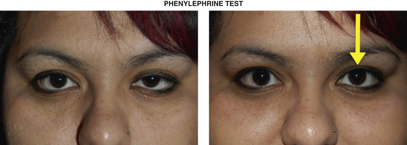
Table 13.3 provides a useful basis for titrating the amount of conjunctival Müller’s muscle resection and the desired amount of eyelid lift. These numbers should be considered as a starting point for one’s surgical decision making. With experience, adjustments of one’s “surgeon factor” can be employed to further optimize the procedure. In contrast to levator ptosis techniques, CMMR can be performed under general anesthesia, as the decision making in terms of amount of resection is determined during the clinical examination and not intraoperatively.
| Amount of ptosis to correct | Amount of resection of conjunctival Müller’s muscle |
|---|---|
| 1.0 mm ptosis | 4 mm |
| 1.5 mm ptosis | 6 mm |
| 2.0 mm ptosis | 8 mm |
| 3.0 mm ptosis | 9–10 mm |
Related posts:


Stay updated, free articles. Join our Telegram channel

Full access? Get Clinical Tree


