Pruritus (syn., itch) is generally defined as an unpleasant sensation that provokes the urge to scratch. There are several caveats that relate to the previous sentence. First, note that pruritus is spelled with an ending of “us” not “is,” and second, “itching” should not be used as a synonym for “scratching” even though patients often erroneously say “I’ve been itching my rash.” Third, notice that pruritus only provokes the “urge” to scratch and that it does not necessarily result in act of scratching. Scratching in the presence of pruritus exists on a spectrum with no scratching at one end (e.g., aquagenic pruritus), a little scratching in the middle (e.g., urticaria), and severe, chronic scratching at the other end (e.g., atopic dermatitis). Whether or not scratching occurs depends on both the nature of the provocation that is involved in the process and the genetic and psychological makeup of the person involved. Pruritus specific to the anogenital region has been recently reviewed (1).
Pruritus is generally classified into four groups based on neuropathophysiology: (a) pruriceptive pruritus (itching arising in the context of recognizable skin disorders); (b) neuropathic pruritus (itching arising as a result of peripheral nerve damage or entrapment); (c) neurogenic pruritus (itching arising as a result of central stimulation secondary to systemic disease or certain medication use and occurring in the absence of skin disease); and (d) psychogenic pruritus (itching arising as a result of psychological factors) (2,3).
Clinical Presentation
Pruriceptive itching is covered with the individual mucocutaneous disorders as they are discussed throughout this book. Neuropathic itching is localized mostly to areas outside the anogenital areas (e.g., notalgia paresthetica, brachioradial pruritus), but pruritus can occur in the genital region with disorders such as postherpetic neuralgia, other small fiber neuropathies, and itching within scars as a result of reinnervation of the scar tissue (4). Neurogenic pruritus tends to be generalized and therefore may affect the anogenital region. This type of pruritus often occurs with medications such as opiates and with disorders such as Hodgkin lymphoma, chronic hepatobiliary diseases (especially in the presence of cholestasis), polycythemia vera, and other myeloproliferative disorders (4). Psychogenic pruritus occurs in patients with conditions such as obsessive-compulsive disorders and those with prurigo nodularis and delusions of parasitosis.
It is difficult to know how to accommodate within the above classification some of the eczematous diseases (specifically, atopic dermatitis, neurodermatitis, and lichen simplex chronicus). All three of these disorders arise from normal-appearing skin, and there is no evidence of a preceding peripheral neuropathy. For this reason, they would not fit well in either the pruriceptive or neuropathic groups. Certainly there are marked psychological aspects present in nearly all of the patients who develop these three conditions, but it remains very controversial as to whether these factors are the cause of, or are the consequence of, these disorders. Perhaps, they might be fit within the neurogenic group based on the commonality with which a genetic tendency for atopy occurs. In any event, we have treated them separately and have placed them within the section entitled “eczematous disorders,” which can be found in Chapter 6.
Regardless of cause, the presence of pruritus (especially if it is accompanied by scratching) leads to a greatly diminished quality of life (QoL) and disturbance of sleep patterns and very often leads to the development of, or worsening of, depression and anxiety (5,6). Scratching while the person is unaware of doing so is common during daytime scratching and is also common during scratching that occurs at night, especially in the lighter stages of non-REM sleep (5).
Diagnosis
Identifying the cause of pruritus occurring in the presence of skin lesions depends on the clinician’s morphologic identification of the associated cutaneous disorder. The general approach to doing so is described in Chapter 3. Once the disease is identified, the clinician can locate it within the pages of this or other similar textbooks and then read about what steps can be taken to confirm the suspected diagnosis.
The situation is quite different and more difficult for pruritus that occurs in the absence of skin disease. In order to identify the cause of the pruritus, it is necessary to take a detailed patient history, carry out a complete physical examination, and obtain appropriate laboratory studies. This process is nicely detailed in the 2016 excellent review article by Pereira et al. (4). Since most of the readers of this text will primarily be treating patients who have skin lesions, and since the material on history, examination, and laboratory testing is long and detailed, I will refer the reader who requires this information directly to their publication.
Pathophysiology of Pruritus
Little is known about the mechanism(s) through which itching at the central level (neurogenic and psychogenic pruritus) occurs. Much more is understood about the pathophysiology through which itching occurs at the peripheral level (pruritoceptive and neuropathic pruritus). For these latter types of pruritus, there are many similarities (and, of course, some dissimilarities) between the pathophysiology of pruritus and pain (2,3). Most of the material on pathophysiology contained in the paragraphs that follow is based on a recent comprehensive review article (3).
Peripheral Nerve Pathways
The cutaneous nerves that receive and transmit pain are termed “nociceptors”; these are made up of narrowdiameter, lightly myelinated type Aδ fibers. Those cutaneous nerves that receive and transmit itching are called “pruriceptors”; they are comprised of very thin diameter, unmyelinated type C fibers. The pruriceptor fibers represent a quite small subset of the nociceptor fibers. Whether or not there are specific C fibers that carry itch, but not pain, remains controversial. The cell bodies of these painand itch-related nerves are located in the dorsal root ganglia of the spinal cord and their axons terminate in the papillary dermis and intercalate in between the cells of the epidermis.
Mediators and Receptors
Approximately 20 mediators and mediator receptors have been identified. Histamine is the best known mediator, and it acts on H1 and H4 receptors. The remainder of the mediators are non-histamine dependent, and some of the better known ones include proteases, substance P, calcitonin gene-related peptide, and bradykinin. The opioids are a special case. They act on µ and κ receptors; opioids that are µ antagonists and those that are κ agonists decrease itching, whereas µ agonists increase itching.
Central Pathways
The peripheral C fibers terminate in the dorsal root ganglia and the itch sensation is then transmitted to neurons expressing gastrin-releasing peptide receptors (GRPR) that cross over to the contralateral side and ascend the spinothalamic tract to the thalamus. Interestingly, there are also itch-inhibiting neurons in the spinal cord. From the thalamus, neuronal signaling is transmitted to cortical and subcortical sensory regions of the brain. Chronicity of pruritus is believed to be related to impairment of the itch-inhibitory pathways as well as to the development of central sensitization in a manner analogous to that which occurs in chronic pain.
Management
The treatment of pruritus is much the same for pruritus occurring from any one of the four types of itching discussed in the second paragraph of this chapter. The basic tenets of this approach are given detailed coverage in the section on eczematous and lichenified disease (see Chapter 6) and will not be repeated here. However, there are a few things that can be added for the treatment of noneczematous cutaneous disorders and for pruritus occurring in the absence of skin lesions (3,4,7). In the pruriceptive group, greater emphasis can be placed on the use on both sedating and nonsedating antihistamines for urticaria and other itching disorders mediated by histamine. In the neuropathic group, the antiepileptics, pregabalin and gabapentin, can be used in the treatment of the itching that sometimes occurs with diabetic and postherpetic neuropathies. In the neurogenic group, mirtazapine, naltrexone, naloxone, aprepitant, and ultraviolet light phototherapy may be considered for the itching associated with chronic renal disease, hepatobiliary disease, polycythemia vera, leukemia, and lymphoma. In the psychogenic group, much can be gained with clinical psychological intervention as well as with placement of earlier and greater emphasis on psychotropic medications.
Genital Pain
Generally, the causes of anogenital pain differ from the causes of anogenital itching. Certainly, some individuals who itch then rub and scratch to the point of pain, but those patients are usually very aware and describe pruritus as their predominant symptom. Many deny pain but describe burning, rawness, irritation, stinging, tearing, swelling, or soreness. For the purposes of this chapter, all of these characterizations of discomfort are termed pain. Itching is generally not included in this differential diagnosis. However, occasionally, some patients experience itching in a setting that usually produces pain.
Most chronic itching with scratching results from dermatoses. Chronic superficial burning, soreness, and pain, as distinct from pelvic pain, can result from a number of conditions. The International Society for the Study of Vulvovaginal Disease (ISSVD) reports that vulvar pain occurs from infection, skin disease, specific neuropathic syndromes such as postherpetic neuralgia, and anogenital pain syndromes of vulvodynia, penodynia, scrotodynia, and anodynia.
These pain syndromes rather than specific, observable abnormalities represent the largest proportion of these patients, and most women with self-reported chronic vulvovaginal pain were found to have vulvodynia on further evaluation (8). Idiopathic vulvovaginal pain is extremely common, with as many as 1 in 20 to 1 in 50 women each year developing unexplained, chronic vulvovaginal pain (9,10). Although there are very few data on pain syndromes in men, one report notes that the second most common condition seen in a male genital dermatology clinic was “dysesthesia” (11).
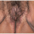
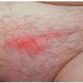
The patient who presents with burning, irritation, and other pain-type symptoms should be evaluated in an organized fashion (see Table 13.1). The skin should be examined carefully, using simple magnification if needed. Many patients who report symptoms of burning, irritation, or rawness also report redness and, often, edema. Redness that is mild, poorly demarcated, and without scaling or thickening is frequently within the range of normal, as genital skin is frequently red in asymptomatic individuals (Fig. 13.1). A patient report of erythema or edema does not indicate skin disease or infection; clinically modest erythema is common in asymptomatic women.
Those patients who have no relevant observable skin disease other than unimportant redness, no infection, and no specific neurologic abnormality fall into the category of genital pain syndrome: vulvodynia, penodynia, scrotodynia, and anodynia. At times, patients present with objective skin disease or infection, but their discomfort is disproportionate to the degree of skin disease noted, the skin disease is in a different location from the pain, or pain persists after the infection or skin disease is cleared. These patients have an underlying pain syndrome that is either unassociated with the skin disease or triggered by it (presented at ISSVD World Congress, Paris, 2011). Therefore, the skin disease or infection should be controlled, but a pain syndrome can be diagnosed and treated concomitantly.
Causes of Genital Pain
Many clinicians and most patients initially assume that genital pain without obvious clinical findings is due to infection, either yeast or sexually transmitted disease (Table 13.1). Chronic, unremitting pain almost never occurs from infection in immunocompetent patients, and negative cultures and a lack of response to antimicrobial therapy also signify a different diagnosis. Yeast, the cause most often implicated by patients and clinicians, usually is a pruritic rather than painful condition, and C. albicans clears, at least briefly, with therapy. Some postulate that an inflammatory response to low levels of yeast may cause pain (12). Either sexually transmitted diseases generally do not cause superficial genital pain (gonorrhea, syphilis, Chlamydia, warts) or they produce intermittent symptoms with visible skin findings (herpes simplex virus infection). However, Trichomonas, particularly in women, certainly produces irritation and burning, although often accompanied by itching. These infections can be ruled out easily by molecular studies.
TABLE 13.1 Causes of chronic genital pain
Infection, especially herpes simplex infection, rubbed/scratched yeast infection, Trichomonas, and fissures associated with infection
Dermatoses (noninfectious skin disease), especially lichen planus, excoriated/eroded lichen simplex chronicus or lichen sclerosus, irritant contact dermatitis, benign mucous membrane pemphigoid, pemphigus vulgaris, desquamative inflammatory vaginitis, atrophic vaginitis, fissures unassociated with infection, and malignancies/eroded tumors
Neuropathy, including diabetic neuropathy, postherpetic neuralgia, multiple sclerosis, pudendal neuralgia, and herniated disc disease
Vulvodynia, penodynia, scrotodynia, and anodynia—multifactorial pain syndromes
The most common skin disease that causes symptoms of burning is irritant contact dermatitis, especially due to overwashing and medications (see Chapters 6 and 11). Allergic contact dermatitis more often produces itching than burning and irritation (see Chapters 6 and 11). Erosive dermatoses most often produce pain and burning as well as itching. Lichen planus is often erosive (Chapter 11), as are other blistering diseases such as pemphigus vulgaris or cicatricial pemphigoid (see Chapter 10). However, dermatoses that are primarily pruritic also can be painful when patients have rubbed and scratched the skin; this is usually clear from the history of itching and the findings of excoriations (see Chapter 6). In women, skin disease causing vulvar burning includes skin disease of the vagina. Even in the face of negative cultures, vaginal inflammation (not infection) can produce introital symptoms. The vaginal skin conditions that most often produce pain symptoms are the atrophic vagina and atrophic vaginitis; erosive lichen planus and desquamative inflammatory vaginitis both produce purulent vaginal secretions that irritate the modified mucous membranes of the vulva (see Chapter 15). Because the vaginal mucosa can be difficult to visualize, especially in the patient with pain, a wet mount can be crucial to rule out inflammatory vaginal disease.
The patient with pain and visible erosions does not present a diagnostic dilemma. However, erosions can be subtle at times; vaginal and introital erosions can be missed in women, and fissures are overlooked easily in both men and women. Only specific skin lesions other than poorly demarcated, nonscaling redness that are not morphologically diagnosable should be biopsied.
Specific neurologic disease can sometimes be identified by history. For example, a diabetic with burning of the feet and genitalia most likely has diabetic neuropathy, and the patient with past herpes zoster of the genital area has postherpetic neuralgia (13,14). Postherpetic neuralgia only follows herpes zoster, not herpes simplex virus infection. Multiple sclerosis is sometimes associated with pain syndromes.
Pudendal nerve entrapment syndrome is an occasional cause of anogenital pain (15). This can be difficult to diagnose, at least partly because there are several types, and there is considerable individual variation in the anatomy and course of this nerve. There is no one standard evaluation and diagnostic regimen. This condition is suggested by sensory abnormalities in the saddle distribution of the pudendal nerve. There is pain or numbness of the genitalia and adjacent buttock, the proximal, medial thigh, and/or rectal area. Classically, the pain is worst when sitting and minimized by standing or lying. The diagnosis can be implicated further by a careful physical examination, magnetic resonance imaging examination performed by specialists in this disease, and magnetic resonance neurography. Management consists of physical therapy, medications for neuropathic pain and nerve block, behavioral modifications, surgical pudendal nerve decompression, radiofrequency, and spinal cord stimulation (16).
Patients with normal genital skin, to include the vagina; negative cultures; and no specific diagnosable neuropathy are diagnosed with vulvodynia, penodynia, scrotodynia, penodynia, or anodynia.
Vulvodynia
Vulvodynia is defined as vulvar discomfort in the absence of objective findings of skin disease, infection, or specific neurologic disease. The discomfort is most often described as burning, stinging, rawness, irritation, aching, soreness, or throbbing. Itching is not a prominent symptom. Earlier contemporary reports referred to this condition as “psychosomatic vaginitis.” In the 1980s, yeast and “subclinical human papillomavirus infection” were implicated. More recently, there have been increasing numbers of studies exploring the potential factors involved in vulvodynia as well as the epidemiology of this condition.
TABLE 13.2 Vulvodynia subsets
Localized
To vestibule (vestibulodynia)
To clitoris (clitorodynia)
Generalized (migratory, or extending beyond the vestibule or clitoris)
Each group can be spontaneous (unprovoked), elicited by touch, friction, or pressure (provoked), or both (mixed)
Adapted and simplified from the ISSVD vulvodynia terminology
Many years ago, the ISSVD divided vulvodynia into subsets, with the premise that different subsets had different epidemiology and different underlying etiologies. This has become increasingly unclear, but dividing vulvodynia into subsets is crucial for two reasons. First, excision of the vestibule is a treatment of choice for women whose pain is localized to that area. Second, this is useful for research purposes to ensure that unlike conditions are not being studied.
In my (Libby Edwards) experience and opinion, the important practical distinction among the subsets is the identification of vestibulodynia, where pain is strictly localized to the vestibule, clitorodynia, where pain is localized to the clitoris, and generalized vulvodynia, where pain extends beyond these areas or is migratory or irreproducible on either examination or careful patient questioning (Table 13.2). Previous synonyms for vestibulodynia include the vulvar vestibulitis syndrome, vestibular adenitis, and infection of the minor vestibular glands. The terminology was changed to eliminate “itis” because there was no evidence of a role for inflammation in vulvodynia. Generalized vulvodynia has been called dysesthetic vulvodynia in the past.
However, the academic subdivision vulvodynia includes not only the location of the discomfort but also the role of touch/pressure/friction (provocation) in the elicitation of pain. Appendix 3 reports the most recently revised classification of recognized patterns of vulvodynia along with associated factors. In this classification, there remain two major patterns of vulvodynia: those with vestibular pain (vestibulodynia) and those patients who have more generalized or migratory discomfort (generalized vulvodynia) with clitorodynia being an uncommon but recognized occurrence. I find that a careful history and examination usually shows overlap among the patterns of vulvodynia in most patients, with pain being primarily vestibular in nearly all patients, but with some degree of discomfort located outside the vestibule. Several studies support this contention (17,18).
Clinical Presentation
Vulvodynia is an extraordinarily common occurrence; why this condition is not widely understood and discussed by women and clinicians alike is a mystery. Several studies report a lifetime incidence of 10% to 17% and a current prevalence of 3.8% to 7% (1,19,20). About one ofin fifty women develops vulvodynia each year. Although initially believed to occur most often in Caucasians, these studies have indicated that patients of African genetic background experience vulvodynia at the same rate as do White patients, and Hispanics may have an increased risk of developing this disease. The risk of Asian individuals is not known. Vulvodynia is most often reported after the age of 20, but this may simply correlate with the increasing numbers of sexually active people about that age. However, the onset of vulvodynia occurs fairly frequently after menopause as well, independent of atrophic changes. Vulvodynia occurs but is uncommon in prepubertal girls (21). Once believed to be a chronic condition, with severity that waxes and wanes, this now is known to sometimes be recurrent, with about one woman in 10 with vulvodynia experiencing remission each year (10).
Women often report that symptoms began with a yeast infection, although idiopathic vulvar symptoms are frequently diagnosed as yeast, and rarely confirmed, so that this assumption is unsubstantiated. The most common and most troublesome symptom for most women is superficial dyspareunia, with burning as the primary quality of pain, although stinging, rawness, irritation, and sensations of tearing are common descriptors. Black women are far less likely to describe burning, and they are somewhat more likely to report aching, so that a diagnosis of vulvodynia may be missed (22). Hispanic women are even more likely than White women to experience burning sensations, and they are more likely to report lifelong rather than acquired symptoms (23). Generally, anything that puts pressure or friction on the vulva, especially the vestibule, produces discomfort; tight clothing, jeans, wiping after urination, tampons, gynecologic examinations, and exercise are common offenders. Most women indicate the vestibule as the area of greatest discomfort, although extension beyond the vestibule, either by touch/pressure in the office or by history, is very common. Less often, pain symptoms generalize to the labia minora, labia majora, and perianal skin, or the area of discomfort is migratory or poorly localized.
A physical examination reveals no skin changes except for variable erythema, especially in the vestibule, and pain to touch with a cotton-tipped applicator that typically is either limited to or worst in the vestibule (Fig. 13.1). Often, patients report redness and swelling that, on examination, is within the range of normal (Fig. 13.2). This is the typical examination for the vestibulodynia subset of vulvodynia. Frequently, patients describe very mild discomfort or “sensitivity” to touch beyond the vestibule but stop short of calling this pain. Less often, patients indicated more generalized or migratory pain; either there is pain to Q tip pressure in the vestibule that is accompanied by pain in other areas as well or, much less often, there is no pain to touch. This is found in patients with generalized vulvodynia.
Only gold members can continue reading. Log In or Register to continue