Fig. 15.1
This 63-year-old man presented with a slow-growing indurated nodule in association with a long-standing ill-defined erosive erythematous plaque in the perianal region
Pathology
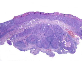
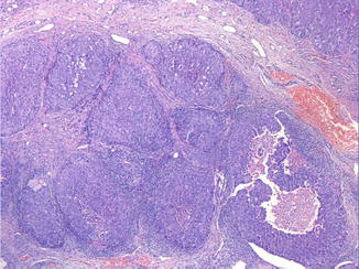
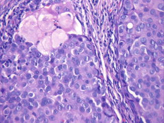
Primary cutaneous apocrine adenocarcinoma is an asymmetrical, poorly demarcated neoplasm with an infiltrating border, situated in the reticular dermis and subcutaneous tissue. Sometimes, the tumor extends into the epidermis in the form of franc extramammary Paget’s disease (Fig. 15.2). The neoplasm grows in different patterns including tubular, papillary, tubulopapillary, cystic, cribriform, diffuse, or solid growth pattern (Fig. 15.3). An “Indian file” growth pattern can also be encountered. The tumor is commonly accompanied by a dense hyaline stroma. Signs of apocrine differentiation such as decapitation secretion and papillary projections into the lumina are almost invariable present. However, these signs maybe lacking in poorly differentiating carcinoma. Areas of tumor necrosis can also be encountered. The neoplastic cells have round or oval vesicular nuclei, prominent nucleoli, and abundant eosinophilic cytoplasm (Fig. 15.4). Signet-ring cell features are sometimes encountered, especially in eyelid tumors with a striking predominance in elderly males. There is a variable cellular pleomorphism and an increased mitotic activity, especially in poorly differentiated neoplasms.


Fig. 15.2
The biopsy of the nodule shown in Fig. 15.1 displays a poorly differentiated apocrine adenocarcinoma with a solid growth pattern located in the reticular dermis. The tumor extends into the epidermis in the form of franc extramammary Paget’s disease

Fig. 15.3
The solid aggregations vary in size and shape and exhibit tumoral necrosis

Fig. 15.4


The neoplastic cells have round or oval vesicular nuclei, prominent nucleoli, and abundant eosinophilic cytoplasm. Some cells demonstrate mucin-laden cytoplasm. Focally, the neoplasm shows ductal differentiation
Stay updated, free articles. Join our Telegram channel

Full access? Get Clinical Tree


