Port-Wine Birthmarks


Port-Wine Birthmarks


Lance W. Chapman

Kristen M. Kelly


BACKGROUND

Vascular anomalies encompass 2 broad categories: vascular tumors and vascular malformations.1 Adopted in 1996 by the International Society for the Study of Vascular Anomalies (ISSVA), this nomenclature allows for the distinction between those vascular lesions with increased endothelial cell turnover and vascular proliferation (vascular tumors) and vascular lesions with a more latent endothelium and abnormal vascular structure (vascular malformations).2

In 2014, the ISSVA updated the Classification of Vascular Anomalies to reflect the additional disease types that have been identified over the previous decade.3 Vascular malformations, which in 1996 were classified as either simple or combined, have been further divided by 2014 ISSVA (Table 3.2.1).

This chapter will focus on capillary vascular malformations know as port-wine birthmarks (PWBs). These lesions were previously known as port wine stains (PWS), but this terminology is no longer preferred because of potential negative connotations.

PWBs are classified as capillary malformations (CMs), but they also contain postcapillary venules.4 The incidence of PWBs in newborns is estimated to be 0.3%, and the vast majority of PWBs are congenital.5,6 Rare acquired PWBs have been reported, but other diagnoses must be carefully considered.7


PRESENTATION

PWBs present at birth as a pink or light red patch. The head and neck region is a common area of presentation; however, they can occur anywhere on the body. Facial PWBs may occur in a dermatomal-type distribution, including the V1 ophthalmic region, V2 maxillary region, and V3 mandibular region. Central facial PWBs also occur. Rather than corresponding to areas of trigeminal innervation, these identified
patterns support the association with somatic mosaicism, which was described by Happle and Assim in 2001.8 PWBs do not follow the lines of Blaschko but rather are mostly localized or segmental in distribution. Multifocal and widespread PWBs do occur but are less common.


TABLE 3.2.1 Vascular Anomalies per the 2014 ISSVA Classification


  1. Vascular tumors


  2. Vascular malformations


    1. Simple


      1. Capillary malformations (CMs)


        1. Cutaneous and/or mucosal CM (PWBs)


        2. Telangiectasia


        3. Cutis marmorata telangiectatica congenita (CMTC)


        4. Nevus simplex/salmon patch/“stork bite”/“angel kiss”


        5. Others


      2. Lymphatic malformations (LMs)


      3. Venous malformations (VMs)


      4. Arteriovenous malformations (AVMs)


      5. Arteriovenous fistulae (AVFs)


    2. Combined


    3. Malformations of major named vessels


    4. Malformation associated with other anomalies


ISSVA, International Society for the Study of Vascular Anomalies.


PWBs tend to grow proportionally with the growth of the child and also thicken over time. The color progressively darkens, and by adulthood there may be a purple-red hue, along with tissue hypertrophy and nodularity (Figure 3.2.1). In a study by Klapman et al,9 of 173 patients with PWBs, thickening was observed in 11% (median age 32 years), nodularity in 24% (median age of 44 years), with 6% having both hypertrophy and nodularity (median 45 years). In another study by Geronemus et al,10 the mean age of hypertrophy was determined to be 37 years and 65% of PWB lesions had either hypertrophied or become nodular by the fifth decade.

Treatment of PWBs should not be viewed from purely a cosmetic perspective. Soft-tissue overgrowth can lead to functional impairment, particularly of the lip. Enlargement of the lips and gums can result in gingival bleeding and lip incompetence. Deeper structural changes can also occur, particularly with overgrowth of the maxilla and surrounding facial bones. This can cause the affected patient to develop a permanent open-bite. Furthermore, several studies have demonstrated that personality development can be severely impacted in patients with PWBs. The negative reaction of others to a “marked” patient can have significant psychological effects.11,12,13


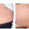
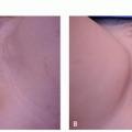
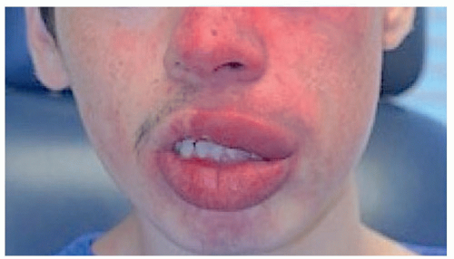
FIGURE 3.2.1 Hypertrophic and nodular port-wine birthmark.


PATHOGENESIS

Vascular malformations encompass a wide array of disorders that involve altered blood vessels or lymphatic channels. Therefore, it is believed that any mutation or dysfunction in any of the processes that signal for migration, differentiation, maturation, and cell survival of vascular anatomy can ultimately cause vascular malformations.23,24,25

Only gold members can continue reading. Log In or Register to continue

Stay updated, free articles. Join our Telegram channel

Jun 29, 2020 | Posted by in Dermatology | Comments Off on Port-Wine Birthmarks

Full access? Get Clinical Tree

Get Clinical Tree app for offline access