and Emir Q. Haxhija2
(1)
Department of Plastic Surgery and Burns, Institute for Mother and Child Health Care of Republic Serbia, University of Belgrade, New Belgrade, Serbia
(2)
Department of Pediatric and Adolescent Surgery, Medical University Graz, Graz, Austria
Keywords
PolydactylyChildrenTreatment8.1 Introduction
Congenital anomalies of the upper extremity vary from a barely noticeable to an absent extremity [1–3]. They are noted in approximately 2 per 1000 live births, with boys affected more commonly than girls (3:2) [2]. The diagnosis is in most cases made by physical and radiological exam, and vascular studies are rarely indicated [1–3]. Patients with hand anomalies can have associated malformations, most often involving the heart, kidneys, or tracheoesophageal complex [2]. Treatment of patients with congenital hand anomalies is multidisciplinary with the primary surgical goals to improve hand function and aesthetic appearance [2, 3].
8.2 Embryology
Around 4 weeks after fertilization, the upper limb bud appears as an oblong ventrolateral bulge on the body wall [1–6]. The emerging limb bud is composed of somatic lateral plate mesoderm covered by the ectoderm [2, 4]. Subsequent limb bud growth and differentiation are controlled by distinctive region-signaling centers: apical ectodermal ridge (AER) (proximodistal growth), Wnt (Wingless type) (dorsoventral growth), and zone of polarizing activity (ZPA) (anteroposterior-radioulnar growth) [1, 3–5]. Signaling pathways critical to limb formation include several other factors such as Sonic Hedgehog (Shh) protein and fibroblast growth factors (FGF) [2, 5, 7, 8]. Digits are recognizable at 41–43 days and fully separate at 53 days of gestation [5].
8.3 Classification
Oberg and colleagues proposed a modified Swanson classification of hand anomalies, and by this classification all hand anomalies are placed within one of three groups: malformations (abnormal cell formation), deformations (insult to cells which have formed normally), and dysplasias (lack of normal cell organization) [2, 3, 8].
- 1.
Malformations
- (a)
Failure in axis formation and differentiation—entire upper limb
Proximal-distal outgrowth (symbrachydactyly, transverse deficiency, intersegmental deficiency)
Radial-ulnar (anteroposterior) axis (radial longitudinal deficiency, ulnar longitudinal deficiency, ulnar dimelia, radioulnar synostosis, humeroradial synostosis)
Dorsal-ventral axis (nail-patella syndrome)
- (b)
Failure in axis formation and differentiation—hand plate
Radial-ulnar (anteroposterior) axis (radial polydactyly, triphalangeal thumb, ulnar polydactyly)
Dorsal-ventral axis (dorsal dimelia, hypoplastic/aplastic nail)
- (c)
Failure in hand plate formation and differentiation—unspecified axis
Soft tissue (syndactyly, camptodactyly, trigger digits)
Skeletal deficiency (brachydactyly, clinodactyly, Kirner’s deformity, metacarpal and carpal synostosis)
Complex (cleft hand, synpolydactyly, Apert’s hand)
- (a)
- 2.
Deformations (constriction ring syndrome)
- 3.
Dysplasias (macrodactyly, limb hypertrophy, tumorous condition)
8.4 Polydactyly
Polydactyly is the presence of extra digits or duplication of digital parts [1, 2, 4–6, 9–11]. The pathogenesis of polydactyly is not known [11, 12]. It is easily detected, most frequently observed congenital hand anomaly that causes cosmetic and functional impairment ranging from minor cutaneous protuberance to complete duplication of a limb [1, 5, 10, 11, 13]. Polydactyly can be detected by ultrasound (US) as early as 14 weeks of gestational age [9].
The true incidence of polydactyly is not known, it depends on the studied population, and it varies from 0.3–3.6/1000 and 1.6–10.7/1000 in general population [7, 9, 10]. The incidence of radial polydactyly varies from 0.08:1000 to 1.4:1000 1ive births, and it represents 90% of all polydactyly cases [1, 2, 5, 8]. Polydactyly affects Blacks more commonly than Whites [2, 9, 13]. Upper limbs are more involved than lower limbs and right hand more than the left [9]. Preaxial polydactyly is the most common (mostly isolated), followed by postaxial (mostly syndromic and bilateral), and meso-axial (central) [7, 10]. Polydactyly has been associated with more than 300 diseases and syndromes (patient with atypical presentation (syndromes) should be referred to genetics) [7, 9, 10].
8.4.1 Classification
Classification of polydactyly proposed by Temtamy-McKusick as preaxial polydactyly, postaxial polydactyly, complex polydactyly, central polydactyly, and mixed polydactyly (syndromic or nonsyndromic) is most widely used (Figs. 8.1a–c, 8.2a, b, and 8.3) [5, 7, 9–12].
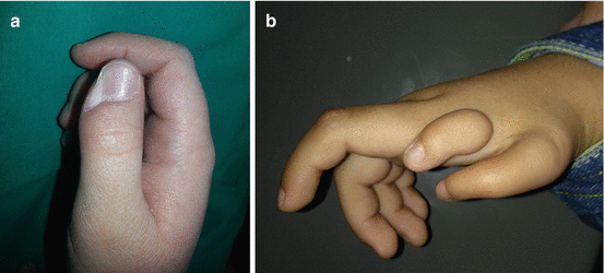
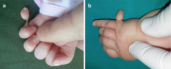
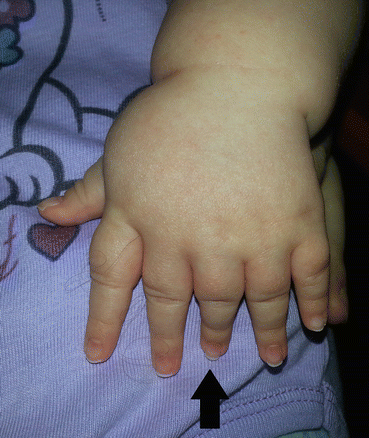
Fig. 8.1
Preaxial polydactyly: (a) barely noticeable deformity; (b) complex radial polydactyly including floating thumb and hypoplastic thumb
Fig. 8.2
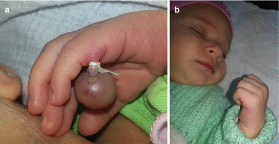
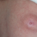
Postaxial polydactyly: (a) rudimentary digit; (b) fully developed digit
Fig. 8.3
Central polydactyly
There are also several classification systems of radial, central, and postaxial polydactyly: Wassel-Flatt classification of thumb duplication, Wood and Miura modification of Wassel-Flatt classification, Stelling and Turek classification of central polydactyly, and Ryan classification of postaxial polydactyly [2, 5, 9, 10, 12].
Wassel-Flatt classification of radial polydactyly is based on the radiological findings, including seven types of splitting of the thumb at different levels [1, 3, 5, 7, 9, 14]. Recently, the classification of thumb polydactyly is named Flatt classification (Harry Wassel was a hand surgery fellow of Adrian Flatt) [4]. Wassel-Flatt types of thumb duplication include type I (distal phalanx), type II (interphalangeal joint, IPJ), type III (proximal phalanx), type IV (metacarpophalangeal joint, MCPJ), type V (metacarpal, MC), type VI (carpometacarpal joint, CMCJ), and type VII (at least on thumb is triphalangeal) [5, 7, 8, 11].
Wood and Miura presented a modification of Wassel-Flatt classification dividing type IV thumbs into three subtypes and type VII into four subtypes [5, 8]. Zuidam et al. proposed the Rotterdam classification system in 2008 that includes Wassel-Flatt classification, with Buck-Gramcko and Behrens intercarpal modification and suffixes to indicate different complex deformities (Tph, triphalangism; T, triplication; S, symphalangism; D, duplication; H, hypoplasia) [4, 8].
8.4.2 Patient Evaluation
8.4.3 Treatment Option
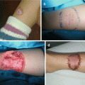
Surgical treatment of polydactyly seeks to remove the least functional component, to reconstruct normal parts, and to allow normal hand function [2, 3, 5, 9, 11]. Anatomic level of duplication, musculoskeletal components involved, stability, developmental stage, and cosmetic outcome have to be considered carefully [1, 2, 5, 7, 9, 11–16]. Floating little fingers with narrow soft tissue stalk <2 mm can be removed early in the newborn nursery by ligation, and simple excision is possible when there is no bone connection and joints are stable [3, 5, 9]. Surgical treatment ideally occurs before the supernumerary elements displace the normal elements and before fine motor skills have developed with the abnormal anatomy (from 6 to 9 months of life, at the end of the first year of life, or during second year of life) [5, 9, 11, 14]. Radiological examination is usually adequate in obtaining additional preoperative information [3].
8.4.3.1 Radial Polydactyly
Regarding radial polydactyly, Wassel-Flatt type II and type IV are the most frequent [1, 7, 8, 14]. In Wassel-Flatt type II, there is a broad proximal phalanx with two (partial) distal phalanges [2, 7, 8]. If there is an unequal duplication, the smaller thumb has to be excised along with the part of articular surface of the proximal phalanx, preserving the collateral ligament [2, 5, 7]. If necessary, tendon augmentation with smaller finger tendons is performed [6]. In case of symmetrical duplication, Bilhaut-Cloquet procedure is performed [5, 9, 14–16].
In Wassel-Flatt type IV, there is often a hypoplastic extra thumb on the radial side [1, 5, 7, 9]. The radial thumb has to be excised similar to the technique for Wassel type II, and the broad MC is partially excised in distal part (with asymmetric tendons realigned in the “ulnar” thumb) (Fig. 8.4a–c) [5, 7, 9]. Deviation at the IPJ should be corrected with a transverse wedge osteotomy [2, 7, 9].
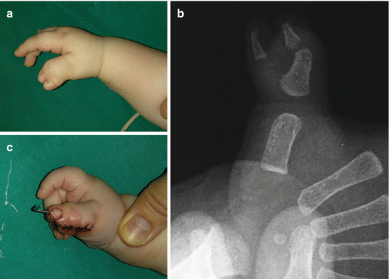
Fig. 8.4
Reconstruction of thumb duplication by excision of the radial part of the thumb: (a) preoperative view; (b) radiography of the thumb; (c) postoperative view
When there is minimal size mismatch between the two thumbs (Wassel-Flatt type I, II, III and occasionally IV), the two outer halves can be joined after excising the two inner halves longitudinally, in the Bilhaut-Cloquet operation (Fig. 8.5a–c) [2, 5, 11, 14, 16]. This procedure can be complicated by joint stiffness, growth arrest, asymmetric growth, and longitudinal nail bed deformities (this can be avoided by Baek modification) [2, 11, 16].
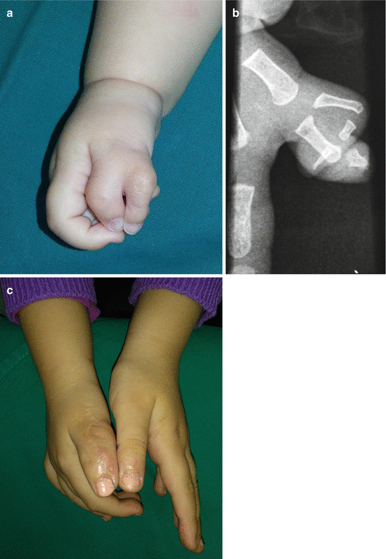
Fig. 8.5
Reconstruction of the thumb duplication (Bilhaut-Cloquet procedure): (a) preoperative finding; (b) radiography of the thumb; (c) postoperative view
8.4.3.2 Postaxial Polydactyly
Postaxial polydactyly includes duplication of small finger of the hand [2, 5]. It has high incidence in African and African-American population [1, 5, 6]. There are two most common used classifications of postaxial syndactyly proposed by Stelling Turek and Temtamy-McKusick [1, 2, 5, 7]. According to Stelling and Turek classification, there are three types of postaxial polydactyly (type I soft tissue, type II partial duplication, and type III complete duplication), and according to Temtamy-McKusick, there are two types of postaxial polydactyly (A, fully developed finger; B, a rudimentary digit) [1, 2, 5, 9, 10]. Postaxial type B digits with narrow stalk can be ligated (revisions may be necessary later in life) (Fig. 8.6a, b) [2, 5, 7, 9]. For more developed extra little finger, surgical excision is recommended [1, 5, 7]. Postaxial type A digits are excised through large ulnar-sided flap with zigzag incisions, spearing ulnar collateral ligament, and with trimming of articular surface of the MC [2, 5].