and Emir Q. Haxhija2
(1)
Department of Plastic Surgery and Burns, Institute for Mother and Child Health Care of Republic Serbia, University of Belgrade, New Belgrade, Serbia
(2)
Department of Pediatric and Adolescent Surgery, Medical University Graz, Graz, Austria
Keywords
Venous malformationChildrenTreatment15.1 Introduction
Vascular anomalies are classified into vascular tumors (characterized by endothelial proliferation) and vascular malformations (present the errors in morphogenesis) [1–3]. Vascular malformations are divided into simple malformation (capillary, lymphatic, venous malformation, arteriovenous malformation, and arteriovenous fistula), combined malformations (including at least two vascular malformations in one lesion), malformation of major named vessels, and malformation associated with other anomalies [1].
Venous malformations (VMs) are the most commonly treated slow-flow vascular malformation with an incidence of approximately 1–2/10,000 births [4–11]. They grow generally in proportion with the child, do not involute, and tend to enlarge disproportionately over time [3, 4, 6, 7, 10–12]. The pathogenesis of VM is unclear (TIE-2 endothelial receptor mutation is found in some of patient with sporadic VMs) [1, 2, 7, 9, 12].
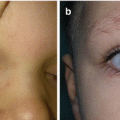
VMs are classified as superficial or deep, focal, multifocal, or diffuse (Fig. 15.1a, b) [1–5]. Superficial VMs are presented as a blue skin discoloration or as a soft, nonpulsatile, compressible subcutaneous mass [2, 4, 6, 7]. Deep VMs infiltrate muscle, bone, and visceral organs and may be unrecognized for until they become symptomatic [1–4, 6, 7, 12, 13]. There is also a classification based on imaging and clinical features, dividing VMs into spongiform (most common type), phlebectatic, aneurismatic, and reticular type [6]. Dubois et al. divided VMs into four types based on the pattern of venous drainage channels, response to treatment, and rates of complication: isolated malformation without drainage, lesions draining into normal veins, lesions draining into dysplastic veins, and lesions consisting primarily of venous ectasia [6, 7]. Histologically, VMs are composed of thin-walled, dilated spongelike channels with normal endothelial lining and abnormal smooth muscle architecture [1–7, 13, 14].
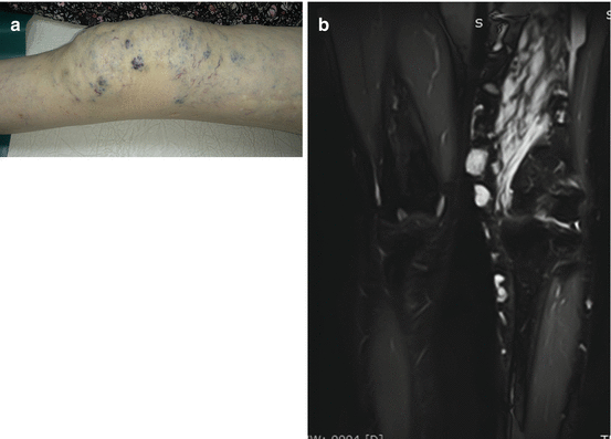
Fig. 15.1
Diffuse venous malformation of the left lower extremity: (a) clinical view of the knee region; (b) magnetic resonance imaging
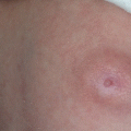
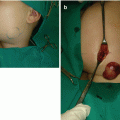
The most common types of VMs are sporadic VM, glomuvenous malformation (GVM), and blue rubber bleb nevus (BRBN) syndrome [1, 4–6]. Most VMs occur as sporadic and solitary (approximately 94% of VM) (Fig. 15.2) [1, 2, 4, 7, 14]. Sporadic VM is usually greater than 5 cm, single, and mostly located on the head and neck and extremities (Fig. 15.3a, b) [2, 8, 13].
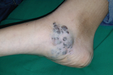
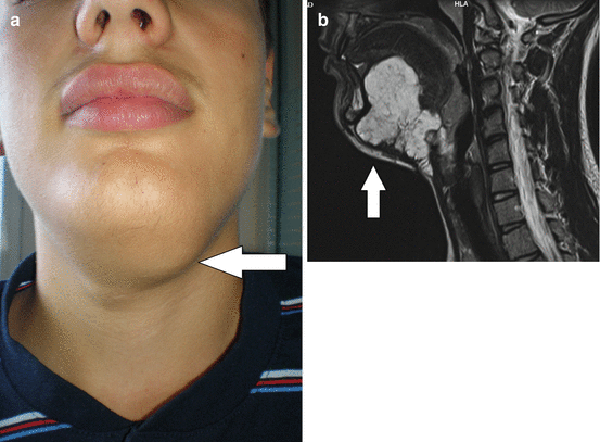
Fig. 15.2
Localized venous malformation of the right foot
Fig. 15.3
Venous malformation of the sublingual region: (a) submandibular leasion; (b) magnetic resonance imaging of the lesion
VMs can be part of the syndromes such as capillary-lymphatic-venous malformation (CLVM) or Klippel-Trenaunay syndrome, congenital lipomatous overgrowth, vascular malformations, epidermal nevi and scoliosis or skeletal and spinal anomalies (CLOVES), mucocutaneous familial venous malformation, Maffucci’s syndrome, and Proteus syndrome [6, 7, 13].
GVM is the most common familial form of venous anomaly, formerly known as glomangioma caused by mutation of the glomulin gene on chromosome 1 [1, 2, 4–6, 14]. They account for about 5% of all VM [4, 5]. GVMs form nodular or plaque-like small lesions (two thirds <5 cm), typically multiple, painful on palpation affecting the skin (rarely mucosa), and involving extremities in most cases [1, 2, 4, 5, 7, 14]. Histologically, GVMs appear as dilated venous channels with abnormal smooth muscle-like glomus cells [4, 14]. Blue rubber bleb nevus syndrome (BRBNS) or Bean syndrome is a rare condition characterized by multiple, small (1–2 cm in size) VMs, involving the skin (typically present on palmar and plantar surfaces), soft tissue, and gastrointestinal (GI) tract (Fig. 15.4) [2, 4, 6, 12, 15]. Lesions within the GI tract may be associated with significant bleeding [2, 4]. Treatment of patients with BRBNS is challenging, involving resection of skin lesions, bowel resection, and ligation of lesions, and recently, sirolimus is used for bleeding control [4, 6, 15].
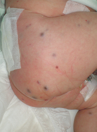
Fig. 15.4
Blue rubber bleb nevus syndrome of the right gluteal region
Differential diagnosis includes verrucous hemangioma (VH) (provisionally unclassified low-flow vascular malformation that is clinically similar to a hyperkeratotic VM); deep (subcutaneous hemangioma), lymphatic malformation; and dermal melanocytic nevi (Mongolian spot, nevi of Ota and Ito, and common blue nevus) [1, 2, 5]. Jugular vein phlebectasia is a congenital fusiform dilatation of jugular vein and is usually asymptomatic (Fig. 15.5a, b) [16].
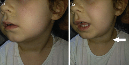
Fig. 15.5
Phlebectasia: (a) normal clinical finding; (b) neck mass enlarged with Valsalva maneuver
15.2 Complications (Symptoms) of VM
The most common symptom of VM is pain due swelling, and other symptoms are usually related to localization of VMs [2, 4, 7, 12]. In head and neck region, VM can cause airway or orbital compromise due to mucosal bleeding [9, 12]. Extremity VM can cause leg-length discrepancy, hypoplasia, pathologic fracture, and hemarthrosis, and muscle VM may result in fibrosis and subsequent pain and disability [2, 12]. Gastrointestinal VM can cause bleeding and chronic anemia [2, 4, 12].
VMs are associated with spontaneous thrombosis and thrombolysis, and they are often proportional to the size of VM [4, 5, 12]. Patients with extensive VMs have been found to have elevated D-dimer levels which are sensitive marker for thrombus formation and fibrinolysis [4, 5, 7, 12, 13]. D-dimers are not elevated in GVM [6]. Stagnation within a large VM results in a localized intravascular coagulopathy (LIC), painful phlebothromboses, and pulmonary embolism [2, 4, 12]. Localized intravascular coagulopathy (LIC) is characterized by an increase in D-dimer level, normal platelet count, and decreased fibrinogen level, and patients usually well tolerate this condition LIC [4, 5]. Severe LIC can aggravate to disseminated intravascular coagulation (DIC) which is a serious life-threatening condition [5].
15.3 Patient Evaluation
VMs are in most cases diagnosed by history and physical examination [2, 4, 5, 7, 11, 14]. VMs are easily compressible and usually swell in the dependent position [2, 6]. Patients with VMs should be screened for a basic coagulation profile including complete blood count, prothrombin time (PT), partial thromboplastin time (PTT), D-dimer, and fibrinogen [4, 5, 12].
Related posts:
Stay updated, free articles. Join our Telegram channel

Full access? Get Clinical Tree


