Fig. 21.1
Solar urticaria on back soon after sun exposure
Management Strategies
General management of solar urticaria includes use of protective clothing, broad spectrum UVA/UVB sunscreens, and avoidance of exposure to the wavelengths of light/UV radiation to which the individuals are sensitive. For those with solar urticaria provoked by visible light or longer wavelength UV-A, exposure avoidance may be challenging. Treatment with H1 antihistamines is helpful, and is generally regarded as first-line therapy. Phototherapy to induce tolerance may also be a useful treatment strategy.
Investigations Recommended
For diagnosis |
Phototesting to UVA (320–400 nm), UVB (290–320 nm), visible light (400–800 nm) |
Consider ANA, anti-Ro, anti-La, plasma and RBC porphyrins to help exclude connective tissue disease and porphyria |
Consider total serum IgE; IgE level may be helpful in treatment-refractory solar urticaria cases for which omalizumab is considered |
This review article provides an excellent overview of the pathogenesis, diagnosis, and treatment of photodermatoses [1].
Table 21.1
First line therapies
H1 antihistamines | C |
In this retrospective case series of 57 patients, which includes 14 patients between the ages of 9 and 20 years, H1 antihistamine daily reduced intensity of wheals and erythema in 63 % of patients. Antihistamine dosing was as follows: terfenadine 60 mg twice daily, astemizole 10 mg once daily, cetirizine 10 mg once daily, and loratadine 10 mg once daily. Patients who did not respond to scheduled antihistamine treatment were treated with oral PUVA as discussed below. Almost half of the patients were disease free within 5 years [2].
Seven patients (mean age 28 years, included one 17-year-old) with documented solar urticaria to visible light were treated with cetirizine 10 mg daily for 4 weeks. During weeks 3 and 4 of treatment, they were encouraged to expose themselves gradually to natural light. Symptomatic relief and increased minimal urticaria doses (MUD) were noted in all participants after 15 days of cetirizine [3].
A 2-year-old girl with solar urticaria on photoprovocative testing to UVB, UVA, and visible wavelengths up to 500 nm had resolution of symptoms while taking loratadine 10 mg daily [4].
Table 21.2
Second line therapies
Phototherapy based hardening (narrow band UVB, broad band UVB, UVA, PUVA) | B |
H1 antihistamines and Leukotriene receptor antagonist | C |
This retrospective case series of 57 cases includes 14 children and adolescents between the ages of 9 and 20 years. Patients who did not respond to scheduled antihistamine treatment were treated with oral PUVA three times weekly × 4 weeks (oral administration of psoralen 0.6–0.8 mg/kg followed by UVA irradiation 2 h later), and 26 % of these patients achieved complete suppression of wheal formation and erythema with phototesting, rendering MUD undetectable [2]
Narrowband UV-B phototherapy controlled solar urticaria in 39 adult patients. Individuals <18 years of age were excluded in this study, however, given that narrowband UVB is a well-tolerated therapy for many childhood skin disorders, and that narrowband UVB is efficacious in solar urticaria in adults, it is reasonable to consider as an alternative to PUVA in children and adolescents, although specific data in the pediatric population is lacking [5].
This review article discusses phototherapy management of solar urticaria [6].
This prospective study treated eight patients with verified solar urticaria to visible light and UVA who were assigned to one of two treatment arms based on disease severity. Those with higher minimal urticaria doses (less photosensitive) were started on combination therapy with desloratadine 5 mg twice daily and montelukast 10 mg once daily. Those with lower minimal urticaria doses (more photosensitive) were started on desloratadine 5 mg twice daily, fexofenadine 120 mg twice daily, cetirizine Hcl 10 mg twice daily, and montelukast 10 mg once daily. The study included four children (ages 5–16 years). Doses were adjusted to standard pediatric dosing for these children. Partial remission was noted in one child (who was in the more aggressive arm), and full remission was noted in the other three children and all four treated adults. The medications were well tolerated [7].
Table 21.3
Third line therapies
Omalizumab | E |
This case report describes a 16-year-old boy with solar urticaria to UV-A and UV-B treated with omalizumab. The patient failed to improve with antihistamines, including loratadine (30 mg/day), cetirizine (20 mg/day), and diphenhydramine (up to 200 mg/day), and was noted to have an elevated serum IgE (851 IU/ml), so he was started on a trial of omalizumab 400 mg every other week for 3 months. He demonstrated partial improvement after six omalizumab treatments, with an increase in the minimal urticaria dose for UV-B, and delayed response time from 1 to 15 min for post-exposure erythema in both UV-A and UV-B spectrum [8].
This case report describes a 16-year-old girl with solar urticaria to UV-B spectrum exposure. She failed to improve with broad spectrum sunscreen use and the combination of cetirizine 10 mg/day, desloratadine 5 mg/day and hydroxyzine 25 mg/day, as well as with the combination of cetirizine 20 mg/day, ranitidine 150 mg/day and montelukast 10 mg/day. Five-day courses of prednisone (25 mg daily) were ineffective. Her IgE was mildly elevated (228 IU/ml). She was treated with 375 mg omalizumab every other week for 6 months, then dosing was reduced. Omalizumab treatment induced remission of her solar urticaria, which remained inactive 4 months after treatment was discontinued. Phototesting at end of therapy and at 4 months after discontinuation was negative [9].
This case report describes a 6-year old boy with recurrent episodes of solar urticaria associated with angioedema. The action spectrum for his solar urticaria was found to be limited to the visible light range on provocative phototesting to UV-A, UV-B, and visible light. He failed to respond to antihistamine treatment of desloratadine 5 mg twice daily, fexofenadine 120 mg twice daily, cetirizine 5 mg in morning and 10 mg at night, and montelukast 4 mg once daily. He had an elevated IgE (2004 IU/ml) and was treated with omalizumab, which was gradually escalated to 300 mg once every 2 weeks. Once remission occurred, dosing was spaced to monthly to maintain remission while antihistamines and leukotriene receptor antagonists were weaned [10].
Non-standard Therapies Not Studied or Reported for Children/Adolescents
Intravenous immunoglobulin (IVIG) | E |
Extracorporeal photopheresis | E |
α-melanocyte-stimulating hormone analogue [Nle4-D-Phe7]-α-MSH | C |
This case series describes two adult women with solar urticaria who were successfully treated with 2 g/kg of IVIG divided over 5 days, and remained in remission post-IVIG for greater than 1 year (13 months and 4 years at time of report) [11].
This case report describes a 54-year-old-man with solar urticaria who failed to improve with oral antihistamines, hardening (with UVA, UVB, visible light, or oral PUVA), and oral cyclosporine. He was treated with nine extracorporeal photopheresis cycles, and his minimal urticaria dose (MUD) increased and symptoms decreased [12].
Five patients with solar urticaria were treated with α-melanocyte stimulating hormone analogue afamelanotide. The mean age of participants was 34 years, and the youngest patient was an 18-year-old. All five subjects had a solar urticarial response to UV-A, and some had overlapping response to UV-B or visible light. Subjects were treated with a single 16 mg afamelanotide implant in winter. In all subjects, increased melanocytic pigmentation was noted, along with a decreased urticarial response and reduced MUD at 30 and 60 days post implant. No serious adverse effects occurred, and no concerning changes in nevi were noted [13].
Polymorphous Light Eruption
Clinical Features
Although more prevalent in young adults, polymorphous light eruption (PMLE) also develops during childhood, and is the most common photosensitivity disorder among children. Pruritic skin lesions develop in photo-exposed areas within hours to days after sunlight exposure, and may have many different morphologies, with the most common being papules or papulovesicles (Fig. 21.2). The eruptions occur most often in late spring to early summer, as “hardening” occurs, and affected individuals experience decreased photosensitivity after gradual sun exposure over time. In some cases, presumably due to the hardening phenomenon, the face and dorsal hands may be spared, whereas lesions develop on areas which were covered in the preceding winter months, such as the upper chest and extensor arms.
Fig. 21.2
Polymorphic light eruption on the face
Management Strategies
Prevention is the first-line therapy for patients with PMLE. Affected individuals should avoid exposure to sunlight, wear protective clothing, and use sunscreen with high sun protection factor UVA/UVB coverage.
In those with recurrent eruption, despite diligent sunlight protection and avoidance, phototherapy to induce hardening in spring can be effective in preventing eruption in up to 90 % of the patients.
In patients with an acute flare of PMLE, topical corticosteroids and oral antihistamines can reduce inflammation and alleviate itch. In more severe cases, treatment with oral prednisone, antimalarial medications, beta-carotene, nicotinamide, cyclosporine, and dietary fish oil can be considered. Further, azathioprine and thalidomide have been reported as possible alternative therapies.
Lastly, several experimental prevention strategies have been reported, including extract of polypodium leucotomos, topical DNA repair enzymes, topical vitamin D, and flavonoids. These approaches may be promising as potential alternative therapies for patients unable or unresponsive to phototherapy treatment.
Investigations Recommended
For diagnosis |
Phototesting |
Skin biopsy |
ANA, anti-Ro, anti-La to help exclude connective tissue disease |
This retrospective study reports successful phototesting in 92 children between the ages of 4 and 16 years. Of the participants, 56 had a photosensitivity disorder and 22 were diagnosed with PMLE. The average age of the PMLE patients was 11 years. Among them, photosensitivity for UVB was found in 32 %, UVA 28 %, and both UVA and UVB in 40 % [14].
Table 21.4
First line therapies
Behavioral sunlight avoidance with use of high-protection level UVA/UVB broad-spectrum sunscreen | C |
Narrowband or Broadband UVB phototherapy | C |
PUVA | D |
Twelve adults with PMLE were tested by photoprovocation after applying placebo or sunscreen with high protection level against UVB/UVA, containing methylene bis-benzotriazolyl tetramethylbutylphenol (Tinosorb M), bis-ethylhexyloxyphenomethoxyphenyl triazine (Tinosorb S) and butyl methoxydibenzoylmethane. PMLE symptoms were elicited after photoprovocation on the placebo-treated arm in 10 of 12 subjects, but were not replicated in any of the subjects on the UVA/UVB sunscreen-protected arm in individual subjects [15].
When applied at a concentration of 2 mg/cm2, all 15 female patients (ages 18–45 years) treated with high UVA protection sunscreen (SPF45, UVA protection factor 25) showed no PMLE skin eruptions upon exposure provocation 15 min after sunscreen application. In comparison, only 4 of 15 (27 %) patients were protected when using lower UVA protection sunscreen (SPF45, UVA protection factor 5) [16].
Among 51 patients (ages 5–82 years) treated with PUVA (three times weekly for 3–4 weeks in the spring), 64 % reported total protection from PMLE symptoms during the summer after treatment completion, 26 % reported partial protection, and 10 % reported no change. Development of the eruption during active treatment occurred in 22 % of the patients, but was usually mild and did not interfere with treatment [17].
In eight of ten patients (ages 18–74), photohardening with narrow-band UVB was successful, even in patients refractory to UVA and broadband UVB phototherapy [18].
Table 21.5
Second line therapies
Oral prednisone | A |
Beta-carotene, lycopene, Lactobacillus johnsonii supplement | A |
Anti-malarial medications | A |
Thalidomide | B |
Nicotinamide | B |
Dietary fish oil | C |
Azathioprine | E |
In eight patients, 25 mg prednisone daily for 7 days was superior to placebo in management of acute PMLE. This suggests that a short course of prednisone is reasonable for patients planning sunny vacations [19].
In 60 patients (ages 18–50 years), treatment with one capsule per day for 12 weeks of a commercially available nutritional supplement (verum) containing 2.5 mg lycopene, 4.7 mg of beta-carotene, and 5.108 cfu of the probiotic L. johnsonii (Inneov Sun Sensitivity, Laboratoires Innéov, Asnières sur Seine, France) resulted in reduced PMLE lesion induction compared to control group [20].
For PMLE, 63 patients (all >18 years) were treated with hydroxychloroquine 400 mg/day for 1 month, and 200 mg/day for a second month, whereas 54 patients received treatment with chloroquine 500 mg/day for 1 month, and 250 mg/day for a second month. Hydroxychloroquine was marginally more effective than chloroquine in managing acute flares [21].
22 of 25 patients (adult and pediatric) treated with 100–200 mg/day thalidomide experienced improvement without notable side effects [22].
25 out of 42 (60 %) subjects (ages 16–56 years) were treated successfully with oral nicotinamide 3 g daily for 2 weeks that led to the absence of symptoms, despite extensive sun exposure. The dose was reduced to 2 g daily after 1 week of treatment, and 12 of the 25 patients developed PMLE relapse [23].
13 patients (ages 21–81 years) were treated for 3 months with fish oil capsule twice daily containing 18 % eicosapentaenoic acid, 12 % docosahexaenoic acid, both w-3 fatty acids, and saturated fatty acids. The patients showed decreased prostaglandin increase after UVA provocation. Nine of 13 patients showed reduced sensitivity to photo induction [24].
Two patients (ages 49 and 50 years) who typically had year-round photosensitivity triggered by as little as 1–2 min of sun exposure, and unresponsive to all standard treatments, were treated with 0.8–2.5 mg/kg of azathioprine daily for 3 months, with complete remission of symptoms and normal sun tolerance [25].
Table 21.6
Third line therapies
Oral polypodium leucotomos extract | B |
Topical DNA repair enzyme | A |
Topical vitamin D3 | A |
Flavinoid antioxidant, vitamin E and sunscreen (SPF 15) | A |
In 20 of 25 patients (80 %) ages 21–68 years, oral daily 480 mg polypodium leucotomos extract provided improvement in tolerance to sun exposure without developing polymorphous light eruptions. Seven (31 %) showed complete normalization, four (13 %) showed clear improvement, and nine (36 %) showed slight improvement [26].
14 patients treated with after-sun lotion containing DNA-repair enzymes from Anacystis nidulans and Micrococcus luteus extract prevented PMLE lesion induction through UV radiation significantly as compared to those treated with placebo lotion. All patients treated with SPF30 broad-spectrum sunscreen completely avoided polymorphic light eruptions [27].
13 subjects (ages 22–50 years) were pretreated with calicipotriol cream twice daily for 7 days before start of phototesting. The pretreatment resulted in a lower severity and frequency of PMLE eruption in up to 83 % of the patients [28].
In 30 patients ages 20–45 years, treatment with preparation consisting 0.25 % alpha-glucosylrutin (a natural, modified flavonoid), 1 % tocopheryl acetate (vitamin E), and a broad-spectrum SPF15 sunscreen was more effective in preventing PMLE compared to sunscreen alone or placebo [29].
Juvenile Spring Eruption
Clinical Features
Juvenile spring eruption (JSE) is a distinct photodermatosis characterized by erythema and itch of light-exposed areas on the ears that begins hours after sun exposure and progresses to nearby erythematous papules and vesicles over 24–48 h. Lesions may crust and tend to heal with minimal or no scarring in 1–2 weeks. Additional extra-auricular skin lesions have been noted on the hands, face, and legs in less than 5 % of reported cases. JSE most commonly occurs in young boys in the early spring, and may recur each year in the same individual. Outbreaks occur on cool, sunny days. Although the pathogenesis has not been completely elucidated, JSE is considered to be a localized variant of polymorphous light eruption.
Management Strategies
JSE typically resolves within 2 weeks with minimal intervention. Topical corticosteroids effectively treat symptoms during flares. In one report, oral antihistamines used concurrently with topical corticosteroids resulted in clinical improvements in two patients. Preventative strategies consist of sunlight avoidance behaviors, wearing longer hair or caps, and application of broad-spectrum sunscreens to reduce frequency and severity of future episodes. As this condition is uncommon and self-limited, there are no large or randomized clinical intervention trials to direct evidence-based treatment.
Investigations Recommended
For diagnosis |
If history and physical exam strongly support diagnosis, no other studies needed. If diagnosis unclear, consider the following tests |
Provocative phototesting |
Skin biopsy |
ANA, anti-Ro, anti-La to help exclude connective tissue disease |
Porphyrin screening to help exclude erythropoietic protoporphyria and other cutaneousporphyrias |
Lesional skin of JSE shows histological features consistent with polymorphous light eruption [30].
All four patients showed normal minimal erythema dose response to UVA and UVB wavelengths. Upon provocative testing with daily 20 J/cm2 UVA dosage on the ears for 3 days, three patients showed no response, while one patient had generalized diffuse erythema without papulovesicles. This differs from polymorphic light eruption and hydroa vacciniforme, which typically produces characteristic lesions upon repeated daily provocative phototesting with UVA [31].
Provocative phototesting is often negative in juvenile spring eruption (JSE). This case report describes a boy with JSE who had a positive phototest reaction 24 h after exposure to a single dose of UV-A radiation (4.8 J/cm2) on the back [32].
Table 21.7
First line therapies
Broad-spectrum sunscreen use and sunlight avoidance | C |
18 patients using prophylactic broad-spectrum sunscreen reported reduction in frequency and severity of attacks [30].
Four patients were advised to use broad-spectrum sunscreens, and no relapse occurred over a year [31]
Two patients managed with broad-spectrum sunscreen reported no recurrence of eruptions in the next 6 months [33].
Table 21.8
Second line therapies
Topical steroids | E |
Case report describes one patient with severe JSE who was treated with topical betamethasone valerate 0.12 % ointment for 6 weeks, with scars upon resolution [32].
Table 21.9
Third line therapies
Oral antihistamines | E |
Two patients treated with oral antihistamine and topical steroids leading to clearing of lesions after 2 weeks [33].
Actinic Prurigo
Clinical Features
Actinic prurigo (AP) is an uncommon, immunologically mediated photosensitivity disorder that mainly affects individuals of Native American descent in North and Latin America. It usually manifests in childhood with photo-distributed pruritic papules, plaques, and nodules which are present year-round, but are more severe during summer months. Fewer similar lesions may be found on areas of skin that are routinely covered by clothing. In contrast to polymorphous light eruption, skin lesions tend to persist more than 4 weeks, and sometimes scar. Prominent cheilitis and ocular findings, including conjunctivitis, photophobia, and pseudopterygium, are often present (Fig. 21.3). AP is diagnosed based on history and physical exam findings. Phototesting and HLA typing may be helpful to provide additional data in cases of diagnostic uncertainty.
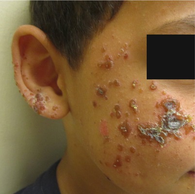
Fig. 21.3
Actinic Prurigo on the face
Management Strategies
Therapy for AP centers around limiting sun exposure, with emphasis on use of protective clothing and broad-spectrum, high sun-protection factor sunscreens. UVA and UVB protective films may be helpful to reduce sun exposure through window glass. High-potency topical corticosteroids may alleviate itch. Phototherapy hardening treatments may offer some benefit. Thalidomide, an anti-TNF−α agent, is very effective for managing symptoms of AP, however, the potential for teratogenicity and peripheral neuropathy may limit its use. Pentoxifylline, which demonstrates some anti-TNF-α properties, has shown promise in one uncontrolled study. Other treatments such as tetracycline, Vitamin E, anti-malarial medications, oral corticosteroids, and beta-carotene may lead to clinical improvement, although the efficacy of these agents is unclear. In patients with ocular manifestations, cyclosporine eye drops have been effective in several cases.
Investigations Recommended
For diagnosis |
HLA-typing |
Provocative Phototesting |
Skin biopsy is not required for diagnosis, but may support the diagnosis; in AP, lip cheilitis shows well-formed lymphoid follicles on histopathology |
This article reviews diagnosis and treatment of actinic prurigo AP. It notes that photoprovocation with repeated exposure to UVA (2.5 J/cm2/day for 10 days) or UVB (3–5 mJ/cm2/day for 15 days) reproduces characteristic actinic prurigo lesions in most cases (75 %–100 %), and that specific HLA types have been noted in patients with AP, including HLA-DR4(DRB*0407), HLA-DR4(DRB1*14), HLA-Cw4, HLA-A24, HLA-A28, HLA-B39(B16). In one study, polymorphous light eruption (PMLE) was not associated with any particular HLA type, suggesting that HLA typing may help distinguish between PMLE and AP [34].
Follicular cheilitis has sensitivity of 74.3 % and specificity of 36.4 % for AP [35].
Table 21.10
First line therapies
Sunlight avoidance – behavioral and environmental avoidance, protective clothing, topical broad spectrum, high sun-protection factor sunscreen | C |
Phototherapy (narrow band UVB, PUVA) | C |
Of 21 patients who completed the clinical trial, 18 had “good to excellent results” after management with broad-spectrum UVA/UVB sunscreen. One patient reported that treatment with PUVA was useful [36].
In this open clinical trial, six patients were treated with weekly narrow-band UVB for 5 weeks in spring. On follow-up, patients reported that treatment was worthwhile and well tolerated, except for transient erythema [37].
One child was treated with systemic PUVA with clearing symptoms; however, improvement was not sustained at 4-month follow-up [38].
Table 21.11
Second line therapies
Thalidomide | B |
Potent topical corticosteroids | C |
11 AP patients (ages 15–59 years) were treated with thalidomide for 1 month; all stopped having active pruritic lesions [39].
In this clinical trial, seven of eight patients (four of whom aged <18 years) were successfully treated with intermittent 3 to 14-day courses of topical 0.05 % clobtasol 17-proprionate cream or ointment, applied once or twice a day. All patients previously failed to improve after treatment with less-potent topical steroids [40].
Table 21.12
Third line therapies
Pentoxifylline | C |
Tetracycline | C |
Vitamin E | C |
Cyclosporine | C |
Cyclosporine eye-drops | E |
Oral corticosteroids | E |
Anti-malarial medications | E |
Cimetidine | E |
Beta-carotene | E |
In this 6-month open-label, uncontrolled study, all ten participants (thrree of whom were <18 years) over 15 years old (>45 kg) received pentoxifylline at a daily dose of 1200 mg (400 mg three times a day) and younger patients (<45 kg) received 800 mg pentoxifylline (400 mg twice a day) for 6 months. Clinical improvement was documented after 1 month of treatment and was maintained at 6 months [41].
Eight patients were treated with tetracycline (1.5 g daily) and another group of eight with vitamin E (100 IU daily). Both drugs used were effective in reducing signs and pruritus in AP, with no significant difference in efficacy between the two medications [42].
In this clinical trial, 18 of 19 patients (mean age of 17 years) showed significant improvement after a 2-month course of cyclosporin A 2.5 mg/kg/day, with effects lasting for at least 6 months after completion of therapy [43].
One 14-year-old girl was treated with 2 % cyclosporine eye drop at 1 drop/8 h. The patient improved 2 weeks later, but relapsed and required maintenance at 1 % cyclosporine eye drop 1 drop every 12 h [44].
Among 21 AP patients (ages 4.5–64.9 years), most had been treated with antihistamines (fexofenadine, loratadine) with little to no improvement. In 11 patients, oral prednisolone (12.5–25 mg/day) was beneficial in providing temporary relief in acute exacerbations. Hydroxychloroquine (200 mg twice daily for 3 months) was moderately effective in one patient, but was ineffective at 200–400 mg daily for 3–6 months in three other patients. Cimetidine 600 mg/day reduced itch, but not skin findings or photosensitivity, in one patient. Of three patients treated with beta-carotene (20 mg twice daily), one had limited improvement while the others had no improvement after a 3-month course [45].
Hydroa Vacciniforme
Clinical Features
Hydroa vacciniforme (HV) is an uncommon, idiopathic photosensitivity disorder (estimated prevalence 0.34 cases per 100,000 per year). It predominantly affects children and frequently resolves spontaneously by early adulthood. Classic HV presents as recurrent pruritic papules and vesicles within hours to days after sun exposure, which appear on the nose, cheeks, and ears. Subsequently, lesions crust then slowly resolve over a period of 1–6 weeks, with characteristic varioliform scarring. In some cases, ocular and oral disease occurs. Ophthalmic complications include photophobia, keratoconjunctivitis, and corneal erosions. Oral complications included aphthous stomatitis and ulcerative gingivitis.
Related posts:

Stay updated, free articles. Join our Telegram channel

Full access? Get Clinical Tree


