Fig. 4.1
This graphic illustrates full-field ablative laser resurfacing where 100 % of the surface area is treated in a single session. The stippled area represents the treated area of the skin. Several passes may be required to achieve the desired treatment depth
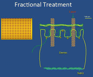
Fig. 4.2
This graphic illustrates non-ablative fractional resurfacing, in which a column of desiccated tissue is created; shown from above (left) and side view (right). The red circles on the left image and stippled area on the right image represent corresponding areas of treated skin. The density of treatment areas can be altered by adjusting the laser settings; typically a single pass is performed at each treatment session
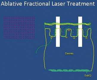
Fig. 4.3
A graphic illustration of ablative fractional resurfacing, where a column of skin (red dots on left; white columns on right) is actually removed. Shown from above (left) and side view (right).
Anatomy
The eyelid skin is the thinnest in the body with epidermal thickness approximating 50 microns (μ) and dermal thickness about 300 μ. The skin thickens as it nears the brow in the upper lid and near the cheek in the lower lid. The subcutaneous layer of fat normally present deep to the skin is sparse in preseptal and periorbital skin and absent beneath the pretarsal skin. However, Asian and Mediterranean patients may have thicker skin and device settings may need to be adjusted accordingly [6].
The sensory innervation to the upper eyelid originates from branches of the ophthalmic division of the trigeminal nerve (V1) which travels through the superior orbital fissure [7]. This nerve provides three branches including the lacrimal nerve to the lateral upper lid and frontal nerve giving off the supraorbital and supratrochlear nerves supplying the medial eyelid. The third nasociliary branch supplies non-eyelid periorbital structures. The lower eyelid sensory innervation is supplied by the maxillary division of the trigeminal nerve (V2) by its terminal branch, the infraorbital nerve via the infraorbital foramen [7].
Indications
For periorbital resurfacing, lasers are used to remove fine lines and wrinkles through epidermal and dermal ablation and to tighten skin via the body’s natural wound-healing response. In comparison to traditional blepharoplasty techniques, laser rejuvenation treats the actual skin elasticity and the eyelid architecture is not affected. Transconjunctival lower lid blepharoplasty and skin pinch blepharoplasty may be performed in combination with laser resurfacing/tightening and will be discussed later. Contraindications may include active infection, history of keloids, and previous deep chemical peel.
Anesthesia
Periocular laser and device procedures may be performed under general anesthesia, intravenous sedation, local anesthesia, topical anesthesia, forced cold air, or nothing depending upon device and setting used. In general, the more aggressive ablation and tightening devices will require a higher level of anesthesia. Often, isolated periorbital laser resurfacing can be performed utilizing regional blocks with local anesthetic agents. Blocking the supra- and infraorbital nerves based on physical examination and basic anatomic principles will typically provide the patient with adequate comfort both intraoperatively and for a few hours postoperatively [8]. It is helpful in situations such as this to also provide conjunctival anesthesia using ocular drops (e.g., tetracaine hydrochloride ophthalmic solution USP 0.5 %, Bausch & Lomb, Inc., Tampa, FL) prior to insertion of lubricated eye shields. However, as with any procedure, the level of required anesthesia will depend upon the overall health of the patient and patient anxiety/pain tolerance.
Laser Safety
It is imperative that laser safety is maintained for all cases, all patients, and all operating staff. There are excellent published guidelines for laser safety requirements including fire risk, emergency equipment needed, and suction devices for plume [9, 10]. Guidelines include nonflammable surgical prep and drapes, moistening towels/gauze with saline, specially coated instruments that do not reflect laser beams, a Laser Safety Checklist, proper signage around treatment areas, and use only by certified personnel. Operating room staff should be educated on laser safety protocols.
Eye protection for staff is mandatory for lasers and goggle choice is dependent on the wavelength being utilized. Additionally, it is paramount that the patient’s eyes be protected from inadvertent laser injury. Most commonly, topical ophthalmic anesthetic is placed within the eye (see section “Anesthesia”), a sterile lubricant is placed on a non-reflective stainless steel intraocular shield, and the shield is carefully placed in each eye beneath the eyelids prior to periocular procedures (Fig. 4.4). The shields are then removed at the conclusion of the case and the eyes are irrigated to remove lubricant. There are numerous suppliers for periocular shields, although we prefer those made by Oculo-Plastik, Inc. (Montreal, QC). Gold shields are sometimes used for light-based heating technologies around the eyes as there is less thermal transmission for these shields. Nonmetal shields (such as plastic) must be avoided for periocular laser procedures. Coverage with specific external (non-intraocular) shields is used when facial but non-periocular procedures are performed.
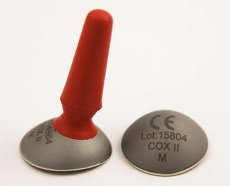
Fig. 4.4
Stainless steel protective eye shields
Full-Field Versus Fractional Resurfacing
Full-field resurfacing implies 100 % of the treated area receives ablation and/or ablation plus tissue coagulation (Fig. 4.1). Healing occurs from tissue appendages deep to the injury (e.g., hair follicles). Depth of treatment and amount of ablation and coagulation depend upon laser used and settings. This is compared to fractional treatment in which only a portion or percentage of the area is treated (Figs. 4.2 and 4.3). Full-field resurfacing is used when more dramatic tightening is needed in one treatment, albeit at the cost of longer healing times. By comparison, fractional treatments usually require a series of treatments with shorter healing times. Three lasers are currently in use for full-field resurfacing: carbon dioxide, erbium, and YSGG.
Carbon Dioxide Full-Field Resurfacing
Carbon dioxide lasers in full-field mode were the first lasers used and popularized for periorbital resurfacing. Their wavelength is 10,600 nanometer (nm) and the chromophore, or tissue target which preferentially absorbs the laser energy, is water. These lasers typically ablate tissue removing about 75 μ and leave behind a layer of residual thermal damage (75–100 μ), but the amount of tissue ablated and amount of thermal damage left behind are ultimately determined by the laser settings used. Initial carbon dioxide systems were used in continuous mode, which generated too much thermal damage and led to unintended sequelae such as scarring. The next generation of carbon dioxide devices was modified to allow shorter amounts of energy to interact with the skin. In the mid-1990s two technologies were in broad use. The Ultrapulse® laser (Lumenis Ltd., Yokneam, Israel) delivered short pulses of light, while the SilkTouch™ and FeatherTouch™ lasers (Lumenis Ltd., Yokneam, Israel) used an opto-mechanical flash scanner to scan a continuous laser beam in a spiral pattern. Both systems allowed short contact time for the laser beam on the skin surface allowing for controlled ablation of tissue and thermal desiccation of a controlled amount of tissue. Current systems use variations of these initial carbon dioxide systems.
Erbium Full-Field Resurfacing
The erbium/yttrium–aluminum–garnet (YAG) laser (2,940 nm) ablates tissue more efficiently and leaves less residual thermal damage (5–10 μ) than the carbon dioxide laser, as it has an absorption coefficient ten times greater. Because of these attributes, there is essentially a linear relationship between energy density (fluence) delivered and tissue ablated, with 3–4 μ of tissue removed per J/cm2. This means that multiple passes create efficient ablation without additive residual thermal injury. For example, two passes at a setting of 10 J/cm2 are equal to one pass at 20 J/cm2 in terms of tissue ablation depth and residual thermal damage. Superficial and deep resurfacing can be performed using erbium/YAG devices with deeper treatments providing correspondingly increasing results but with longer recovery times.
Variable pulse erbium/YAG systems (Sciton, Inc., Palo Alto, CA) vary the manner in which the laser delivers energy by providing a short ablative erbium pulse followed by longer sub-ablative pulses. The ablative pulse removes tissue while the sub-ablative pulses create controlled thermal damage. These devices can achieve more skin tightening than non-variable pulse width erbium lasers. They allow a varied clinical response with variations in depth of ablation and depth of thermal damage.
YSGG Full-Field Resurfacing
Similar to the Er/YAG laser at 2,940 nm, the 2,790 nm yttrium–scandium–gallium–garnet or YSGG (Pearl™ Cutera, Inc., Brisbane, CA) has less affinity for water. In full-field mode this device ablates approximately 20–30 μ of tissue and causes residual thermal damage of approximately 20 μ. Multiple passes may be performed to achieve increasing depth of treatment.
Plasma Resurfacing
Plasma resurfacing systems use radiofrequency energy to convert nitrogen gas into plasma to create tissue ablation and thermal damage. These devices differ from laser resurfacing in that the plasma system creates a coagulated eschar that remains in place as a biological bandage until the skin is reepithelialized. Recovery after a single pass with this device is approximately 7 days. This device was recently reintroduced to the marketplace and may improve mild or moderate facial rhytids, skin color, and skin texture [11–13].
Fractional Resurfacing
Fractional resurfacing implies that a percentage of the treated area receives laser treatment and a percentage is left untreated. Healing occurs from uninjured tissue lateral and deep to the injured area. As previously described, fractional resurfacing is classified as “non-ablative” in which the device creates a column of desiccated tissue (Fig. 4.2) or “ablative” in which the device removes a column of tissue with or without thermal damage (desiccated tissue) lateral or deep to the hole created (Fig. 4.3). Depth of treatment and amount of ablation and coagulation depend upon the laser used and its settings.
Non-Ablative Fractional Resurfacing
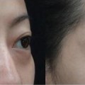
Non-ablative fractional resurfacing was pioneered by Manstein in 2004, with a 1.5 μm prototype laser creating an array of laser micro-exposures called microscopic treatment zones (MTZ) [14]. The original paper discussed treatment of periorbital skin with linear shrinkage of 2.1 % and wrinkle improvement of 18 %. This original research led to development of a number of lasers by varied manufacturers now called non-ablative fractional lasers (Fig. 4.2). The chromophore of these lasers is also water. Wavelengths of these lasers include 1,440, 1,470, 1,540, and 1,550 nm. The devices vary in the width of the thermal zone created and range from 100 to over 400 μ. Overall device power and settings determine the maximum depth of the thermal zone created. The density of treatment is defined by the number of MTZ per cm2. The density and spot size determine the treatment area and may be from under 5–70 % depending upon device, settings, and number of passes performed. When depth is factored in, an idea of the three-dimensional area treated can be ascertained. Epidermal healing occurs very quickly—within 24–72 h—and an inflammatory response is created in the deeper tissue that creates collagen remodeling that lasts weeks to months. Multiple treatments are needed for optimal improvement and usually are performed on a monthly basis, providing skin tightening of 25–62 % or more for most patients [15–17].
Ablative Fractional Resurfacing
Ablative fractional resurfacing is similar to non-ablative fractional resurfacing in that an array of tissue injury is created by the laser device. The difference being that instead of creating an area of tissue damage, ablative fractional lasers create an area of tissue removal (Fig. 4.3). Three lasers are currently in use for fractional ablative resurfacing—carbon dioxide, erbium, and YSGG. As with full-field resurfacing, these lasers differ in the amount of tissue ablated and amount of residual thermal damage. When comparing periocular fractional treatment of rhytids with carbon dioxide versus erbium, for both modalities wrinkle depth was reduced by 20 % with only one pass [18]. Other authors have reported over 50 % improvement in eyelid rhytids and 42 % reduction in eyelid skin redundancy using slightly more intense laser settings or treatment passes in a single treatment session, and treatments have been found to have long-lasting results [19–21] (Fig. 4.5).
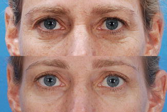
Fig. 4.5
Patient images showing skin quality before (top) and after (bottom) fractional variable pulse width erbium laser resurfacing
Radiofrequency
Nonsurgical treatment of the periorbital area with radiofrequency has been shown to improve skin quality and texture [22]. One device on the market, Pellevé® (Ellman International, Inc., Oceanside, NY), is expressly advertised and sold for periorbital rejuvenation (Fig. 4.6). The theory is that the sustained heat from this device causes an inflammatory response and subsequent collagen remodeling [23, 24]. In patients with less photoaging and shallower wrinkles, use of this device in the periorbital region provided 3.5 mm of eyebrow lift, 1.84 mm increase in upper eyelid crease (suggesting tightening), as well as a subjective improvement in periorbital rhytids at 8 weeks [25]. After a series of six treatments, patients were noted to have an average of 50 % improvement which was maintained at 1 year at an average of 30–46 % [26].
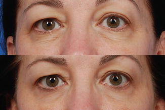
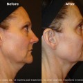
Fig. 4.6
A 52-year-old patient before (top) and 1 year after (bottom) a series of four Pellevé® treatments (radiofrequency) to the lower eyelid
Micro-fractional Ultrasound
Micro-fractional ultrasound is a device that offers both diagnostic and therapeutic ultrasound treatment for skin tightening. This noninvasive device is called Ulthera® and the treatment is Ultherapy® (Ulthera, Inc., Mesa, AZ). The device causes fractional thermal injury to a precisely set depth using ultrasound technology with minimal discomfort and transient erythema [27] (Fig. 4.7). Currently, transducer options are of different depths—4.5 mm, 3 mm, and 1.5 mm. Studies are in progress to evaluate use in the periorbital area. There is a suggestion that the thermal area can be used to either remove fat or perhaps tighten the orbital septum through controlled heat application.
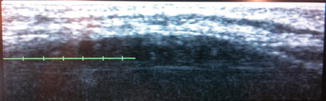
Fig. 4.7
An example of an ultrasound image of the mid-lower eyelid fat pad utilizing the Ulthera® device during a skin tightening treatment. Note the boney orbital rim (dark) with overlying layers of the lid soft tissue (muscle, fat, and skin)
Laser Treatment Alone for Eyelid Rejuvenation
In patients who suffer from periorbital aging, evidenced by crow’s feet, thinning crepey lid skin, and mild to severe rhytids but without a significant amount of excess skin, laser treatment alone can be a very effective treatment modality, offering at least 2.5 years of age reduction [28]. This treatment will tighten skin and improve the appearance of rhytids [29].
The question is—what device to use and what settings for that device? As described above most of the early work on periorbital resurfacing was with full-field carbon dioxide devices. The results were very good, but interest in these devices waned after 2000 due to long healing periods and complications including hypopigmentation.
We decide upon device and procedure to be used depending upon pathology and healing time of the patient. For patients with lower eyelid tissue laxity and crepiness, minimal excess skin, skin types I–IV, and enough “downtime” for healing, we prefer full-field resurfacing and have extensive experience with the Sciton, Inc. (Palo Alto, CA), variable pulse width erbium/YAG 2,940 nm laser for eyelid resurfacing [30, 31]. Settings vary from patient to patient, but our typical settings are two passes of 80 μ of ablation with 50 μ of coagulation. We treat both upper and lower eyelids with the same settings and treat full upper and lower eyelids to the lash line (Figs. 4.8, 4.9, and 4.10).
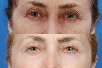
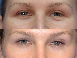
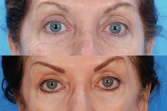
Fig. 4.8
A 64-year-old patient before (top) and 2 years after (bottom) full-field resurfacing with variable pulse width erbium laser. Note the improvement in periorbital rhytids as well as skin texture and color
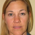
Fig. 4.9
A 39-year-old patient before (top) and 3 years after (bottom) laser resurfacing. Note the more youthful appearance to the lower lid skin and tone in this patient
Fig. 4.10
A 58-year-old patient before (top) and 5 years after (bottom) full-field resurfacing of the periorbital area with the erbium laser
For those patients with lower eyelid laxity and crepiness, skin types I–IV, and limited downtime, we prefer an ablative fractional device—either carbon dioxide or variable pulse width erbium.
Laser Treatment as a Surgical Adjunct
Related posts:
Stay updated, free articles. Join our Telegram channel

Full access? Get Clinical Tree


