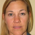
Fig. 7.1
A youthful brow is contoured. The anterior vector or position of the brow needs to be understood and positioned in order to re-create an attractive eyelid
We have long assumed that our faces and periorbital area, in particular, are victims of age that degrades our skin and subcutaneous muscles to the point that gravitational forces can stretch and lengthen these elements. We now know that there are significant volume losses, particularly from subcutaneous fat, that are mostly responsible for this apparent downward descent of the tissues as well as the visualization of “pseudo-herniated fat pads.” It’s important to understand that fat cells have a finite length of survival (probably all cells do). Based on research by Yoshimura and others, it appears fat cells survive for 7–10 years. Once they succumb to death, neighboring stem cells are signaled to replace them. At some point, one must also run out of these regenerative cells. This is seen most notably on the face where subtle volume losses can effect millimeters of change that can significantly impact one’s appearance. By understanding the process of fat cell death, depletion of regenerative replacement cells, and the necessity to restore the three-dimensional nature of the facial skin, one can see that fat, replete with regenerative (stem) cells, is the optimal physiologic choice for the most natural replacement tissue [13]. Of course, it requires a surgical procedure and generally some form of sedation to be best accomplished; however, because of the volume requirements and the long-lasting nature of the material, it remains ideal as a subcutaneous filler.
Many will argue that the hyaluronic acid fillers are superior because they are not long lasting and they can be reduced with hyaluronidase in the event of over corrections; however, one generally needs much more than a few cc’s of solution and no face naturally contains hyaluronic acid. Likewise, this can be said for the other nonpermanent filler substances. And, while there are those that argue they prefer permanent implants or permanent fillers for long-lasting results, as long as the patient is alive, they will continue to metabolize fat and eventually the permanent materials will become apparent and they will require further camouflage—probably with fat grafting [14].
Arguably, many doctors are concerned about placing fat around the eyelid area because they fear it will leave disfiguring lumps and fail to correct the physical appearance as well as traditional excisional procedures. While one can easily find ways to disfigure a patient with fat, the list of complications that can occur with traditional procedures should provide some solace for one considering the procedure. Fundamentally, there are only two significant complications that could occur from fat grafting—a poor result or an infection.
A poor result is simply due to artistic ability or inability. Because we are literally sculpting the patient from the inside out, while we mark the patient prior to surgery as a guide to where volume is needed, we are dependent upon our visual senses to decide that the proper reshaping has been accomplished. So while there is some technical skill required and an understanding of where you must and must not place graft material, one is dependent upon their artistic judgment for optimal outcomes—something that concerns most physicians and surgeons. While most surgeons develop and subsequently possess good technical skills, far fewer are truly artistic.
It’s probably better to under correct than try to over correct. Simple examples of problems occur from too much placement of fat, though, generally, most problems are simply related to not enough fat “taking” or the need for additional grafting. Patients should be advised to maintain a normal body weight. Significant increases in body weight could show up as significant changes in one’s face. This is rare but I’ve seen it occur in a couple patients.
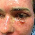
As for infection, it is largely avoidable. Since we are performing a sterile procedure with autologous materials, it’s unlikely that we are providing a contaminant to the graft. However, we are causing trauma in multiple areas. This results in bleeding into the traumatized area. Of course, we’ve introduced a great culture medium (i.e., fat). So, from where does the bacteria come? I submit that one must be certain that your patient is free from any obvious source of blood-borne infection, particularly via the dental route. As such, I admonish all of my patients to care for any dental issues prior to surgery, not right after the operation. Any bacteria introduced into the blood stream can thus be introduced into the traumatized area and seed around the fat. If one picks up an atypical mycobacteria from the oral cavity or via dental intervention, this could lead to a very difficult infectious situation. Further, should a patient have a history of herpes or shingles in the area of injection, then prophylaxis with an anti-herpetic should be recommended and the patient advised of the possible risk of a “break-out” (Fig. 7.2).
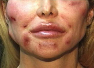
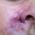
Fig. 7.2
While rare, bacteria can be introduced into the graft site via dental infection or other blood-borne sources. Advise your patients to fix any dental problems prior to surgery
Autologous fat transfer is not a matter of simply placing a layer of fat and hoping the body reconnects with the material. Thanks to the work of several plastic surgeons, we have a much better understanding for how this graft material actually works. Further, these principals have helped us to understand many concepts about adipose-derived stromal (stem) cells. Most body fat deposits are incredibly abundant with these regenerative or stem cells. The same hemopoietic stem cells found in bone marrow can be found in far greater abundance within fat tissue. Based on studies by Yoshimura and his group, we now understand that when you graft fat, you are actually doing a stem cell/fat graft. The fat cells suffer from hypoxia and as they die, they thus signal the stem cells to differentiate into supporting tissues, e.g., new fat cells and blood vessels, to connect and support the cells. Some of the fat cells survive. Some cells die and are eventually resorbed. It’s a dynamic process.
How I Do It
In general, most aging patients and even some younger ones, lack fat around the orbit and need to reposition their skin in a finite dimension away from the brow or cheek. The upper eyelid generally shows sagging of the brow secondary to volume loss, but it may present with deep hollowing, also secondary to volume loss. Often as periorbital and brow fat is lost, the appearance of bulges of intraorbital fat appear. This is generally explained to be pseudo-herniated fat, but this is simply a visualization of normal intraorbital contents made visible by the lack of fat lifting the skin away from these structures. Removing this fat (the generally accepted procedure) only serves to make the eyelids appear more hollow and affectively decompress (regardless to what extent) the intraorbital compartment. Decreasing fat within the eye socket simply allows the eye to start a retrograde descent into the orbit, something that really makes one look older.
Thus, for most upper lid changes associated with age, the basic plan is to restore the position of the brow. In the simplest situations, one only needs 1–3 cc of fat placed around the upper brow over the periosteal tissues and deep to the orbicularis oculi muscle of the upper eyelid. With losses of temporal fat, there is a lateral descent of the brow and this fat should likewise be replaced—typically over the superficial temporal fascia. And for more aggressive descent of the brow due to additional volume loss in the forehead, one must consider lifting the forehead anteriorly with placement of fat there as well. The skin over the forehead is too adherent to the frontalis muscle to be able to place fat in between the skin and the muscle without seeing lumps; therefore, it is safer to place fat deep to the frontalis muscle. Technically, one is placing the fat in a rather avascular plane, the galea aponeurosis, but due to the ability of the stromal cells to form blood vessels, it can actually survive.
In some cases, particularly older individuals, the upper eyelid skin may become so lax that a small excision of skin and/or a strip of muscle may be necessary. Of course, any pathology, such as true ptosis or other conditions, may need further operative attention or, depending upon your level of competency, referral to an ophthalmic plastic surgeon.
The lower lid generally presents with the appearance of increasing “bags” with advancing age. In some cases, there is increased hollowing under the lower lid, not infrequently due to iatrogenic removal of fat. In order to correct these defects, it’s important to understand the basic cause. The bag does not appear because of increased fat volume in the orbit and pseudo-herniation as we’ve generally been taught. While there may be circumstances where there is an actual increase in orbital volume resulting in additional fat bulging against the lower orbital septum, this is infrequent. In the extreme, one may see this with Grave’s exophthalmos. In such cases, not only is the fat protruding, but because of the constriction of the orbital septum, the fat can only protrude so much and then the entire globe starts to protrude, thus causing the severe bulging appearance of the eyes familiar with this condition. Treatment of this condition requires removal of fat and often opening of the adjacent sinuses in order to decompress the orbit. Traditional blepharoplasty with fat removal likewise causes decompression of the orbit, although to a smaller degree that largely goes unnoticed, though not always by the patient.
Freeing the arcus marginalis and allowing lower orbital fat to slide inferiorly will often remove the demarcation of the lower lid bag or hollow. This can also be done in combination with fat grafting, but quite often just filling the cheek/lid tissues gets the same result without the additional surgery.
Patient selection criteria are fairly straightforward. Your patients generally are coming to you because they don’t like the appearance of the area around their eyes. While this is most frequently seen with signs of aging, it’s not unusual to see young people with bags or hollows that bother them as well.
A general history and physical exam will determine your patient’s suitability for the potential surgical procedure. The procedure is most often done under intravenous sedation and local anesthesia.
For older individuals that want to look like they used to, ask them to bring a youthful photograph that they would like to resemble. Use it as a guide. Younger patients with other problems can bring photos of the kind of eyes they would like to have on their own face. This helps bridge any communication gaps. You may think you know exactly what would improve your patient’s appearance and yet, be completely surprised by the youthful photo they’d like to duplicate. Their best image inside their head could be fairly different from what you might expect. It happens.
Preoperative photographs are essential. The key is to take them with consistent lighting from an overhead position in order to see all the necessary shadows. With direct flash lighting you can essentially wipe out the annoying shadows that the patient would like to eliminate. Direct flat lighting should be avoided.
Mark you patient in an upright position. Around the eyelids you may need to place fat into the forehead, brow, glabellar area, temporal area, and the cheek. I’ll go over the proper placement in each of these areas. During the operation, you may prefer to keep your patient in a semi-seated position so you can more easily observe and judge the level of correction as you progress. In time, you may be comfortable with the patient in a supine/recumbent position, perhaps raising their head from time to time toward the end of the procedure.
Related posts:
Stay updated, free articles. Join our Telegram channel

Full access? Get Clinical Tree


