26 Perioperative Care and Rehabilitation Specific to the Lower Limb
Summary
The lower extremity is subject to unique stresses and pathophysiologies that make reconstruction, particularly with free tissue transfer, a challenging but rewarding process. In order to ensure a successful outcome, the reconstructive surgeon should optimize the patient’s comorbidities in the preoperative period and adhere to tried and true perioperative management principles. Postoperative rehabilitation is crucial for a patient to achieve her ultimate functional goal, which is typically ambulation. Important preoperative considerations include hardware/prosthesis presence, diabetic and vascular disease, oncologic resection, thrombophilia, biomechanical optimization, and malnourished states. In the perioperative period, accurate fluid intake and output management is paramount. In addition to clinical flap monitoring, there are various adjunctive measures that have shown to increase salvage rates. Though anticoagulation differs among microsurgeons, aspirin should be included in the regimen. Postoperatively, it is important to ensure the patient is still maintaining his or her cardiovascular health and prehabilitation prior to surgery can set the patient up for success. In addition to physical therapists, occupational, vocational, and mental health experts should be included in the care team. With regard to rehabilitation, it is important to consult colleagues in pedorthotics and podiatry so custom inserts and pressure-relieving footwear can be designed. Finally, the clinician should keep an eye out for pain conditions, such as neuroma pain. In the case a patient has had a partial or total amputation, chronic phantom limb pain can preclude the patient from ambulating again.
Keywords: lower extremity reconstruction, free tissue transfer, free flap, flap monitoring, lower extremity rehabilitation, perioperative management, perioperative optimization
26.1 Introduction
In many cases, free tissue transfer is required to reconstruct complex, composite defects and wounds with exposed vital structures (i.e., vessels, nerves, tendons, bones, joint space, etc.) when simpler or local options for coverage are either unavailable or inadequate to achieve structural and/or functional restoration.
In the lower extremity, reasons for transfer include large segmental bone loss, muscle and/or tendon loss resulting from low- and high-energy injuries, osteomyelitis, bony nonunions, tumor excision, exposed prostheses, irradiated wounds, burns, diabetic ulcers, and peripheral vascular disease.
Grafts, local flaps, and regional options may be inadequate due to infection, inflammation, trauma, irradiation, insufficient volume or surface area, insufficient vascular pedicle length, and/or unsuitable morbidity at the donor site.
Successful lower extremity reconstruction requires successful patient optimization as well as a multidisciplinary team, including plastic surgeon, podiatrist or pedorthotist, vascular surgeon, physical therapist, and occupational therapist. Additionally, orthopaedics, endocrinology, cardiology, hematology, rheumatology, dermatology, psychiatry, and/or pain management consultation may be needed (▶ Table 26.1).
26.2 Perioperative Optimization
26.2.1 Managing Comorbidities Preoperatively
Pertinent comorbid diseases include cardiac disease, renal disease, infection, vasculopathy, diabetes, neuropathy, venous hypertension, lymphedema, immunodeficiency, hypercoagulability, connective tissue disease, malnutrition, autoimmune disease, neoplasm, and psychiatric illness.
Exposed Hardware/Prostheses
Traditional methods to manage exposed hardware included irrigation, debridement, antibiotics, and removal of hardware. Prognostic factors for hardware salvage are location, duration, and type of infection, duration of exposure of hardware, and hardware loosening.1 Orthopaedists separate prosthetic infections according to chronologic criteria: early (within 12 weeks of surgery), delayed (within 2 years), or late (after 2 years, usually via hematogenous spread).2
If hardware is clinically stable, time of exposure is short, and infection is controlled, there is a possibility that hardware can be salvaged. If the hardware is secure and used for fracture fixation, an attempt should be made to salvage until radiographic evidence of bony healing. However, delayed or late infections require one- or two-stage reimplantation.
Alternatives for chronically seeded/late-presenting infected hardware are external fixation (for fractures), minimally invasive plating, and antibiotic spacers (particularly for joint arthroplasties).3,4,5 Antibiotic spacers can be in either a block form or an articulating form. Increased duration of intravenous antibiotics, positive initial wound cultures, chronic osteomyelitis on initial pathologic evaluation, and number of chronic morbidities are a predictor of failed hardware salvage in microsurgical lower extremity reconstruction.6
Exposed vascular prosthetic grafts are limb- and life-threatening and should be managed with early debridement and muscle flap coverage.
Diabetes and Peripheral Vascular Disease
In a retrospective study by Oh et al, 66 of 71 patients with diabetic lower extremity wounds were functionally salvaged using a microsurgical approach.7 A separate meta-analysis of 528 diabetic patients with lower extremity wounds showed a limb salvage rate of 83.4% at 28 months in patients otherwise needing amputation.8 Hong and Oh showed that microvascular transfer increased survival in the diabetic patient with lower extremity wounds.7,9,10
Table 26.1 Members of the Multidisciplinary Care Team Specific to Rehabilitation
Team Member | Role |
Plastic Surgery | Patient and wound evaluation, selection of reconstruction in light of functional goals |
Podiatrist | Biomechanical and gait evaluation |
Prosthetist/Pedorthotist | Biomechanical evaluation and design/planning of adjunctive footwear/devices for ambulation, activities of daily living |
Vascular Surgery | Evaluation and treatment of impaired perfusion |
Orthopaedic Surgery | Fracture and hardware management in addition to biomechanical evaluation |
Infectious Disease | Antibiotic stewardship; culture-driven antibiotic selection; minimization of iatragenic drug injuries |
Medicine | Management of patient comorbidities |
Physical Therapist | Prehabilitation and rehabilitation |
Occupational Therapist | Rehabilitation focused on activities of daily living |
Nutrition | Improvement of patient’s protein intake in light of his/her comorbidities |
Endocrinology | Perioperative and postoperative management of blood glucose in setting of Diabetes Mellitus |
Hematology | Management of thrombophilia |
Rheumatology | Evaluation and treatment of rheumatologic disorders contributing to disease process |
Dermatology | Evaluation and treatment of unusual wound etiologies |
Cardiology | Optimization and clearance for surgery in the setting of coronary artery disease or congestive heart failure |
Psychiatry | Evaluation and management of depression, anxiety, and other disorders perioperatively |
Pain Management | Management of acute and chronic pain conditions, minimize opioid requirements |
Diabetic patients with poor glycemic control (blood glucose > 200 mg/dL or hemoglobin A1c > 6.5%) have been noted to have an increase in dehiscence rates of closed wounds,11 although the effect of tight glycemic control on free flap outcomes has not been studied.
In patients with peripheral vascular disease, free tissue transfer provides increased venous drainage from, supplements blood flow to, and augments angiogenesis of hypoxic areas.12 The vascular status must be evaluated to ensure success.13 Vascular assessment and treatment has been discussed earlier in this textbook.
In patients with severe vessel disease and soft-tissue defects, improved rates of limb salvage have been reported combining bypass and free tissue transfer in staged or simultaneous fashion.14 Endovascular techniques allow for direct and indirect revascularization with angioplasty dilatation and atherectomy to recanalize stenosed or obstructed arteries in patients who are poor candidates for open vascular bypass.15
Usually additional concerns ranging from chronic renal failure, nutrition, and blood sugar control are best managed by a multidisciplinary team.16,17,18 These conditions lead to a predisposition to chronic bacterial colonization, osteomyelitis, complex wounds, bone deformity, local wound ischemia, and vascular disease.
Oncologic Reconstruction
Surgeons should have close coordination with the oncologists and must acquire adequate knowledge of tumor characteristics, behavior, and adjuvant treatment in order to plan and choose the appropriate reconstructive procedure. For patients requiring postoperative radiation or for wounds over joints and high-friction regions, skin grafts should be avoided with a preference for durable flaps.19
Flaps should be carefully chosen in patients with preoperative radiation therapy where local tissues would become fibrotic and ischemic around cancer, and thus may not allow local coverage. Free flap procedures will not interfere with chemotherapy nor will chemotherapy have an impact on free flap survival, though such treatment may have an impact on local wound healing. Following reconstruction, recommended delay prior to initiation of adjuvant chemotherapy and/or radiation is 3-4 weeks or until adequate wound healing has occurred per the reconstructive surgeon.20,21
Thrombophilia
Patients with factor V Leiden mutation, protein C deficiency, hyperhomocysteinemia, antiphospholipid antibody syndrome, prothrombin gene mutation, factor VIII elevation, anticardiolipin antibody syndrome, and essential thrombocytosis trend toward higher rates of microvascular thromboses, though patients with microvascular transfer have comparable overall success rates when compared to nonthrombophilic patients. Thrombophilic patients also trend toward delayed thrombotic complications and nonsalvageability in the setting of postoperative thrombosis.15,22
It is important to screen patients for a procoagulant condition, including asking about a history of deep venous thrombosis, pulmonary embolism, history of multiple miscarriages, family history, and exogenous estrogen use. It is also important to probe for patient accounts of acquired thrombophilia risk factors (i.e., venous thromboembolism, myocardial infarction [MI], cerebrovascular accident, and miscarriage) and/or known hereditary thrombophilia (i.e., people who know they have a genetic predisposition). A hematology consultation may be needed if patient is positive for thrombophilic state. This may miss patients who are thrombophilic without a discernible history, which can still affect free flap outcomes. Universal screening is still a source of controversy.22,23,24
Biomechanical Optimization
A sensorimotor examination is essential to ensuring the success of any reconstruction (▶ Table 26.2). Sensibility is evaluated with a 5.07 Semmes–Weinstein filament that represents 10 g of pressure. If the patient cannot feel the filament, protective sensation is absent, and the risk of breakdown and/or ulceration is significantly increased. Motor function is assessed by looking at the resting position of the foot and by testing the strength and active range of motion of the ankle, foot, and toes.
Gait analysis will provide an assessment of personal areas of high stress. High-stress areas include the plantar surface, mobile ankle, and knee (▶ Fig. 26.1). These are subject to pressure as well as shear forces. Prolonged weight-bearing on a single spot will lead to ulceration regardless of type of reconstruction.
Preoperative assessment of gait may include an F-scan analysis or pressure mapping as well as a measure of ankle dorsiflexion. F-scan analysis uses multiple pressure-sensing probes that record pressures on multiple areas of the sole of the foot during all phases of gait. One should evaluate for equinus deformity by assessing ankle motion when the knee is in complete extension and then flexion (which reduces stretch and tension on the gastrocnemius) to evaluate for gastrocnemius tightness (during knee flexion) or both gastrocnemius and soleus tightness (during both knee flexion and extension). If percutaneous release of the relevant portion of the Achilles tendon fails to improve dorsiflexion, then a posterior capsular release of the ankle joint should be performed. These will reduce pressure and lead to high ulcer resolution rates.25
Table 26.2 Preoperative Biomechanical Assessment
• Evaluate sensibility with a 5.07 Semmes Weinstein filament • Evaluate motor function by evaluating both resting position and active range of motion of ankle, foot, and toes • Evaluate location of current ulceration representing areas of increased pressure • Gait analysis includes F-scan analysis or pressure point-mapping of the foot • Evaluate for equinus deformity secondary to tight Achilles tendon |
Prior midfoot amputations can cause ulceration from musculotendinous imbalance within the foot, which leads to abnormal posturing and weight distribution. Patients often require Achilles tendon lengthening and/or tibialis anterior tendon transfer. Metatarsal head prominences can be remedied with “floating” neck osteotomies and internal fixation. The fifth metatarsal head can be resected. However, the first metatarsal head should rarely be resected as this is the base of the medial column of the leg. Resection of the plantar flare of the first metatarsal head is preferred in this instance.
During reconstruction, it is important to reestablish bony stability and eliminate any bony prominences. Such prominences occur particularly with collapsed midfoot bones (e.g., Charcot), osteophyte formation, or abnormal biomechanical forces (hallux valgus, hammer toe). Charcot arthropathy is a neuroarthropathy that is a destructive process of the bones, joints, and soft tissues of the foot. Reconstruction is futile when residual bone spike leads to subsequent ulceration. Soles can be difficult to reconstruct because of generalized scarcity of glabrous skin. Only the instep area of the foot provides a vascularized glabrous skin donor site.
Authors have found a higher rate of flap ulceration in muscle flaps with skin grafts, so fasciocutaneous flaps are preferred.26 Ducic et al emphasized that sensation is not essential for durability, but sensory nerve reconstruction improves surgical outcomes, quality of life, and patient satisfaction due to earlier sensory recovery in fasciocutaneous flaps. There is earlier return to normal footwear and daily activities.27,28
Typical postoperative rehabilitation is similar to that of diabetic patients, including custom-made insoles that facilitate load-sharing away from high-pressure areas on the heel and forefoot. Good custom therapeutic insoles relieve and distribute plantar pressure.29,30 Casting can be utilized for pressure ulcers.
Nutritional Optimization
Markers of nutritional status include albumin, prealbumin, C-reactive protein, and hemoglobin A1c. Albumin has a half-life of 21 days and prealbumin has a half-life of 48 hours, though the latter is considered an acute-phase reactant and arguably decreased in the setting of acute inflammation. Trending these markers can give critical information on host inflammatory response and if patients’ health is improving. Goals should be albumin greater than 3.5, prealbumin greater than 12, and hemoglobin A1c less than 6.5 (▶ Table 26.3).
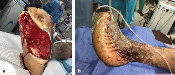
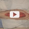
Fig. 26.1 Anterolateral thigh free tissue transfer used to construct weight-bearing portion of the foot. (a) Preoperative and (b) postoperative pictures.
• Albumin greater than 3.5 g/dL • Prealbumin greater than 12 mg/dL • Hemoglobin A1c less than 6.5% |
Placing surgical patients on protein-rich diets decreases levels of inflammatory cytokines postoperatively.31 Also, a diet rich in omega-3 fatty acids has also been shown to reduce inflammatory cytokine levels. Many times, patients, even if obese, are malnourished with low protein levels required to fight infection and promote wound healing. Nutritional support decreased infectious complications and noninfectious complications, and shortened length of hospital stay—immunomodulatory nutrition diets reduced infectious complications even further.32
26.2.2 Perioperative Optimization
Basic perioperative principles should be regarded (▶ Table 26.4). This includes the maintenance of normothermia to reduce complications. There is debate of what may be the optimal hematocrit level, particularly in free tissue transfer; blood transfusions are associated with longer hospital stays, higher rate of vascular thrombosis, and higher rates of major surgical and medical complications. Restrictive strategies (hemoglobin level < 7 g/dL or clinically symptomatic) for blood transfusion minimize medical complications.33 Fluid replacement is also controversial, though this is dependent on comorbidities of the patient. The surgeon can discuss with anesthesia whether a set maintenance infusion rate or replacement of calculated fluid losses is appropriate.
Postoperatively, it is important to monitor hemodynamic and pulmonary function as adequate hydration and oxygenation are critical to flap survival. Input and output of fluids should be closely monitored—distal perfusion is affected by hypotensive episodes. Fluid management can be complex for patients with end-stage renal disease who are on dialysis, which usually removes large volumes of fluid.
26.3 Immediate Postoperative Management
26.3.1 Flap Monitoring Protocols
The patient and flap should be closely monitored. Clinical examination is the gold standard; and the surgeon at the bedside can evaluate edema, turgor, color, capillary refill (too brisk can indicate venous congestion), and temperature. An experienced and knowledgeable staff is key to obtaining flap retention rates above 95%, as reported at high-volume centers.
It is important to utilize standardized flap monitoring protocols to enhance early detection of impending anastomotic complications. At the authors’ institution, serial Doppler assessments are performed every 15 minutes for the first 4 hours postoperatively, then every 30 minutes for the subsequent 8 hours, and then hourly until the second postoperative day. For patients with uncomplicated recoveries, the interval between Doppler checks increases to every 2 to 4 hours until postoperative day 5.
Table 26.4 Perioperative Patient Optimization
Intraoperative • Maintenance of normothermia (36°C to 38°C) • Transfuse only if clinically symptomatic or hemoglobin < 7 g/dL • Adequate discussion with anesthesia regarding goals of fluid resuscitation • Adequate discussion with anesthesia regarding use of vasopressors |
Postoperative • Monitor hemodynamic and pulmonary function • Strict inputs/outputs of fluids, particularly in end-stage renal disease • Standardized flap monitoring protocols; an experienced clinical exam provides the best assessment |
The threshold for surgical take-back should be very low, as microvascular compromise may be devastating. Early identification and re-exploration of a compromised flap dramatically increases salvage rate. Salvage rates are higher with arterial compromise compared with venous compromise.34
The first 24 hours are crucial for flap monitoring, as most thromboses will occur in this time. Up to 85% of compromised flaps can be salvaged when the first sign of vascular compromise is noted during the first 3 days.35 Available monitoring techniques include (1) conventional clinical monitoring, (2) handheld Doppler probes, (3) implantable Doppler systems, (4) color duplex sonography, (4) near-infrared spectroscopy, (5) microdialysis, (6) laser Doppler flowmetry, and (7) glucose monitoring.
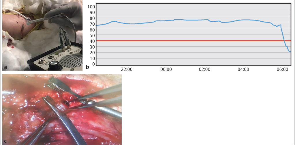
A recent review showed implantable Dopplers, near-infrared spectroscopy, and laser Doppler flowmetry were most effective.36 The implantable Doppler uses a 20-MHz ultrasonic probe to monitor anastomosis, typically on venous segments because arterial signal will persist for hours after a venous thrombosis. The implantable Doppler around the vein can be used during the original operative flap inset to ensure the pedicle isn’t kinked or compressed. Previous studies have demonstrated 100% flap salvage with a negative predictive value of 81 to 93%.37
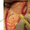
Near-infrared spectroscopy detects change in oxygenated and deoxygenated hemoglobin through optical spectrometry38 (▶ Fig. 26.2). It has the advantage of being independent of clinical experience. It detects signs of both arterial and venous compromise before experienced clinical examination, with positive and negative predictive values approaching 100%.39,40
The trend is the most important consideration; a gradual decrease in perioperative (over 4–16 hours) does not usually indicate flap compromise. Oxygen saturation usually returns to normal levels within 12 hours.41 Criteria for surgical exploration are a rapid 20-point drop from baseline in 1 hour or an absolute recording less than 30%. Flaps with less than or equal to 30% tissue oxygen saturation or greater than 20% drop per hour over 30 minutes were predictive of vascular compromise.42 Currently commercial devices are available only for flaps with cutaneous paddles, but use has been described for buried flaps in the literature.41
Related posts:
 General Wound Preparation and Timing
 The Pertinence of the Reconstructive Ladder and the Reconstructive Elevator
 Supermicrosurgery Approach to the Lower Limb
 Lower Limb Vascularized Composite Allotransplantation
 Using the Flap and Angiosome Concepts to Optimize Functional Lower Leg and Foot Amputations
 Procurement of Thin Flaps as Indicated in the Lower Extremity
Stay updated, free articles. Join our Telegram channel

Full access? Get Clinical Tree


