Pediatric otoplasty is generally considered to be a “simple” procedure, but an astute surgeon recognizes the challenges of this operation and is mindful of the degree of detail involved in its planning and execution. The vast number of described otoplasty methods, which are ever evolving, is a testament to the complexity of this procedure. In this article, the authors’ methodology with respect to preoperative analysis and planning, surgical technique, and postoperative care, including management of complications and potential pitfalls, are highlighted.
Key points
- •
Otoplasty is a thinking-surgeon’s operation, much like rhinoplasty, that requires assiduous planning and execution.
- •
Meticulous attention to detail is required during initial patient evaluation, including less commonly appreciated features such as asymmetries, cartilaginous contours, and abnormalities of the scapha and lobule.
- •
Suture techniques provide a more predictably natural auricular contour compared with cartilage-cutting otoplasty but potentially at the expense of diminished stability of the correction over time.
- •
Conchal setback sutures should be placed before antihelical contouring, because much of the medialization desired can be achieved in this manner, while obviating over-tightening the antihelical sutures.
- •
A single triangular fossa–temporalis fascia suture can help address persistent overprojection of the superior pole.
Introduction/Overview
Auricular deformities in children are a frequent source of ridicule and ruthless taunting by peers, beginning at an early age. “Bat ears,” “elephant ears,” “Dumbo ears,” and “donkey ears” are only some of the unflattering names heard in association. As such, cosmetic ear problems, none more common than protruding ears, or prominauris , frequently impose developmental psychological problems on young children, including behavioral disturbances such as aggression and petulant behavior, social phobias, neurosis, and feelings of insecurity. Such issues may impact social development and persist in later stages of life. One particular study demonstrated that 40% of adolescents with problem behaviors had auricular deformities. Adults with auricular deformities frequently continue to suffer from varying levels of insecurity and may contemplate corrective surgery for years while attempting to hide their ears with camouflaging hairstyles. Thankfully, there are techniques today that allow for correction of these deformities with minimal pain and require limited time away from school and extracurricular activities.
Surgical techniques for correction of auricular deformities have evolved considerably over time. The expansive history is detailed in other works of the senior author. Despite inventive and varied contributions to esthetic correction of the malformed auricle by surgeons over the last century, modern-day “cartilage-sparing” techniques have only evolved since the 1960s. Notable contributions include those of Mustarde and Furnas, who influenced the shift in philosophy away from cartilage-cutting otoplasty techniques. The more aggressive excisional techniques can result in contour irregularities, auricular instability, and an operated appearance. It should be noted that cartilage-cutting techniques are still more commonly applied in certain parts of the world such as Europe. Cartilage-sparing surgery involves reshaping techniques using sutures; these have been largely adopted in North America. These more conservative techniques, which attempt to re-create and strengthen the antihelical fold by folding scaphal cartilage using permanent transcartilaginous sutures (Mustarde), and setback the concha using tacking sutures to the mastoid periosteum (Furnas), provide a more predictably natural auricular contour. They also help eliminate unsightly cartilage ridging, which commonly results from resection techniques. These advantages are, arguably, at the expense of diminished stability of the correction over the long term.
Furnas later described additional suture methods, including fossa triangularis–temporalis fascia sutures to medialize a protruding superior crus and lobule-mastoid sutures to medialize a prominent cauda helicus. Webster is credited with assimilation of many of these available techniques to provide a comprehensive approach to otoplasty, including posterior skin and soft tissue excision, circumspect conchal resection, anterior cartilage scoring, and application of suture techniques as described. The senior author’s current philosophy and approach to pediatric otoplasty have largely evolved as an adaptation of the historical techniques already mentioned, principally relying on suture techniques with adjunctive cartilage scoring or shaving performed in rare cases as required. The authors’ most updated methodology is shared in this article.
Clinical assessment
As is the case with all facial plastic surgical procedures, pediatric otoplasty requires meticulous attention to detail including careful patient evaluation during consultation and astute preoperative planning to optimize outcomes. The surgeon must have an appreciation for facial esthetics, which is expected of the facial plastic surgeon but, naturally, less of a focal point for the pediatric Otolaryngologist. Extensive knowledge of ear anatomy and a firm understanding of the rationale for the various techniques applied are required.
As of the 1990s, nearly two-thirds of the senior author’s otoplasty cases had been performed on the pediatric age group, with 50% of patients falling between the ages of 5 and 9 years of age. Since that time, most cases have been performed on adults, many of them revisions, which reflect the author’s transition to a mostly noninsured private practice.
In general, the multimodal peaks in demand for otoplasty coincide with early school years, adolescence, and early adulthood, when social pressures reach their pinnacle. Patients should be considered for otoplasty no earlier than age 5 when the auricle’s size and strength approximates its mature form but remains pliable and elastic. These features diminish with age, necessitating more aggressive treatment in older patients. Five is also the approximate age when children begin to notice abnormalities in others, and teasing may begin. As this also happens to be a key childhood stage of social growth and identity development through interaction with peers, surgical correction at this stage is almost a way of “protecting” children from senseless bullying.
During the initial patient evaluation, it is extremely important to elicit both the child’s and their parents’ specific concerns about their ears. Needless to say, young children will often be unable to voice specific cosmetic concerns and are more likely to share their general distress imposed by their esthetic disadvantage. In other instances, the decision to proceed to surgical consultation might be solely the parents’ initiative, with the best interest of their child in mind. Parents should be asked about school performance, self-esteem, and potential bullying and teasing within the classroom. A medical history should be elicited, including associated medical conditions and fitness for surgery, developmental history, allergies, and medications. As the inheritance of auricular deformities is autosomal-dominant with variable penetrance, and close to 60% of otoplasty patients have a family history, an extended family history of auricular deformities and associated syndromes should be investigated. Potential familial concerns, such as bleeding tendencies, pathologic scar formation, and potential anesthetic concerns such as pseudocholinesterase deficiency, should be elucidated.
On physical examination, each ear must be examined in isolation and in relation to each other. Although both ears tend to share similar characteristics, they are not infrequently affected to varying degrees by deformities, and in some instances, only one ear is affected. Asymmetries in contour, projection, and size must be noted and brought to the attention of the parents, as some of these elements may be difficult or impossible to correct. The individual anatomic features of the auricle should be noted and recorded in a systematic way, effectively taking note of each anatomic constituent in an orderly fashion. In the authors’ practice an itemized template is used to be as comprehensive as possible. Although more obvious and common deformities, such as a deficient antihelix or superior crus, are likely to be apparent to the surgeon at first glance, secondary deformities such as excessive vertical height of the concha wall, excessive angulation between the triangular fossa and squamosal of the temporal bone, or unfurling of the helical rim require more focused inspection to be noted. The mastoid prominence should be examined, because it is occasionally hyperpneumatized and may contribute to lateral displacement of the auricle. Unlike in older adults, the lobule is less commonly redundant or elongated, but may be outstanding. Cartilaginous contours should be inspected for prominence of the tragus or antitragus and for the presence of a Darwinian tubercle. If missed on consultation, these issues are unlikely to be addressed at the time of surgery and may result in either undercorrection of the auricle or overcompensation in other parameters.
During the evaluation, measurement of auricular protrusion from the temporal bone is mandatory, and each side should be measured and compared at 3 points: (1) the most cephalic aspect of the helical rim (ideal = 10–12 mm from the temporal bone), (2) the most laterally prominent part of the rim (generally at the midpoint of the helix, ideally 16–18 mm), and (3) caudally at the intertragal incisura (ideal = approximately 20 mm). The helical rim should be roughly 2 to 5 mm lateral to the antihelical ridge on the frontal view. The auriculocephalic angle should measure from 25° to 35° and typically exceeds 40° in prominauris cases. The conchal-mastoid angle should be between 45° and 90°, whereas the chonchal-scaphal angle should measure less than 90°.
The auricular cartilage should be evaluated for its pliability and density by manually simulating correction of the deformities. This approach also helps instill confidence in the patient/parents by demonstrating to them a general sense of the expected result. For instance, the antihelix can be re-created by applying gentle posterior pressure on the helical rim. Pressure with a cotton-tip applicator applied at the concha bowl can simulate conchal setback sutures. Greater strength in the cartilage and its tendency to recoil may confer a higher risk of unfurling following suture correction and necessitate adjunctive scoring or weakening of the cartilage. Anticipated redundancy in posterior soft tissue and skin can be gauged through this process as well. Depending on the age, disposition, and degree of cooperation of the child, these elements may be hard to elucidate during consultation and can only be appreciated once under sedation at the time of surgery. Although most school-age children will be sufficiently collaborative, those that are uncooperative can potentially be poor candidates for otoplasty considering the postoperative care involved.
Finally, standard preoperative photography should be performed, including frontal view, both lateral views (with close-ups), and a posterior view. A birds-eye (cranio-caudal) view can also be used to demonstrate lateral projection. If applicable, hair should be held out of the way with a concentric elastic band. Photography is later repeated at 6 and 12 months postoperatively.
Surgical goals
Much like rhinoplasty, otoplasty is a thinking-surgeon’s operation that requires detailed planning and execution, frequently involving the orchestration of a combination of techniques to achieve desired results. It also commands recognition of the balance and harmony among the various elements of the auricle and the interplay between these elements, in addition to the relationship to the face, in contributing to the overall esthetics. It is an operation with no predefined steps but rather a compendium of employable techniques. Otoplasty has to be adapted and tailored on a case-by-case basis. Although the pathway is certain to vary, the desired endpoint is generally a shared theme, namely, the achievement of a natural, unoperated, and durable postoperative appearance. This natural appearance is constituted by gracefully arcing curvatures to the helical and antihelical contours and the absence of any obvious interaural asymmetries to the observer. Similar to the nose, the auricles are not commonly considered to be hallmarks of facial beauty; however, they can certainly detract from an otherwise beautiful face if these objectives are not met, and a “normal” appearance is not obtained.
More specific goals should include the following : (1) precise anatomic defects and contour abnormalities should be corrected, most commonly an unfurled antihelix and a high conchal wall; (2) auriculocephalic angles and distances should fall within normal limits, as detailed earlier; (3) the helical rim should project slightly more lateral than the antihelix, at least down to the level of the midauricle, to avoid a “stuck-on” appearance; (4) the posterior sulcus should be maintained (facilitated by avoiding incision placement directly in the furrow and by trimming only the redundant soft tissue), (5) interaural “approximate symmetry” should be coveted, and the lateral protrusion of the helices should be within 3 mm of one another at the 3 points of measurement; (6) both the anterior and the posterior surfaces should be devoid of sharp notches, edges, creases, and unfavorable scars; and (7) the superior and inferior poles should be aligned with the concha. A “no-frills” set of guidelines for the otoplastic surgeon has been proposed by McDowell and elaborated by Mallen. These guidelines include absence of protrusion in the upper one-third (although slight residual protrusion of the lower two-third is permissible), helix visible to mid ear, smooth helix, postauricular sulcus preservation, appropriate auriculocephalic distances, and symmetry.
The senior author uses a postauricular approach and relies primarily on cartilage-sparing techniques to help achieve these goals consistently. Again analogous to rhinoplasty, a logical stepwise progression through the case is used. The exercise is anatomic, with constant attention to the combination of deformities. With respect to suturing, some degree of trial and error is at play. This process typically begins with conchal setback sutures through which most of the medialization desired can be achieved and proceeds to antihelical contouring. As a general rule, slight overcorrection is required because cartilage memory and elastic recoil will result in as much as 40% loss of correction, especially in the upper one-third. Particularly in the early postoperative period, slight overcorrection is likely to be perceived by patients and their families as a successful outcome, and undercorrection is likely to be perceived as a surgical failure. Even in the setting of a unilateral deformity, bilateral surgery is often advantageous if there is even minimal deformity because it can account for the overcorrection-recoil cycle and help achieve a balanced outcome from the very outset.
Surgical technique
Preparation and Incision
The goals of the operation should be reviewed briefly with the patient before the induction of anesthesia. The senior author usually prefers general anesthesia for young children, but adolescents and young adults can elect for local anesthesia or conscious sedation if preferred. Preoperative marking is occasionally indicated at the level of the lobule if slated for reduction or to highlight previous scars requiring revision.
The patient is positioned in slight reverse Trendelenberg position. A conservative amount of hair may be trimmed around the auricle superiorly. Circumferential autoclave tape is used to keep the hair out of the operative field. Both ears are prepared and draped simultaneously and should be visible at all times for the purpose of comparison on the fly. The more involved auricle is selected first for correction. A measured amount of postauricular skin for excision should be measured and marked before distortion with local anesthesia. This redundancy created by the conchal setback is usually in the 10-mm to 12-mm range. The auricular and mastoid soft tissues are widely infiltrated with equal parts lidocaine 1% with 1:100,000 epinephrine and bupivacaine 0.5% with 1:200,000 epinephrine. If placed in the appropriate supraperichondrial plane, the injection will provide some hydrodissection.
Postauricular Skin Excision
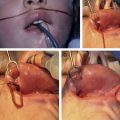
An eccentric fusiform excision based around (but not incised within) the postauricular sulcus is made ( Fig. 1 ). Greater extension is made onto the posterior concha than onto the mastoid. In this way, forces acting on the medialized cartilage are distanced from the soft tissue closure, which can help prevent wound complications and suture extrusion. This incision placement also allows for adequate cartilage exposure and placement of sutures. Furthermore, an anterior bias will allow the incision to fall back into the sulcus as the ear is set closer to the skull, rather than falling into visible postauricular skin. The ends of incision should be kept at least 1 cm away from the superior and inferior edges for camouflage. Skin and soft tissue excision (usually 10–12 mm at its widest point) is performed en bloc down to the level of the perichondrium/periosteum, encompassing a variable amount of mastoid soft tissue to allow for later retro-displacement. More soft tissue is excised superiorly, because this is where the most correction is desired, and to avoid the neurovascular bundles at the inferior pole. Wide undermining is then performed. Frequently a superolateral releasing incision is made 1 cm from the superior apex of the ellipse, resulting in a “Y” or “T” shape. This releasing incision will aid exposure for Mustarde suture placement. Hemostasis is achieved with bipolar electrocautery.