Head and neck tumors requiring large composite resections are rare in pediatrics. Large soft tissue and/or bony resections are usually the result of a neoplastic, traumatic, or infectious process. Sarcomas are the most common malignancy. Surgical resection is usually recommended after chemotherapy and/or radiation therapy. Free tissue transfer is safe and effective in this population, which has continued craniofacial growth and development. The surgeon must know the anatomic location of the growth centers and facial skeletal relationships because disruption results in abnormal development. Free tissue transfer can restore normal maxillomandibular occlusion and condylar-cranial articulation.
Key points
- •
Pediatric free tissue transfer is safe and reliable.
- •
Primary consideration must be for long-term restoration of mastication, deglutition, and cosmesis.
- •
Long-term functional and cosmetic results require reestablishment of the normal maxillomandibular occlusion and condylar-cranial articulation for normal craniofacial development to occur.
- •
Fixed dental prosthodontic rehabilitation is favored in the pediatric population because it results in maintenance of the normal occlusal relationships and promotes normal craniofacial development.
Head and neck tumors, craniofacial trauma, or infections are the most common indications for large composite oromandibular resections. When encountered, the surgeon’s primary focus should be on optimal restoration of function and cosmesis. In contrast to the adult population in which most oromandibular resections are due to malignancies, most pediatric conditions are benign, allowing for narrow margins and minimizing the need for complex reconstruction.
Common neoplasms within this population include osteosarcoma, rhabdomyosarcoma, ameloblastoma, neuroblastoma, lymphoma, germ cell tumor, and teratoma. The two most common head and neck cancers involving the maxilla and mandible are osteosarcomas and rhabdomyosarcomas (through direct bone invasion). Treatment algorithms have been difficult to create due to the low incidence combined with the wide variety of rare conditions. However, sarcoma studies have shown the importance of obtaining negative surgical margins as evidenced by local failure and poor survival if positive margins remain. Five-year survival dropped from 80% to 55% in pediatric sarcoma patients who had a positive surgical margin. In addition, surgical resection is usually recommended after the patient has been treated with radiation therapy, chemotherapy, or both, resulting in compromise of the recipient bed and limiting the use of adjacent tissue transfer or bone grafts.
Free tissue transfer has been shown to be safe and effective in the pediatric patient population. Unique to the pediatric population compared with adults is their continued craniofacial growth and development. It is essential that the pediatric reconstructive surgeon know the anatomic location of the growth centers and the normal oromandibular relationships. Disruption of the normal maxillary, mandibular, and cranial relationships results in abnormal midface, mandible, or skull base development, resulting in profound functional and cosmetic consequences. The use of free tissue transfer can restore normal maxillomandibular occlusion and condylar-cranial articulation resulting in normal craniofacial development. In addition, free tissue transfer options, including the fibula, iliac crest, and scapula, provide sufficient bone volume to allow for osseointegrated implants.
Craniofacial development
Normal craniofacial development, including facial symmetry, requires the reestablishment of the normal maxillary and mandibular relationships. Growth of the mandible occurs through two mechanisms: epiphyseal proliferation and remodeling ( Fig. 1 ). Epiphyseal proliferation is the dominant method of growth in both bone length and projection during the first 18 years of life. The mandibular epiphysis is located just beneath the condyle in the proximal subcondylar ridge. This growth center is what allows the intercondylar distance to widen as the skull base expands with growth. Therefore, if surgically feasible, it is preferred to preserve the condyle and this subcondylar growth center. Girls reach mature mandibular height and depth at a mean age of 13, whereas boys’ maturation lags behind approximately 2 to 5 years.
Even after the fusion of this growth center, bony remodeling continues to shape the mandible. Bone deposited at the posterior margin of the ramus (combined with anterior margin resorption) results in forward projection and buccal bone deposition (along with lingual bone resorption) increases mandibular width. The muscles of mastication create mechanical forces that promote bony remodeling throughout adult life. Therefore, limiting the amount of dissection on the native mandible (ie, restricting detachment of the masseter muscle) may promote normal growth and contouring.
The maxilla grows in both vertical height and width. The vertical height increases as the maxilla is displaced inferiorly and bony remodeling occurs along the suture lines. The maxilla does not contain any endochondral growth centers. Mature vertical height of the maxilla is achieved by girls at approximately 14-years-old and boys at approximately 16-years-old. Fusion of the palate occurs at age 18.
Finally, normal maxillomandibular occlusion is essential for normal craniofacial development. Without normal occlusion, such as in cases were defects are left unreconstructed, midface and mandibular growth halts, resulting in facial asymmetry. This is unfavorable cosmetically and produces inferior functional outcomes. Therefore, to preserve facial growth, symmetry, and oromandibular function it is essential to restore the patient’s baseline occlusion.
Recipient considerations and outcomes
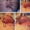
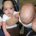
In these complex composite surgical resections it is essential that the surgical teams work together to facilitate complete surgical excision while maintaining the best possible functional and cosmetic reconstruction. As described above, if the condyle and subcondylar epiphyseal growth center can be preserved without compromising the surgical margins, they should be. Consideration of the patient’s current dentition and permanent dentition are also important with the goal of complete surgical resection while maintaining as much of the normal maxilla or mandible as possible. Based on the age of the patient and the current state of their dentition, it may not be possible to place the patient in mandibulomaxillary fixation. Furthermore, the disease burden or bony involvement may prevent preplating of the mandible. In these cases, craniomaxillomandibular relationships can be maintained by a temporizing external fixator, which can then be removed once the permanent plating is applied. An emerging option in these difficult cases involves the use of computer-aided prefabricated bone plates and surgical cutting guides. Regardless of the plate size or type, it is essential that the head and neck surgeon place the plates on the native mandible as low as possible to limit any injury to permanent dentition. The reconstructive surgeon may prefer to place the osteocutaneous bone flap flush with the superior edge of the mandible to assist with future osseointegrated implants; however, this may be impossible based on the location of the dental roots or permanent dentition buds. The head and neck surgeon should also consider if distal mental nerve and proximal inferior alveolar nerve can be preserved, allowing for possible nerve grafting. Fig. 2 details an 8-year-old girl with a right mandibular alveolar osteosarcoma who underwent fibula osteocutaneous free flap reconstruction with greater auricular nerve graft to connect the proximal inferior alveolar nerve to the remaining distal mental nerve stump. Every effort should be made to maintain the marginal mandibular nerve while performing the primary resection and/or neck dissection. In addition it is paramount that these structures be maintained on the contralateral side to avoid oral incompetence. When dissecting around the condyle and temporomandibular joint, care must be taken to ensure safety of the facial nerve, which may require complete identification and dissection.
Related posts:

Stay updated, free articles. Join our Telegram channel

Full access? Get Clinical Tree


