Although papular eruptions are not always listed as a separate clinical category in dermatology texts, there are some diseases that present with a decidedly papular quality. Moreover, whereas some diseases remain papular over time (e.g., insect and arthropod bite reactions), other skin conditions may present in a polymorphic fashion, with papules that coalesce into plaques.
- •
Bedbug bites
- •
Chiggers
- •
Fox-Fordyce disease
- •
Gianotti-Crosti syndrome
- •
Miliaria
- •
Mite bites (or bites of other insects or arthropods)
- •
Papular eczema (see atopic dermatitis)
- •
Papular drug eruptions
- •
Scabies
Important History Questions
How long have the papules (bumps) been present?
Acute presentations are typical of viral exanthems, id reactions, and acute drug eruptions, whereas a more chronic course favors papular atopic dermatitis, repetitive or chronic insect or arthropod bite reactions, or a prolonged or chronic drug reaction.
Did all the papules (bumps) start at the same time?
Some papular conditions present as a single wave of lesions (e.g., Gianotti-Crosti syndrome), whereas others present as crops of lesions in different phases (insect, arthropod reactions), and yet others present as continuous or seemingly random eruptions (e.g., papular atopic dermatitis).
Have you had a similar rash in the past?
An affirmative response favors exacerbation of a chronic condition (e.g., atopic dermatitis) or a repeat exposure to an exogenous agent or external trigger (e.g., insect or arthropod bite reactions, miliaria).
Where did the rash start?
Determining the area that was first involved may suggest a cause. The involvement of extremities that protrude from the bedsheets may suggest bedbugs, and involvement of the lower extremities after outdoor activities may suggest chiggers; a photodistribution may suggest polymorphous light eruption.
Have you or your immediate relatives had eczema, asthma, or hay fever (seasonal allergies)?
An affirmative response may suggest an atopic diathesis, leading to the consideration of papular atopic dermatitis.
What sort of work do you do?
Certain occupations predispose a person to insect or arthropod bite reactions (e.g., grocers, animal handlers or groomers, veterinarians, hotel and hospitality industry workers).
How do you feel?
Systemic symptoms such as fever, malaise, sore throat, or others may suggest a viral infection.
Have you started any new medications in the last month?
Papular drug eruptions are uncommon but are reversible with discontinuance.
How are you treating this rash?
Over-the-counter treatments, home remedies, or even some prescription medications may improve or worsen a papular dermatitis. This important information will guide the diagnosis and immediate care.
What do you think caused your rash?
This is an obvious question that is often overlooked—some patients may have keen insight into their condition.
Important Physical Findings
What is the distribution of the papules?
Some papular lesions have distinct distributions. Scabies preferentially affects the wrist, intertriginous areas, and genitalia, whereas chiggers preferentially occurs beneath areas of the skin where clothing is in close contact. Gianotti-Crosti, on the other hand, tends to affect acral areas.
Are any linear lesions present?
Scabies burrows are short linear lesions that can be a diagnostic clue in an otherwise enigmatic pruritic papular dermatitis.
Scabies
ICD10 code B86
ARTHROPOD INFESTATION
Pathogenesis
Scabies is caused by the human scabies mite, Sarcoptes scabiei var. hominis . It is acquired chiefly by skin to skin contact, usually between family members and sexual partners. Although mites remain viable in bed linens for up to 96 hours, most infestations are not acquired from fomites. The pruritus of a scabies infestation is caused by an allergic response to mite antigens in their saliva, eggs, and feces (scybala).
Clinical Features
- •
Symptoms occur within 1 to 6 weeks after a first infestation and 1 to 3 days after a repeat infestation.
- •
Patients typically complain of marked generalized pruritus that is more pronounced at night.
- •
Primary lesions are usually found in the axillae, wrists, interdigital spaces, and groin. It has been found that 80% of all cases present with evidence on the wrists and interdigital webs of the hands.
- •
Other commonly affected areas include the nipples, beltline, umbilicus, and buttocks.
- •
The lesions of scabies often appear as follows:
- •
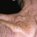
Papules—1- to 3-mm erythematous papules; these are typically numerous ( Figs. 6.1 and 6.2 ).
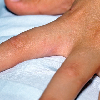
Fig. 6.1
Hand of a patient with scabies. There are small erythematous papules of the intertriginous space, a characteristic location.
(From the William Weston Collection, Aurora, CO.)
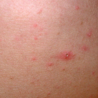
Fig. 6.2
Patient with scabies. There are small erythematous papules of the trunk, with one lesion demonstrating an excoriation.
- •
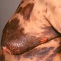
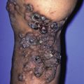
Nodules—5- to 10-mm red or red-brown nodules that are often found on the penis ( Fig. 6.3 )
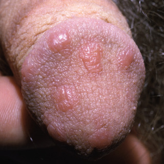
Fig. 6.3
Patient with so-called nodular scabies. The name is somewhat of a misnomer because most lesions are large papules. These diagnostic lesions are almost exclusively seen on the head of the penis.
- •
Burrows—1- to 5-mm threadlike linear or serpiginous forms, with varying erythema and scale. The adult mite may appear as a gray fleck at the end of the burrow ( Fig. 6.4 ).
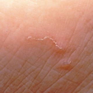
Fig. 6.4
Scabies burrows. There is a small, new linear burrow on the bottom and an older burrow on the top.
(From the Fitzsimons Army Medical Center Collection, Aurora, CO.)
- •
Atypical lesions—pustules may occur on the acral surfaces of infants and toddlers or as psoriasiform hyperkeratotic plaques in immunocompromised and/or mentally impaired individuals (crusted, Norwegian scabies).
- •
Diagnosis
- •
Scabies should be in the differential diagnosis for any patient with pruritus and numerous papules.
- •
The diagnosis is substantiated by recognition of a burrow and performance of a scabies prep (see Chapter 2 ) to identify the mite, eggs, or scybala. A skin biopsy (usually a shave) can be performed in an attempt to recognize the same structures, but the results will be delayed by days.
- •
Even though a patient may have hundreds of papular lesions, in a typical case there are only 10 to 15 adult mites on the entire body, and a skin scraping or biopsy may not encapture diagnostic evidence.
- •
If microscopic evidence cannot be demonstrated by a skin scraping or skin biopsy, a response to appropriate empiric treatment may be diagnostic.
Treatment
- •
Topical permethrin, 5% cream, is a first-line agent for treating scabies. It is applied to the whole body, from the neck down, left on the skin for 8 hours (overnight), and then washed off in the shower. A second treatment a week later may be needed to eradicate all mites.
- •
Precipitated sulfur (10%) may be applied to the whole body, from the neck down, for 3 consecutive days. The use of sulfur was once advocated for pregnant women, although many literature reviews on the subject have expressed no reservations regarding the use of permethrin, 5% cream.
- •
Topical 1% lindane lotion is now a second- or third-line agent but should not be used in children or pregnant women. It is banned entirely in California.
- •
Crotamiton, 10% cream, is not as effective as other therapies, with resistance reported.
- •
Oral ivermectin use has been increasing, with 200 µg/kg of body weight given as a single dose. Because it is not an ovicide, many experts advocate giving a second dose 1 week later. Oral ivermectin may be used with topical permethrin 5% cream in recalcitrant cases. The safety of oral ivermectin use during pregnancy has not been established.
- •
Although there are little data to support it, all clothing and linens should be decontaminated by machine washing in hot water and drying using a hot cycle.
Chiggers (Trombiculosis)
ICD10 code B88.0
ARTHROPOD REACTION
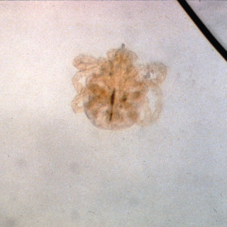
Pathogenesis
Chigger bites are caused by the larval stage of certain species of trombiculid mites ( Fig. 6.5 ). In the United States, the predominant species is Trombicula alfreddugesi, and chiggers occur chiefly in the humid portions of the Midwest and Southeast. The red six-legged larvae lie in ambush on blades of grass or other short vegetation (typically, <15 inches off the ground), leap onto a passing host, and attach and feed for 3 to 4 days before dropping to the ground. The chigger is smaller than 1 mm in size and can barely be seen by the human eye.
Many patients have been taught by their families and friends to treat chigger bites with nail polish or various glues to suffocate the mite. In many cases, the mite has already dropped off. Occlusion actually increases heat retention at the site of the bite, which will increase pruritus. The only potential benefit is that the polish or glue will reduce skin damage from excoriations.
Clinical Features
- •
The primary lesion is a red papule that may demonstrate a small, central, punctum-like area.
- •
Marked pruritus is a near-constant feature.
- •
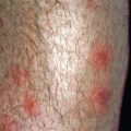
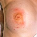
Lesions occur in crops and are notoriously grouped on the ankles, behind the knees, or along points of pressure or contact of clothing ( Figs. 6.6–6.8 ).
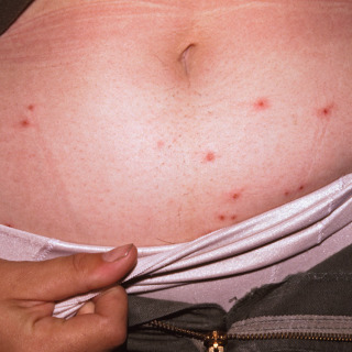
Fig. 6.6
Patient with typical papular lesion of chigger bites acquired in Georgia (United States), with accentuation under the elastic of the underwear.
(From the Fitzsimons Army Medical Center Collection, Aurora, CO.)
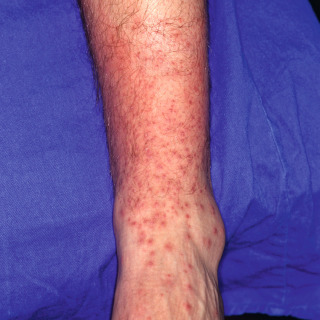
Fig. 6.7
Patient with innumerable pruritic papular lesions of chiggers, with marked accentuation under the elastic of the sock.Related posts:

Stay updated, free articles. Join our Telegram channel

Full access? Get Clinical Tree