Keywords
Psoriasis, Plaque psoriasis, Clinical subtypes, Pustular psoriasis, Nail psoriasis, Comorbidities
Key points
- •
Today, psoriasis is a global disease causing significant impairment in quality of life, disfiguring morbidity, and increased mortality.
- •
Psoriasis affects men and women equally. Two-thirds of patients are thought to have mild disease, and one-third of patients are thought to have more severe involvement.
- •
Psoriasis is found in practically all racial groups and is thought to affect approximately 2% of the world’s population.
- •
Psoriasis may appear at any age, but it most commonly presents between the ages of 15 and 30.
- •
Over the last 20 years, many of the immunologic drivers of psoriasis have been uncovered, and this research has been translated to the clinic with the development of increasingly powerful but selective biologic drugs targeting these immune pathways.
Psoriasis was first described by the ancient Greeks, Hippocrates and Galen, and grouped together with leprosy. The term psoriasis was derived from the Greek “psora” which means an, “itch, mange, scab.” In the early nineteenth century, Robert Wilan and Hebra refined the description of psoriasis and distinguished it from leprosy. In 1879, Koebner described the appearance of psoriatic lesions at sites of injury, called the “Koebner phenomenon.” Today, psoriasis a global disease affecting 125 million people across the world and causing significant impairment in quality of life, disfiguring morbidity, and even mortality. In the late twentieth century, basic science research demonstrated that psoriasis is an immune-mediated polygenic disorder that can be triggered by various environmental triggers. Over the last 20 years, many of the immunologic drivers of psoriasis have been uncovered, and this research has been translated to the clinic with the development of increasingly powerful but selective biologic drugs targeting these immune pathways. This increased understanding of the immunology of psoriasis has led to remarkable improvements in the treatment of psoriasis and makes it an exciting time to care for patients with psoriasis.
Epidemiology
Psoriasis affects male and female patients equally. Two-thirds of patients are thought to have mild disease, and one-third of patients are thought to have more severe involvement. Psoriasis is found in practically all racial groups and is thought to affect approximately 2% of the world’s population. Although psoriasis is a global disease, there are some regional variations in prevalence, with rates varying from 0.4% in Asians to 2.6% in the United States and 2.9% in Denmark. In Africa, there is a higher prevalence in East Africans as opposed to West Africans, which may help explain racial differences within the United States. Interestingly, a study of 26,000 South American Native Americans did not document a single case of psoriasis.
Age of onset
Psoriasis may appear at any age, but it most commonly presents between the ages of 15 and 30. An earlier age of onset and family history have been associated with particular HLA class I antigens, most notably HLA-Cw6 . Based on this finding, Henseler and Christophers proposed that 2 different forms of psoriasis exist: type I psoriasis, with onset before the age of 40; and type II psoriasis, with age of onset after the age of 40. Both types of psoriasis respond similarly to treatment.
Cause and Pathogenesis
The root cause of psoriasis remains unknown. However, research is beginning to link the complex genetic, biochemical, and immunologic abnormalities that underlie the disease. These changes can be seen in both psoriatic lesions and normal-appearing skin of psoriatic patients.
There is strong evidence suggesting at least a partial genetic basis in psoriasis. Genome linkage studies in the 1990s identified a locus termed psoriasis susceptibility 1 (PSORS1) in the major histocompatibility complex (chromosome 6p21.3), home of the HLA genes. Many HLA markers have been associated with psoriasis, but HLA-Cw6 has constantly demonstrated the highest relative risk for psoriasis in Caucasian populations. HLA-Cw6 is also strongly associated with early onset, guttate psoriasis, and psoriatic arthritis. However, only about 10% of HLA-Cw6 carriers develop psoriasis and the PSORS1 may account for only one-third of the genetic liability to psoriasis. In recent years, additional genetic risk variants have been identified for psoriasis, with more than 60 genetic loci identified to date. These findings confirm the polygenic nature of psoriasis and also its heterogeneity because most patients carry different combinations of these risk variants, and this may influence clinical course of the disease as well as therapeutic responses.
Development of a lesion
Psoriasis is characterized clinically by red and scaly plaques sharply demarcated from normal skin. Histologically, it is characterized by marked proliferation of keratinocytes, altered epidermal differentiation, and proliferation of endothelial cells accompanied by an influx of a variety of inflammatory cells. The development of a psoriatic lesion is a complex and multicellular process that involves keratinocytes, T cells, dendritic cells, macrophages, mast cells, endothelial cells, and neutrophils. Cytokines and growth factors initiate and sustain inflammation in this process through pathways that involve cells of both the innate and the acquired immune systems. Initial pinhead-sized macular lesions show edema and mononuclear cell infiltrates within the upper dermis. The overlying epidermis becomes spongiotic, and there is a focal loss of the granular layer. As the plaque matures, the epidermis becomes thickened in the center with increasing parakeratosis, capillary elongation, and perivascular infiltration of various types of immune cells. Mature psoriatic lesions contain elongated and uniform rete ridges with thinning of the epidermis overlying the dermal papillae. The tips of the rete ridges may become clubbed with dilated, tortuous capillaries in the dermal papillae. There is typically confluent parakeratosis and hyperkeratosis. More inflammatory cells are present with CD4+ T cells and dendritic cells in the upper dermis and CD8+ T cells in the epidermis. Neutrophils are commonly seen in psoriatic lesions and form characteristic collections in the spinous layer (spongiform pustules of Kogoj) and in the stratum corneum (Munro microabscesses). Eosinophils are not seen in psoriasis, unless the disease is drug induced.
T cells
The role of T cells in psoriasis was first documented in the 1980s, and the last 30 years of scientific and clinical research on psoriasis have further highlighted their role in the pathophysiology of the disease. The latest biologic medications for psoriasis target T-cell immune pathways, and their ability to clear plaques underlines the importance of the T-cell pathways these medications target.
In 1984, Dr Baker and colleagues were the first to show a correlation between the eruption of psoriatic skin lesions and the epidermal influx and activation of T cells. Subsequently, deletion of epidermal T cells was shown to predate resolution of psoriatic plaques in patients on phototherapy. In 1986, cyclosporine was shown to be highly efficacious in treating psoriasis due to its blockade of T-cell function. Ten years later in 1996, activated autologous T cells initiated psoriatic lesions when injected into uninvolved psoriatic skin transplanted onto severe combined immunodeficient mice. This showed that T cells were sufficient to induce a psoriatic process. More recently, using xenograft models where human skin is grafted onto immunodeficient mice, trafficking of T cells to the epidermis, particularly CD8+ T cells, was shown to be critical for development of psoriatic plaques, highlighting the importance of these cells in psoriasis.
Psoriatic lesions are typified by T helper 1 (Th1) polarized CD4+ cells and T cytotoxic 1 (Tc1) polarized CD8+ T cells producing interferon (IFN)-γ, which is the dominant cytokine profile of psoriatic lesions. IFN-γ drives the production of interleukin-12 (IL-12) and IL-23 by dendritic cells. IL-23 supports and expands CD4+ T cells, and likely CD8+ T cells, that produce IL-17 and/or IL-22, whereas IL-12 promotes development of Th1 and Tc1 cells. The secretion of IL-17 and IL-22 by these cell types likely maintains the chronic inflammation in psoriasis, but the exact role of IFN-γ in this process is still unclear. T cells also contribute to the production of tumor necrosis factor-α (TNF-α), but TNF-α is a potent proinflammatory cytokine, the main role of which may be to amplify the effect of other cytokines, including IFN-γ and TNF-α. Biologic medications targeted at inhibiting these inflammatory mediators, including TNF-α, IL-12/IL-23, and IL-17, have shown great efficacy in treating psoriasis, underlining the important role this molecule plays in driving the disease.
Macrophages and dendritic cells
Macrophages are important phagocytic cells that reside under the basement membrane, adjacent to proliferating keratinocytes, and are important in the early development of psoriatic lesions. They express Factor XIIIa and secrete the chemokine MCP-1 (CCL2). They are an important source of TNF-α, inducible nitric oxide synthase, and IL-23. It has been shown in mouse models that the selective elimination of macrophages leads to prompt improvement of psoriatic lesions.
Dendritic cells have an important role in both priming the adaptive immune response and inducing self-tolerance. Subtypes of dendritic cells, such as Langerhans cells, dermal dendritic cells, and myeloid dendritic cells, help drive the Th1, Th17/Th22 polarization of psoriatic plaques. In particular, myeloid dendritic cells help make IL-12 and IL-23 cytokines that promote Th1 and Th17 differentiation and responses, respectively. In a psoriatic plaque, myeloid dendritic cells can be increased up to 30-fold as compared with uninvolved skin and make up about 80% to 90% of the dendritic cells.
Uninvolved psoriatic skin
Normal-appearing skin of patients with psoriasis has been shown to have subclinical biochemical changes that lead to subtle histologic findings. Lipid biosynthesis is predominantly affected with measurable changes in the levels, constitution of phospholipids, free α-amino acids, and hydrolytic enzymes. These changes lead to histopathologic findings that can be identified on microscopic examination and have been termed “histochemical parakeratosis.”
Clinical findings
History
In approaching a patient with psoriasis, it is important for the clinician to obtain a thorough personal, family, and social history because it often will influence the choice of therapeutic agent. Relevant information includes the age of onset of psoriasis and whether psoriasis is present in any close relatives, because both a younger age of onset and a positive family history have been associated with more widespread and recurrent disease. In addition, the prior course of disease and frequency of relapses should be recorded, because there is significant variability in the clinical presentation of disease and the disease may change from one clinical phenotype to another ( Box 1.1 ). In some patients, the disease frequently relapses, and this has been associated with more severe disease with rapidly enlarging lesions covering significant portions of the body surface. Other patients have more chronic, slowly developing lesions with only occasional recurrences.
- •
Chronic plaque psoriasis (psoriasis vulgaris)
- ○
Psoriasis geographica
- ○
Psoriasis gyrata
- ○
Annular
- ○
Rupioid
- ○
Ostraceous
- ○
Elephantine
- ○
- •
Guttate psoriasis (eruptive psoriasis)
- •
Small plaque psoriasis
- •
Flexural (Inverse) psoriasis
- •
Erythrodermic psoriasis
- •
Sebopsoriasis
- •
Napkin psoriasis
- •
Linear psoriasis
The presence or absence of joint symptoms should be recorded—such as painful, warm, or swollen joints. Any of these complaints are concerning for psoriatic arthritis and prompt a more thorough evaluation. It is important to remember that osteoarthritis is common and frequently coexists with psoriasis.
Psoriasis is associated with several comorbidities, including increased incidence of myocardial infarction, stroke, and death, particularly in patients with moderate-to-severe psoriasis. Psoriasis has been shown to be an independent risk factor for cardiovascular disease, and it is important to screen patients for other cardiovascular risk factors in their social and medical histories, because modification of these can help offset their increased risk. Cardiovascular risk factors include smoking status, diet, and any previous diagnoses of hypertension, diabetes, dyslipidemia, or obesity.
Treatment history should also be recorded. Although the ability to predict treatment responses to a given agent are still very limited, patients that have been on previous biologics and failed generally respond less well to other biologic agents, even when these are in a different class. The nature of this decreased therapeutic response is still unclear. There is a broad differential diagnosis for psoriasis that physicians should consider ( Table 1.1 ), and treatment failures may prompt a reconsideration of the diagnosis.
| Psoriasis Vulgaris | Guttate | Erythrodermic | Pustular |
|---|---|---|---|
| Common | |||
|
|
|
|
| Consider | |||
|
|
| |
Cutaneous lesions
Psoriasis Vulgaris
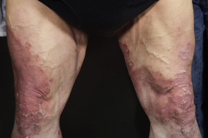
Psoriasis vulgaris, or chronic plaque psoriasis, is the most common clinical manifestation of psoriasis, affecting approximately 90% of psoriasis patients. Psoriasis vulgaris is characterized by well-demarcated, erythematous, raised plaques with white micaceous scale. Lesions vary in size from pinpoint papules to large plaques and tend to be symmetrically distributed on the scalp, postauricular skin, elbows, gluteal cleft, and knees. These lesions produce significant amounts of scale and removing the scale produces pinpoint bleeding (the Auspitz sign), which is a sign of the dilated capillaries below the epidermis and thinned suprapapillary plate. Disease presentation is impressively variable among patients, and the clinical findings can change quickly even within the same patient.
Psoriatic lesions can be induced by trauma, and this is known as Koebnerization or the isomorphic response. This phenomenon is more likely to occur when the disease is flaring and is an all-or-nothing response (meaning if psoriasis appears at one site of injury then it will appear at all sites of injury). The isomorphic response typically appears 7 to 14 days after injury, and the lifetime prevalence of the phenomenon is estimated to be 25% to 75%. The isomorphic response is not specific to psoriasis.
Historically, psoriasis vulgaris has been subclassified by the shape and scale of the plaques. Today, the terms have little significance clinically, but they connect the modern disease to its long history. Psoriasis geographica describes plaques that resemble a land map. Psoriasis gyrata consists of confluent, connected plaques with a circinate appearance. Rupioid lesions present in the shape of a cone or limpet. Ostraceous plaques have a circular, hyperkeratotic concave lesion resembling an oyster shell. Elephantine psoriasis refers to large, thick, scaly plaques on the lower extremities. Annular lesions have partial central clearing, giving a ring-shaped appearance, and are associated with a good prognosis because the annular shape suggests clearing ( Fig. 1.1 ). Finally, a hypopigmented ring on the periphery of an individual plaque, or Woronoff ring, may be seen after treatment with UV radiation or topical steroids. Woronoff rings are thought to be caused by inhibition of prostaglandin synthesis and are associated with lesional clearing and a good prognosis.
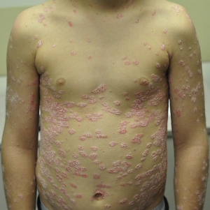
Guttate Psoriasis
Derived from the Latin gutta , “a drop,” guttate psoriasis is distinguished by the eruption of small psoriatic papules across the upper trunk and proximal extremities ( Fig. 1.2 ). Guttate psoriasis typically presents in children, adolescents, and young adults. It is often preceded by a streptococcal throat infection, and less commonly, perianal strep infections, and more than half of patients will have molecular evidence of a recent streptococcal infection, such as an elevated antistreptolysin O, anti-DNase B, or streptozyme titer. Despite this association, antibiotics are not helpful in the treatment of guttate psoriasis and do not alter the course of the disease. One-third to one-half of patients who develop guttate psoriasis will later develop chronic plaque psoriasis. As mentioned above, guttate psoriasis shows the strongest association with HLA-Cw6.
Inverse Psoriasis (Flexural Psoriasis)
Inverse, or flexural, psoriasis is distinguished by psoriatic lesions appearing in the major skin folds, such as in the axillae, inguinal creases, intergluteal cleft, umbilicus, and inframammary folds. Lesions are erythematous and sharply demarcated, with a glossy appearance and little to no scale. The lesion may contain a central fissure. The sharp demarcation and glossy appearance help distinguish the lesions from other diseases of the skin folds. Sweating is decreased in affected areas and localized fungal or bacterial infections may be a trigger.
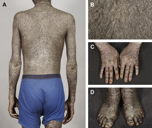
Erythrodermic Psoriasis
Erythrodermic psoriasis is the generalized form of disease and affects all body sites: face, trunk, extremities, hands, and feet. Erythema is more prominent and the scaling is finer, more superficial and diffuse, as compared with psoriasis vulgaris. Patients lose their autonomic control of body temperature and may present with systemic symptoms. They lose excessive heat from generalized vasodilation and so may shiver to try to compensate. In warm climates, there is a risk for hyperthermia because patients do not sweat from their psoriatic lesions. The generalized vasodilatation also puts patients at risk for high-output cardiac failure, impaired hepatic and renal function, and lower extremity edema. Erythrodermic psoriasis is thought to have 2 general presentations: first, as a chronic form that is thought to be a slow progression of psoriasis vulgaris ( Fig. 1.3 ). A second form is distinguished by its sudden onset and may be a generalized Koebner reaction to treatments such as phototherapy or anthralin. Finally, other generalized forms of psoriasis may give an appearance of erythrodermic psoriasis as they heal, such as generalized pustular psoriasis.
Related posts:

Stay updated, free articles. Join our Telegram channel

Full access? Get Clinical Tree


