Outcome Assessment of Breast Distortion Following Submuscular Breast Augmentation
Scott L. Spear
Jaime Schwartz
Introduction
Animation deformities or breast distortions during pectoralis muscle contraction following subpectoral breast augmentation are known entities, but their prevalence and significance are only beginning to be elucidated. There have been very few reports describing possible correction of such animation deformities, and, even more remarkably, there have been a few comprehensive reviews describing the frequency or severity of the problem (1,2,3,4).
While undoubtedly there are patients in whom distortion may be clinically significant, there is little information about how many patients are affected, how much the distortion bothers most patients, and with which specific activities the distortion is a problem. The purpose of this chapter is to review the frequency of animation deformities, how such deformities affect patients, how many patients have objective evidence of animation deformities, with the activities with which such deformities occur, and how to measure, quantify, or grade the degree of distortion.
Technique for “Dual-Plane” Subpectoral Breast Augmentation
Our preferred technique for breast augmentation involves a “dual-plane” approach in which the implant is placed beneath the pectoralis major muscle superiorly and in a subglandular plane inferiorly (4,5). This concept allows the parenchyma of the breast to redrape over the implant inferiorly and is particularly useful in patients with ptosis. It helps to avoid irregularities between the inferior border of the parenchyma and the inferior border of the implant, which can produce a “double-bubble” deformity. The procedure begins with an abbreviated subglandular dissection exposing the lower border of the pectoralis muscle. The amount of subglandular dissection is dependent on the degree of preexisting glandular ptosis and laxity. Patients with minimal or no breast ptosis may only require a couple of centimeters of dissection, while those with more significant ptosis may require a subglandular dissection up to the level of the nipple or as high as the superior border of the areola to allow for more redraping of the parenchyma over the implant. The subpectoral pocket is then developed by grasping the lower edge of the pectoralis major muscle with an Allis clamp and dividing its attachments along the inframammary fold under direct vision, leaving the sternal attachments largely intact. This prevents the pectoralis muscle from retracting superiorly while allowing the implant to fill out the lowermost portion of the breast parenchyma. The implant is placed in the subpectoral pocket but now lies in a “dual-plane” space, partly subglandular and partly subpectoral. The dual plane is particularly advantageous in thin patients with glandular ptosis or a constricted inferior pole, in whom purely subglandular placement would provide more control in the initial contour of the breast but at the expense of increased implant visibility and palpability. The dual-plane technique maintains the benefit of added soft tissue camouflage in the superior pole while providing greater contact between the implant and the lower aspects of the breast gland for better overall contour and redraping.
Results
In order to gauge patient satisfaction with the procedure, a survey of a 195 consecutive patients operated on by the senior author who underwent primary subpectoral breast augmentation (without mastopexy) was conducted. The minimum time after breast augmentation for patients to be surveyed was 6 months. The questionnaire involved a self-evaluation of the degree of breast distortion, impact on various activities, and overall satisfaction.
There were 69 responses from the 195 questionnaires that were sent (35% response rate). Fifty-six patients (82%) rated their breast distortion as none to mild, 7 patients (10%) rated their distortion as moderate, and 5 patients (7%) felt that they had severe distortion (Table 126.1.) One patient did not answer the question regarding severity of breast distortion. When asked if the muscle-related breast distortion was a problem, the most common affected activities reported were weight lifting and exercising (24% and 19%, respectively). None of the respondents reported any interference from animation deformities with normal activities of daily living (Table 126.2.)
Overall, with subpectoral implant placement there was an 86% satisfaction rate, 3% of patients were neutral, and 10% felt somewhat unsatisfied, and one respondent was entirely unsatisfied. When asked if they would choose subpectoral implant placement again, 70% responded affirmatively, 28% were unsure, and 3% said they would not choose subpectoral implant placement. When asked if they would recommend subpectoral positioning, only one patient stated that she would not recommend subpectoral breast augmentation.
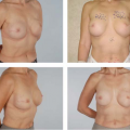
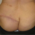
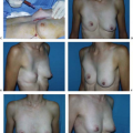
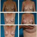
To improve the objective evaluation of breast distortion as seen in photographs in repose and actively flexing, a grading system was developed for breast distortion using a four-point scale (1): grade I, no distortion and unable to discern whether or not the implant lies in front of or behind the pectoralis muscle; grade II, one is able to tell that the implant is subpectoral, but there is minimal distortion, with an aesthetically pleasing result; grade III, moderate distortion but still an aesthetically acceptable result; and grade IV, severe distortion with an unattractive result during muscle contraction. Photographs of the
patients are shown both at rest and with the pectoralis major muscles aggressively contracted (Figs. 126.1 to 126.4).
patients are shown both at rest and with the pectoralis major muscles aggressively contracted (Figs. 126.1 to 126.4).
Table 126.1 Patient Self-assessment of Implant-Related Breast Distortion
Related posts: Follow-Up After Surgery for Primary Breast Cancer: Breast-Conserving Therapy and Mastectomy Follow-Up After Surgery for Primary Breast Cancer: Breast-Conserving Therapy and Mastectomy
 Breast Implants: Materials and Manufacturing Past, Present, and Future Breast Implants: Materials and Manufacturing Past, Present, and Future
 Reconstruction of the Irradiated Breast Reconstruction of the Irradiated Breast
 Perforator Flaps in Breast Reconstruction Perforator Flaps in Breast Reconstruction
 Lipomodeling of the Reconstructed Breast Lipomodeling of the Reconstructed Breast
 The Inframammary Approach to Augmentation Mammaplasty The Inframammary Approach to Augmentation Mammaplasty
Stay updated, free articles. Join our Telegram channel
Full access? Get Clinical Tree
 Get Clinical Tree app for offline access

|
|---|