Clinical Anatomy
The orbit is conical and has a volume of about 30 cm3. The anterior orbital rim has horizontal and vertical diameters of 4.0 cm and 3.5 cm, respectively; however, the orbital dimension widens posteriorly, with a maximum diameter 1 cm posterior to the rim. The distance from the anterior orbital rim to the apex is approximately 4.5 to 5 cm along the medial orbital wall, whereas the distance from the lateral orbital rim to the superior orbital fissure is about 4 cm.1,2
The medial orbital walls are parallel to each other and approximately 2.5 cm apart. The lateral wall forms a 45-degree angle with the ipsilateral medial wall. The two lateral walls form a 90-degree angle (Fig. 13.1).
Orbital Bones
Orbital Rim
The orbital rim is composed of the frontal, zygomatic, and maxillary bones that adjoin at suture lines (frontozygomatic, zygomaticomaxillary, and frontomaxillary, respectively). The lateral and superior rims are the strongest. The orbital rim creates the anterior lacrimal crest and then spirals posteriorly to end at the posterior lacrimal crest. The lacrimal sac fossa is found between the two crests (Fig. 13.2a).
Orbital Walls
Seven orbital bones compose the four walls of the orbit (Fig. 13.2a).
Orbital Roof
The orbital roof, which is the floor of the anterior cranial fossa, consists of the frontal and lesser wing of sphenoid bones. The supraorbital notch or foramen, through which the supraorbital nerve (CN V1) and vessels travel, divides the medial one-third and lateral two-thirds of the superior orbital rim. The lacrimal gland is found laterally in the lacrimal gland fossa.
Lateral Orbital Wall
The lateral orbital wall, separated from the roof by the superior orbital fissure, consists of the zygomatic and greater wing of the sphenoid bones (Fig. 13.2b). The zygomaticofacial and zygomaticotemporal canals transmit vessels and zygomatic nerve branches. Occasionally, a meningo-orbital foramen is identified lateral to the superior orbital fissure and transmits a branch of the middle meningeal artery.3,4 Whitnall’s tubercle is a protuberance on the inner aspect of the lateral orbital rim approximately 4 mm posterior to the rim and 10 mm inferior to the frontozygomatic suture. The lateral canthal tendon, lateral horn of the levator aponeurosis, Lockwood’s ligament, and check ligament of the lateral rectus muscle attach to the tubercle.
Orbital Floor
The orbital floor, separated from the lateral wall by the inferior orbital fissure, consists of the zygomatic, maxillary, and palatine bones. The floor forms the roof of the maxillary sinus. The infraorbital groove or canal, through which the infraorbital nerve (CN V2 branch) and artery travel, divides the floor. The infraorbital nerve and artery then exit through the infraorbital foramen, which is approximately 1 cm inferior to the orbital rim on the anterior maxillary bone face (Fig. 13.2c).
Medial Orbital Wall
The medial orbital wall consists of the maxillary, lacrimal, ethmoid, and sphenoid bones. The anterior and posterior ethmoidal foramina, with ethmoidal vessels passing through, are found on the medial wall along the frontoethmoidal suture. Along the medial wall, the distance between the anterior lacrimal crest, anterior ethmoidal foramen, posterior ethmoidal foramen, and orbital apex is approximately 24 mm, 12 mm, 6 mm, respectively (Fig. 13.2d).
Additional Fissures, Canals, and Foramina and Contents
The superior orbital fissure (SOF) is located between the greater and lesser wings of sphenoid. The annulus of Zinn, a tendinous ring at the orbital apex formed by the extraocular rectus muscles, divides the SOF. The annulus also encircles the optic foramen, which is medial to the SOF. Above the annulus, the lacrimal and frontal nerves (CN V1 branches), trochlear nerve (CN IV), and superior ophthalmic vein pass through the SOF. The superior and inferior branches of the oculomotor nerve (CN III), abducens nerve (CN VI), and nasociliary nerve (CN V1) pass through the annulus. The inferior ophthalmic vein may pass below the annulus (Fig. 13.3).
The optic foramen is located medial to the SOF in the lesser wing of the sphenoid and separated by a bony optic strut. It extends posteriorly as the optic canal that has an approximate diameter of 6 mm and a length of 10 mm. The optic nerve and ophthalmic artery pass through the optic canal.
The inferior orbital fissure (IOF) is located inferior to the SOF between the greater wing of sphenoid (lateral orbital wall) and palatine and maxillary bones (orbital floor). The infra orbital and zygomatic nerves (CN V2), infraorbital artery, inferior ophthalmic vein, and pterygopalatine ganglion autonomic branches pass through the IOF.
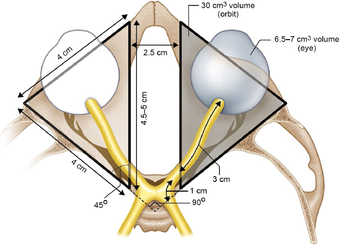
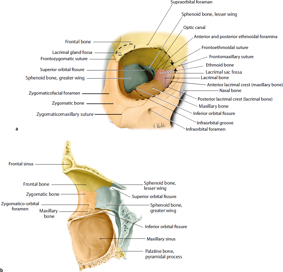
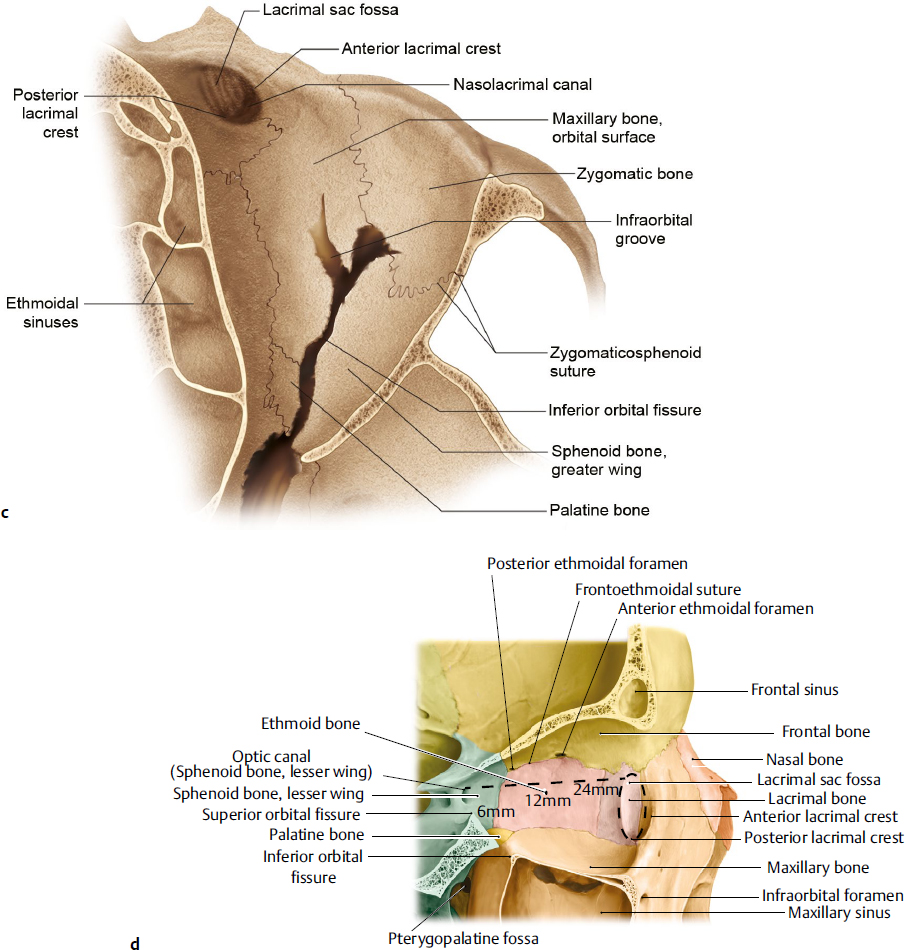
Fig. 13.2 (a) Orbital bones, sutures, foramina, and fissures. Anterior view. The orbital rim is composed of the frontal, zygomatic, and maxillary bones that adjoin at suture lines (frontozygomatic, zygomaticomaxillary, and frontomaxillary, respectively). (b) Lateral wall, right orbit. The lateral orbital wall, separated from the roof by the superior orbital fissure, consists of the zygomatic and greater wing of sphenoid bones. (c) Orbital floor, right orbit. The orbital floor, separated from the lateral wall by the inferior orbital fissure, consists of the zygomatic, maxillary, and palatine bones. (d) Medial wall, right orbit. The medial orbital wall consists of the maxillary, lacrimal, ethmoid, and sphenoid bones. (Modified from THIEME Atlas of Anatomy, Head and Neuroanatomy. © Thieme 2010, Illustrations by Karl Wesker.)


