Introduction
The oral tongue and mandible act as an inseparable functional unit of the lower face. Embryologically, tongue, mandible, and muscles of mastication share a common origin from the first branchial arch (BR). There is an interdependent developmental relationship between tongue and mandible, and the architectural malposition of one can lead to deformity of the other. The classic example is Pierre Robin Sequence, the syndromic condition that includes respiratory obstruction, glossoptosis, mandibular hypoplasia, and cleft palate. The challenges of tongue reconstructions are chiefly due to its multidimensional roles in prehension, mastication, deglutination, speech articulation, and airway protection. The optimal tongue reconstruction strategy depends on the size and the location of the defect, as the precise reconstruction goal varies from tip to base of the tongue. The complexity of mandibular reconstruction is to provide sturdy construction for its characteristic functions, including mastication, swallowing, speech, and smile. Esthetically, the mandible outlines the shape, contour, and height of the lower face. A well-defined jaw line is considered to be a sign of youth. The mandible reconstruction options are principally based on the location and tissue components of the defect (isolated bone, compound, composite, or extensive composite defects). Simultaneous reconstruction of tongue and mandible may be required, and careful planning of relative anatomical positions is the key for satisfactory reconstruction. Oral tongue and mandible reconstruction signifies the classical indication for the “reconstructive escalator”, where locoregional options are limited and free flaps are required.
Normal Anatomy and Function of Oral Tongue and Mandible
Anatomy of the Tongue
The tongue is a mass of skeletal muscle covered by mucous membrane, derived from two separate muscle halves that fuse into the midline fibrous septum. It is supplied by a pair of lingual arteries coursed above the greater horn of the hyoid bone, deep to hypoglossus. It branches into the dorsal lingual artery posteriorly and supplies the sublingual gland and floor of mouth anteriorly. Additional arterial supply comes from the tonsillar branch of the facial artery and the ascending pharyngeal artery. The venous drainage of the lingual vein follows the course of arterial tributaries. Its dorsal branches drain directly into the internal jugular vein, whereas the tip of the tongue is drained by the sublingual vein and ends with the lingual, facial, or internal jugular vein. Anatomical parts include the dorsum, tip, inferior surface (ventral tongue), and the base. The tongue is composed of both intrinsic and extrinsic muscle groups; each group consists of four muscles from each half of the tongue. The intrinsic muscle is comprised of superior longitudinal, transverse, inferior longitudinal, and vertical muscles. The multidimensional muscle fibres deliver great mobility as well as volume and shape transformability, thus allowing the tongue’s performance of both gross and fine movements. The extrinsic muscle is comprised of genioglossus, hypoglossus, styloglossus, and palatoglossus, and each is attached to the mandible (and hyoid bone), hyoid bone, styloid process, and hard palate, respectively. Extrinsic muscles alter the relative position of the tongue and provide the stability of the intrinsic muscle. The orchestrated muscle function allows intelligibility of speech, and precise execution of the oral process and transport phases of swallowing. Based on functional and structural differences, the tongue can also be described as the anterior two-thirds (oral or presulcal), and posterior third (pharyngeal/postsulcal/tongue base), separated by sulcus terminalis. The mucosa appearances and function of these two parts are distinctive. The anterior two-thirds in general has a rougher surface covered by various forms of papillae (filiform, fungiform, and vallate) and taste buds within; this character assists the transportation and processing of the food bolus during mastication. The posterior third of the tongue is part of the oral pharynx; it has a smoother surface, with abundance of glandular tissues to facilitate swallowing.
To appreciate sensory and motor supply of the tongue is to understand their embryological derivations. The epithelial layer of the tongue derives from the first, third, and fourth BR. The epithelium of the presulcal part of the tongue derives from the midline tuberculum impar, and a pair of lateral lingual swellings (tongue primordium) from the first arch. The postsulcal part derives from the midline hypobranchial eminence of the third arch and, to a lesser extent, the fourth arch (vallecula). Respectively, sensory supply of first, third, and fourth arch are the lingual nerve (CN V 3 and chorda tympani nerve of CN VII), glossopharyngeal nerve (CN IX), and internal laryngeal nerve (CN X).The musculature, with the exception of palatoglossus, derives from suboccipital myotomes and migrates into the primordium, carrying its motor supply from hypoglossal nerve (CN XII) for voluntary movements. Palatoglossus, conversely, is part of the oropharynx and is supplied by the pharyngeal plexus from the vagus nerve. Lymphatics of the tongue drain into the floor of the mouth; the tip drains into sublingual nodes, the rest of the presucal part drains into submandibular nodes then to both the upper and lower cervical chain. The posterior part, on the other hand, drains directly into deep cervical nodes. Most clinically relevant feature of the tongue’s lymphatic drainage is its bilateral drainage pattern, especially when the ipsilateral system is obstructed. This feature is more prominent in the posterior tongue, and prompts bilateral neck dissection in oncological resections. Although not part of the tongue, the floor of the mouth is its continuum and frequently involved in radical resection of the tongue. It contains a pair of sublingual glands in the sublingual folds, and is composed of a pair of mylohyoid muscles inferiorly and geniohyoids superiorly. The floor of the mouth is structurally analogous to the pelvic diaphragm; it separates the mouth from the neck, supports the weight, and thrusts the tongue. The muscles attachment between mandible and hyoid bone further enhances the mobility of the tongue. Mylohyoid is supplied by the myohyoid branch of the inferior alveolar artery and the submental artery of the facial artery. It is innervated by the mylohyoid nerve from the inferior alveolar branch of CN V 3 . Geniohyoid is supplied by the lingual artery and directly innervated by C1 fibres via the hypoglossal nerve.
Function of the Tongue
To understand the goals of tongue reconstruction, it is essential to appreciate its functions in deglutition, airway protection, and speech articulation. Deglutition has three distinct phases: oral, pharyngeal, and esophageal phases, where tongue movements are important for both the oral and pharyngeal phases. The oral phase is subdivided into: (1) stage I transport, (2) processing, (3) stage II transport, and (4) bolus formation. In stage I transport, it represents the moment of food reception, where the anterior third of the tongue surface is hollowed and the posterior third is sealed against the soft palate (posterior oral seal). In the processing phase, the anterior two-thirds of the tongue rotates at the posterior–anterior axis against a sealed posterior margin. This allows constant food bolus mixture with saliva, food grinding against hard palate, and continuous mastication. Stage II transport is defined by the movement of the food bolus through the posterior oral seal prior to the pharyngeal phase of deglutition. It starts with sealing of the anterior hard palate with the anterior third of the tongue and propulsion of the food bolus posteriorly in sequence into the oral pharynx by other parts of the tongue. In the pharyngeal phase of the deglutition, the tongue base retracts then moves superoposteriorly, thus directing the food bolus toward the pharynx. The orchestrated movements of tongue, floor of mouth, and larynx protect the upper airway from aspiration. The closure of the velopharyngeal port by the tongue during food bolus propulsion prevents food or fluid regurgitation into nasal cavity.
Tongue movements that are associated with speech and articulation can be categorized into four types based on the findings of Stone and Lundberg. They are: (1) “front raising,” where the anterior and middle tongue are raised (“t,” “d”); (2) a complete midline groove with elevated lateral tongue margins (“she,” “th”); (3) “back-raising,” the reciprocal tongue movements of the first category (“k,” “g”); and (4) “two-point displacement,” where the tongue has an elevated anterior and posterior segment with a small central groove in the middle segment (“l”). The understanding of commonly used tongue movements during speech and articulation aids in preoperative patient assessments and consultation regarding the expected postoperative speech deficit.
Anatomy of the Mandible
The mandible is a strong, semicircular bone of the lower face. It is composed of two symmetrical halves that fuse at the symphysis, and a pair of condyles that articulate with temple bones. Morphologically, the mandible is described in parts, including the processes (condylar, coronoid, and alveolar process), ramus, angle, body, and symphysis. The sophiscated design of the mandible offers a stable root for teeth, and a robust platform to withstand forces of mastication. Angle and ramus of the mandible consist of thick cortices and are designed to resist excessive unidirectional force, whereas trabeculae in the alveolar process and condylar head possess the ability to endure greater stress from multidirectional forces. There are a total of 16 teeth on the mandible, providing opposing occlusion to the 16 teeth on the maxilla. The force on dentition is transmitted to the underlying trabecular bone in the alveolar process; this constant force serves as a remodeling stimulus to the mandible. Loss of dentition, altering mastication habits, and being edentulous thus alter or regress the mandibular strength and thickness. The mandible is supplied by a pair of inferior alveolar arteries with additional vascular tributaries from the periosteum and the attached muscles. The pair of inferior alveolar nerves supplies the sensation of the mandible and transmits sensation from the dentition, gingiva, and sulcus for protective feedback. The mandible also provides the attachments for the muscles of mastication, muscles of oral continence, and facial expression, floor of mouth, genioglossus, digastric muscle, and platysma.
Etiology and Classification System
Etiology
The majority of oral cavity defects derive from oncological resections, and post-reconstruction complications. Oropharyngeal cancer is ranked 11th most common cancer worldwide, with incidence of 11.3/100,000 per year and 5-year survival of 64.8%. Oral cancers are predominantly squamous cell carcinoma (86%), whereas verrucous carcinoma, sarcoma, melanoma, and lymphoma account for less occurrence. Prototypical risk factors are long-standing history of cigarette smoking and heavy alcohol consumption. Betel nut, in addition, is an established independent risk factor for oral cancer, with an adjusted odds ratio of 58.4%. Statistically, there is a significant rise of human papilloma virus-associated oral tongue cancers affecting younger males and females less than 40 years old. Similar to skin cancer, field cancerization in the oral cavity is common; it is associated with a complex defect, high incidence of recurrence, and second primary tumors. Reconstructive surgeons therefore need to have a close working relationship with the ablation team, and be prepared for repeated major reconstructions.
The majority of mandibular defects are the result of soft tissue tumor ablations. Odontogenic tumors represent uncommon heterogeneous lesions. Among them, ameloblastoma, a group of benign yet locally invasive tumors, is frequently associated with major mandibular resection and reconstruction. Segmental excision and vascularized bone reconstruction is considered the treatment of choice to achieve long-term successful tumor control. Osteoradionecrosis (ORN) of the mandible occurs uncommonly, with an estimated incidence of 8.2%. It is seen following radiotherapy of the oral cavity. The risk of ORN increases in the presence of insufficient vascularized tissue coverage, aggressive muscle detachments, stripping of periosteum, marginal mandibulectomy, and mandibulotomy. Although conservative management is frequently employed, the definitive management of ORN remains segmental mandibular excision and free tissue transfer. A similar approach is recommended for other forms of osteonecrosis induced by infection, trauma, glucocorticoids, and high-dosage bisphosphonate. Trauma of the mandible is rarely associated with segmental mandibular loss, unless caused by ballistic wound or barotrauma. Patient stabilization should be the first priority prior to the consideration of mandibular reconstruction.
Classification of Oral Tongue Defects
Tongue reconstruction is defect-dependent, and a widely adopted classification system is currently unavailable. Most published classification systems are based on the defect percentage across the medial–lateral axis. The defects are generally subdivided into small glossectomy defect (less than one-third), hemiglossectomy, subtotal, and total glossectomy. Composite and compound defects involving the surrounding soft tissue and mandible are frequently incorporated in the classification to guide the reconstructive options. , These tongue classifications are useful in deciding simple reconstructions from complex options, but do not accentuate the regional anatomical differences for the guidance of reconstruction selections. The key deciding factors for complex reconstructive options of the tongue are the location (presulcal versus postsulcal), involvements of floor of the mouth, and the presence of bony defect. Thus, in addition to size, categorization of tongue defects into presulcal and postsulcal (tongue base) with or without involvements of floor of the mouth and mandible should be considered. These variants enhance clinical relevance of the classification, facilitate logistic defect reconstruction planning, and allow comparative studies to be conducted.
Classification of Mandibular Defects
The mandible is a semicircular structure that delivers unified and coordinated actions to its attached musculatures. Disruption of its continuity drastically reduces the power of mastication, speech articulation, deglutition, and protective mechanisms against upper airway obstruction and aspiration. Segmental bone defect is thus the most common indication for mandibular reconstruction. In contrast, marginal mandibulectomies are generally not reconstructed. Nevertheless, in the presence of insufficient residual mandibular height (less than 1 cm) and the necessity for mandibular height augmentation for dental rehabilitation, onlay nonvascularized or vascularized bone flap is recommended. In our practice, the classification system formulated by Daniel is the most clinically relevant tool in dictating options for defect reconstructions. It accounts for soft tissue involvement and the need for its synchronous reconstruction. The system classifies the defect into: (1) isolated bone; (2) compound defect (bone, oral lining or skin); (3) composite defect (bone, oral lining, and skin); (4) extensive composite (bone, oral lining, skin and intervening soft tissues). In dentulous patients, teeth are powerful landmarks to describe defect locations within teeth-bearing segments. On many occasions, defect distance can also be estimated based on the documented number of resected teeth. To simplify the descriptive terms for mandibular defects, Jewer’s classification is commonly used. The system signifies letter “L,” “C,” and “ H” to correspond to lateral segments, central segments (between canines), and condylar defects. Combined use of the letters enables the description of complex and extended defects; for instance, using “LCL” represents an angle-to-angle defect of the mandible. Although soft tissue defects were added in the modified Jewer’s classification, using “O,” “M,” and “S” to represent osseous, mucosa, and skin defects, this new addition does not distinguish small composite defects from extensive soft tissue loss.
Reconstruction Principles
Patient Evaluation
Detailed medical assessments including social status and reconstruction expectation should be evaluated. With best intention, both tongue and mandibular reconstruction can still have devastating outcomes due to the complexity of their respective functions and esthetic importance. Allowing sufficient consultation time for emotional response from both the patient and their family is essential. Presenting clinical photos of similar cases during the consultation decreases the risk of miscommunication and expectation disparity.
Multidisciplinary assessments are mandatory for surgical planning and streamlining the corresponding adjuvant therapies. It is indispensable to have a thorough understanding of the patient’s present physical status, disease progression, and life expectancy. These factors can affect the final tactics of the reconstruction. Physical optimization and surgical risk minimization with emphases on cigarette cessation, anticoagulation management, and strategy of deep vein thrombosis prophylaxis reduce unnecessary complications for patients.
Reconstruction Preparation and Logistics
Surgical ablation and reconstruction are better done simultaneously, in a two-team model. The concept of a two-team approach needs to be utilized to shorten the anesthetic period, lessen physiological demand, and facilitate the recovery process. Nevertheless, a two-team approach is donor-site dependent, and inapplicable in locoregional reconstructions. Although preoperative assessments of the presumed defect provide guidance for the reconstructive approach, the ideal assessment occurs intraoperatively after ablative surgery. This allows the defect to be measured accurately, and permits the formulation of flap choice, flap design, vessel positions, and insets. Clear communication between the ablation and resection teams perioperatively is imperative; although a drastic change of intraoperative plan is unlikely, good communication permits a dynamic surgical approach to ensure ultimate reconstruction is achieved.
Tongue Reconstruction Goals and Surgical Pearls
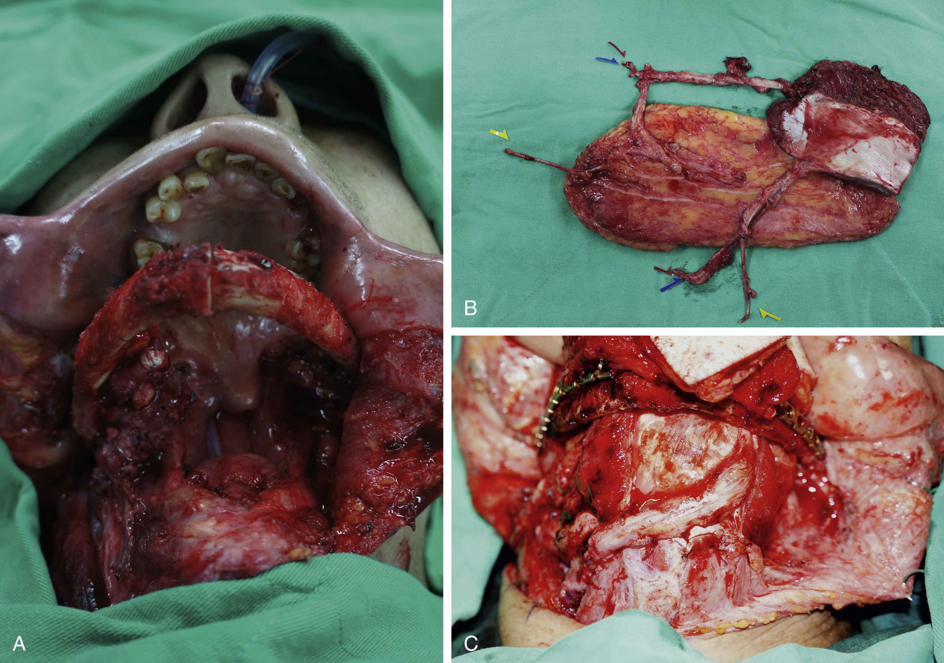
Tongue reconstruction requires the restoration of both mobility and loss of tissue bulk against weakened native propulsive power of the tongue. Therefore, meticulous execution of surgical margins to retain maximum intrinsic and extrinsic muscles effectively reduces the loss of tongue function. Similarly, preservation of pharyngeal muscles and minimization of their use in regional reconstructions maximizes their coordinated intraoral functions with the tongue. The thickness of the flap should appropriately match the residual tongue. If the flap is thicker than the native tongue, the mobile part of the tongue (anterior two-thirds) may be overpowered. Conversely, if the flap is thinner than the native tongue, it results in tongue asymmetry. The length of neo-tongue should also match the native tongue if the tip is not affected. In the resected tip, the length of the neo-tongue should extend no shorter than the location of the incisors. If the length of the tongue is insufficiently reconstructed, the overall mobility is reduced and affects “front raising” speech articulation, as well as the process and transport II phases of oral deglutition. Insufficient length of the neo-tongue and inadequate tissue replacement at the junction between floor of mouth and mobile tongue (anterior two-thirds) can cause tongue tethering, and will be worsened post radiotherapy.
When the floor of the mouth is resected or neck dissection is performed, complete separation of the oral cavity from the neck is crucial. Complete separation of the oral cavity from the neck avoids fistula formation. Subsequent wound infection, necrosis, and abscess of the neck may develop, leading to delayed pedicle thrombosis of the free flap and possible arterial or venous blowout. In the posterior third of the tongue, volume replacement is the most important. As the tongue normally seals against the soft palate during the transport and process phase of oral deglutition, lack of volume replacement will, therefore, increase the risk of aspiration. The tongue base propulses fluid and food boluses towards the laryngopharynx during transport II phase to initiate pharyngeal deglutition. Failure of its superoposterior movements due to loss of its bulk post reconstruction, reduces overall swallowing efficiency and increases the likelihood of nasal regurgitation via the velopharyngeal port. The incomplete contact of the posterior tongue will increase the difficulties of “back raising” articulation as the result of insufficient volume replacement. In an extended defect that involves the floor of the mouth and buccal sulcus, the contouring of the flap is commonly inadequate to reconstruct the concavity of the buccal sulcus. The sulcus functions as a food reservoir during mastication and helps in directing the saliva and food toward the oropharynx during deglutition. To avoid bulkiness and formless reconstruction of the buccal sulcus, an origami technique can be utilized. The steps of the procedure are: (1) pre-fixing the flap to the defect; (2) marking the area of concavity at the sulcus on the flap; (3) de-epithelialization of the triangulated concaved edge; (4) edge-to-edge repair of the deepithelialized skin. However, the technique is restricted in bulky flaps or bulky insets, as skin-deep folding may be difficult.
Sensation is complementary to muscular function of the tongue for prehension, speech, and deglutition. The tongue possesses a significant portion of the cortical homunculus, and sensory feedback allows constant adjustments of the tongue position and movements. This coordinated function is the key to the high functioning tongue articulations that can be observed among trumpet players. Evidently, sensory preservation from surgical ablation and reinnervation is generally recommended. Tongue reconstruction with sensory reinnervation results in superior sensory recovery compared with those left with spontaneous resensitization. Coaptation to lingual and inferior alveolar nerve provides statistically superior sensory function than other nerves. The overall tongue function is enhanced in those with sensory recovery post reconstruction compared with those without. However, sensory reconstruction of the tongue should proceed with caution. The potential development of hyperalgesia and allodynia during axon regeneration may outweigh its benefit, especially among those who are planned for postoperative radiotherapy. Motor innervation is sought after functioning muscle transfer, and the common donor nerve is the hypoglossal. Motor nerve reinnervation reduces the impact of muscular atrophy, and the reanimated flap provides much needed protective function of the neo-tongue.
Mandibular Reconstruction Goals and Surgical Pearls
The goal for mandibular defect reconstruction is both functional and esthetic. Specifically, it aims to restore sturdy continuum of the mandible, reinstate accurate occlusion, preserve or reestablish condylar articulation of the temporomandibular joint (TMJ), achieve sensory reconstruction, and provide adequate bone width and height for dental osseointegration and esthetics. To achieve functional and esthetically pleasing reconstruction of the mandible, adequate restoration of occlusion is crucial. Occlusion provides a platform for constant intraoral force transmission from mandible to maxilla. The force provides constant stimuli for mandibular remodelling, soft tissue positioning, and dentition alignments. Malocclusion induces unopposed force of the mandible, and is associated with dental caries, masticatory muscle contracture and shortening, mandibular recession, as well as dentition loosening and shift. It can lead to significant anterior subluxation of TMJ with mandibular swing, thus worsening the malocclusion and facial asymmetry. Sensory reconstruction is important, yet difficult to achieve in complex resections. Where possible, inferior alveolar nerve should be reinnervated with sural nerve graft to restore sensation of the lip, especially in benign cases such as ameloblastoma reconstructions. Protective mechanism of the oral cavity and maintenance of oral continence will be impaired if numbness and/or dysesthesia of the lip persists. The optimal segmental reconstruction of the mandible is with a free vascularized bone flap that has durable soft tissue. Alternatively, segmental defects can be reconstructed with vascularized soft tissue flap with/without a reconstruction plate. The second option is typically reserved for patients who present with advanced disease and short life expectancy. It is worth noting that a mandibular reconstruction plate with soft tissue coverage is associated with significant risk of plate-related complications, that include osteomyelitis, screw loosening, stress fracture of the plate, osteocutaneous fistula, and plate exposure. It is reported that up to 46% of these patients will present with plate exposure, most commonly in the combined lateral and central segments (L-C segment) and patients who received postoperative radiotherapy.
Surgical Options
Tongue Reconstruction with Non-Flap Options
Primary closure can generally be achieved in small and isolated presulcal tongue defects up to a third of tongue resection. As a structure that consists of pure skeletal muscle, postoperative functional compensation of the tongue can be expected. Primary closure is contraindicated in similar defects that involve the floor of the mouth, particularly if the remaining floor of mouth mucosa is less than 1 cm away from the gingiva. Healing by secondary intention is indicated when the defect cannot be reconstructed by primary closure without substantial distortion, or as the result of postoperative dehiscence. Oncologically, the primary site of the tumor is more difficult to monitor for recurrence if allowed to heal secondarily. In small defects among suitable patients, primary closure or healing with secondary intention can be utilized; the resultant speech and swallowing is superior not only because more muscle was preserved but also due to the avoidance of the potential overpowering effect of the flap to the native tongue.
Full-thickness/split-thickness skin grafts and mucosa grafts are rarely used for tongue reconstruction. Within the narrow spectrum of skin graft application in tongue reconstruction, to ensure its success, maneuvers such as overgrafting and quilting technique can be used. Tie-over dressing, on the other hand, may be poorly tolerated, and is a potential risk of upper airway obstruction. These techniques should be reserved for candidates who are unfit for any other forms of reconstruction and present with a well-vascularized noninfected wound bed that is shallow and flat.
Tongue Reconstruction with Intraoral Flap Techniques
Intraoral local flaps, in theory, are the optimal donor tissues for tongue reconstructions. These options, however, are restricted due to limited donor tissue and potential risk of trismus at the donor sites. They are generally unable to provide the bulk that is required for tongue reconstruction; they are, nevertheless, important options for defects that are small and thin.
Facial artery myomucosal flaps (FAMM) and the buccinator myomucosal island flap resurface small to moderate size defects that require thin tissue for reconstruction among edentulous patients. FAMM flaps are based on the facial artery anterior to Stensen’s duct, and can be based superiorly or inferiorly for anterior third tongue reconstruction. In contrast, the buccinator myomucosal island flap is based on the buccal artery (from the internal maxillary artery) posteriorly, or branches of the facial artery anteriorly. The buccinator myomucosal flap has wider flap reach, is comparatively more versatile, and able to cover the anterior two-thirds of the tongue. Flap orientation should be carefully designed, using the flap sparingly to prevent postoperative trismus.
Mandible Reconstruction with Nonmicrosurgical Techniques
Mandible reconstruction with nonmicrosurgical technique is generally reserved for small benign defects that do not require subsequent radiotherapy or patients who are unsuitable for prolonged microsurgical reconstructions. These techniques include the use of nonvascularized bone graft or bone substitutes, and prosthetic implants. Nonvascularized bone graft and bone substitute are suitable for small to moderate size bony defects up to 6 cm. They require both well-vascularized wound beds without pre- or postoperative radiotherapy. Durable vascularized tissue coverage is essential for eventual union to occur. These techniques are commonly used for enucleation procedures for benign lesions such as primary ameloblastoma, and usually covered by local gingival flaps. Nonvascularized bone has a more significant role in marginal defect reconstruction for height augmentation prior to dental osseointegration. It may still be indicated for segmental defects in biomechanically favorable locations such as body or ramus of the mandible. The durability of nonvascularized bone is frequently in doubt as it atrophies significantly compared with vascularized options. Alternatively, a mandibular reconstruction plate is used to replace the need of bony reconstruction. The use of a reconstruction plate invariably requires durable soft tissue reconstruction for its coverage with a pedicle flap.
Regional Flap Reconstruction for Oral Tongue and Mandibular Defects
In modern day reconstruction, regional flaps have a limited role, but remain valuable options, especially in critically ill patients or patients with a vessel-depleted neck.
Soft Tissue Regional Flaps
Submental Artery Flap
The submental artery (SMA) flap is a fasciocutaneous flap located in the submental region. The flap is based on submental artery located 5–6.5 cm from the origin of the facial artery. The artery courses deep to the submandibular gland via myelohyoid muscle inferior to the mandibular angle and extends medially deep to anterior digastric muscle. Once it reaches the mandible margin, it branches to supply platysma and overlying skin. It is a reliable flap with pliable tissue; the pedicle length enables it to reach to ipsilateral tongue, floor of mouth, and oropharynx. Oncologically, this flap is raised over common regions of nodal metastasis. Therefore use of this flap should proceed with caution, and the flap is better dissected off the regional lymph nodes to prevent metastasis.
Pectoralis Major Myocutaneous Flap
The pectoralis major myocutaneous (PMC) flap is classified as a type V muscle flap by Mathes and Nahai, with one dominant artery and several secondary segmental arteries. The dominant artery originates from the pectoral branch of the thoracoacromial artery, whereas secondary segmental arteries derive from the superior thoracic artery, clavicular branch of the thoracoacromial artery, lateral thoracic artery, and internal mammary artery. A PMC flap is more commonly used as a salvage procedure in the modern reconstruction era. It is a thick flap with a bulky pedicle. The muscle is based on its axial blood supply, whereas the skin is based on a random pattern. A PMC flap typically requires larger tunneling to breach the oral cavity from the neck for intraoral reconstruction, which invariably produces an unsightly bulge at the recipient site, and is less suitable for anterior tongue and oral cavity reconstruction. It can also be raised as an island flap and the pedicle skeletonized to counteract the undesirable cosmesis. It is a durable flap to cover a mandibular reconstruction plate, and is applicable to cover vascularized or nonvascularized bony reconstruction of the mandible. A two-stage operation is frequently planned for mandibular coverage, where the pedicle is commonly divided after 3–4 weeks for superior contouring results. A major drawback of the PMC flap is donor site morbidity. Reduction in adduction and internal rotation of the arm is expected. The bulk of the flap invariably shrinks as muscle atrophies, which can produce a sagging appearance and intraoral pooling. Surgical refinement or secondary operation may be needed for these corrections. Regaining protective sensation post reconstruction is unlikely.
Supraclavicular Artery Island Flap
Supraclavicular artery island flap is a recent addition to regional flap options for head and neck reconstruction. It is a fasciocutaneous flap over the tip of the shoulder and extends to midlateral deltoid muscle. The flap is based on the supraclavicular branch of the transverse cervical artery. Anatomical studies have revealed the flap is a combination of axial flap proximally and random pattern distally, demarcated at the origin of the deltoid muscle. It provides thin sizable hairless skin with good reach up to 30 cm; distal necrosis may occur in flaps with reach greater than 20 cm. Level V neck dissection can damage the course of the supraclavicular pedicle, however, it is usually preserved and thus not contraindicated. It is advisable to use hand-held Doppler intraoperatively to ensure the patency of the pedicle prior to its dissection. Flap prelamination with skin graft, and delayed procedure can be applied to extend its versatility.
Bone-Bearing Regional Flaps
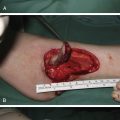
Regional bone-bearing flaps are convenient options with creative use of regional osseous constructs and shapes. Nevertheless, none of the described options has achieved popularity due to poor bone quality, fragile osseous blood supply, inadequate cortical thickness, restricted flexibility in bone insetting, and significant donor site morbidity. Although general consensus recommends pedicle-based or downgraded reconstruction in failed primary microsurgery reconstructions, however, head and neck reconstruction with regional flap consists of higher subsequent complication rates than those who received second free flaps. The success rate for a second free flap reconstruction was 94.1% in the published series. Regional options of bone-bearing flaps for intraoral reconstruction include the use of vascularized bone from the ribs, clavicle, and scapula. They can be harvested with pectoralis major, sternocleidomastoid, and trapezius, respectively, as pedicle osteomusculocutaneous flaps. In addition, a temporalis osteomuscular flap can also be used for simultaneous soft tissue and bony reconstruction of the intraoral cavity.
Pectoralis Major Osteomusculocutaneous Flap
Intergraded harvesting of fifth rib into a traditional PMC flap added further diversity to the flap for composite oromandibular defects. The blood supply to the fifth rib is based on a vascular plexus derived from its periosteum and pectoralis muscle. The blood supply to the osseous component is small and occasionally unreliable. The drawback of this technique is the difficulty of inset, and insufficient cortical thickness for functional mandibular reconstruction and dental osseointegration. Hemothorax and pneumothorax are possible donor site morbidities. They should be actively monitored, and promptly treated. Similarly, sternum can be harvested in conjunction with a PMC flap. It shares similar drawbacks with ribs, and a long-term result is not yet available. There is also reported success of a latissimus dorsi flap incorporating rib for extensive mandibular reconstruction.
Microsurgical Reconstructions of Oral Tongue and Mandible
Microsurgical reconstruction for tongue and mandibular reconstruction comes with a greater than 95% success rate. Different from local regional flaps, free flaps are not restricted by defect size and location. The flexibility of free flap positioning is unrestricted from its pedicle, thus liberating the imagination and creativity of free flap inset and design. The phenomenon of mucosalization popularized the use of free fasciocutaneous flaps for intraoral reconstruction over intestinal flaps (jejunal flap). Muscle flaps can adequately replace the volume deficit, and may also enhance the overall function if reanimated. When using a muscle flap or flap with a muscle component, the operating surgeon needs to be mindful of total ischemia time; the completion of inset and arterial anastomosis should be kept within 4 hours to prevent irreversible muscular injury.
For head and neck reconstruction in general, the use of distant free flap donor sites such as lower extremities is preferred. This principle permits a two-team approach, and shortens overall operative time. An abdominal donor site is generally avoided as disruption of internal viscera may induce ileus, and possible bowel obstruction. This can significantly reduce the patient’s nutritional status postoperatively, prolong healing time, and increase overall bed-bound related complications. In addition, increase in intraabdominal pressure and pain reduces inspiratory effort, increases atelectasis of the lung, and escalates the risk of postoperative pneumonia. Single free flap reconstruction is suitable for most tongue and mandibular reconstruction. In extended oromandibular defects, a combined technique of locoregional flaps with free flap may be necessary. If volume replacement is necessary, double free flap reconstruction should be considered. The commonly used recipient vessels are superior thyroid artery, transverse cervical artery, and facial artery. The lingual artery is uncommonly used for head and neck reconstruction, if preserved, however, it can be a reliable option. The following represent common soft tissue and bone-bearing free flap options in oral tongue and mandibular reconstructions.
Soft Tissue Free Flaps
Radial Forearm Free Flap
The radial forearm free flap (RFFF) is also known as the Chinese flap and was introduced in 1978. It is one of the most commonly used flaps in intraoral reconstruction today. It is easy to raise, provides thin and pliable tissue, allowing large skin paddle harvest with a lengthy pedicle, and good vascular caliber. It is usually raised as a fasciocutaneous flap by suprafascial or subfascial dissection, however, it can also be harvested as an adipofascial flap, or osteocutaneous flap incorporating radius. It is a type C fasciocutaneous flap, based on the radial artery and its perforators. The artery is drained preferentially by a pair of accompanying venae comitantes, and cephalic vein to a lesser extent. Allen’s test should be performed in the case of a radial dominant forearm.
The flap planning starts with locating the radial artery by palpation; the skin paddle border should be designed within the radial border of the forearm without extending to the extensor surface. A tourniquet should be used to minimize bleeding while optimizing the surgical field to prevent unnecessary vascular injury. The dissection starts at the radial edge in the suprafascial plane as described by Wong et al. The distal pedicle is ligated at the wrist, followed by careful dissection proximally preserving paratenons and superficial radial nerve. Once the dissection is completed, the tourniquet is released and flap and distal circulation is observed for 15–20 minutes. This is to ensure the integrity of the pedicle is preserved, circulation of the flap is adequate, and distal circulation of the hand and fingers is unaffected. If circulation of the hand is compromised, interposition vascular anastomosis should be performed to restore its perfusion. If innervation is planned for tongue reconstruction or as soft tissue coverage for mandibular reconstruction, antebrachial cutaneous nerve can be included during harvesting.
As there are nine fasciocutaneous branches of the radial artery in the forearm, multiple skin paddles can be designed for multiple defect reconstruction. This concept is commonly used for trismus release where two skin paddles were designed based on the same vascular system. Similarly, double skin paddle design is useful to separate the oral tongue from the floor of the mouth to prevent tethering of the tongue. If tissue volume is required, a proximally-based flap can be designed, however, this will significantly shorten the pedicle length. Alternatively, volume augmentation can be achieved by harvesting a flap with excess, and invaginating the excess flap for volume augmentation after de-epithelialization.
As an osteocutaneous flap, the quality of the radius is inadequate to replace segmental mandibular defects, as it lacks cortex thickness, is brittle, and unsustainable for osteotomies. It is an unsuitable option if full functional reconstruction is anticipated. It may be indicated for marginal mandibular defect reconstructions, nevertheless, the bone quality is inappropriate for subsequent osseointegration for full dental rehabilitation.
Donor site morbidity is the major drawback of the RFFF. The donor site is commonly closed with full- or split-thickness skin grafts. The donor site may present with contracture, stiffness of the wrist and fingers, recurrent wound breakdown and tendon exposure. These complications are predominately related to failure of the skin graft, representing integral disadvantages of skin graft reconstructions. Suprafascial technique reduces the incidence of tendon exposure, and lessens risk of graft failure. Preferentially using a thick split-thickness skin graft for a larger defect, and a full-thickness skin graft for a smaller donor site defect may reduce the contracture from the skin grafts. However, in selected cases, a proximally-based local flap may be used successfully for donor site closure and prevent the above mentioned complications. If an osteocutaneous flap is used, the risk of distal radius fracture is clinically significant. In high-risk patients, prophylactic plating may be necessary ( Fig. 24.1 ).