and Ziv Gil2
(1)
Division of Otolaryngology Head and Neck Surgery and Maxillofacial Surgery, Tel Aviv Sourasky Medical Center, Tel Aviv, Israel
(2)
The Head and Neck Center Department of Otolaryngology Head and Neck Surgery, Rambam Healthcare Campus, Haifa, Israel
Keywords
Operating roomSetupSkull baseSurgeryReconstructionEndoscopicAnesthesia3.1 Introduction
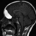
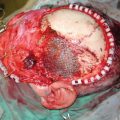
Surgery has become a routine procedure for management of neoplasms arising in the skull base and paranasal sinuses. The main objective in skull base surgery is ablation of the tumor while minimizing morbidity and preserving the patient’s quality of life. Decisions regarding the goals of surgery are made according to factors involving the tumor and the patient. Tumor-related factors include histology, tumor size, extension, differentiation, and the presence of locoregional or distant metastases. Patient-related factors include, age, sex, family history, previous operations, and prior radiotherapy. Many of these surgeries require multidisciplinary treatment by otolaryngologists, neurosurgeons, and plastic surgeons.
To allow safe ablation of the tumor with minimal morbidity, the operating room (OR) setup should be arranged accordingly. This chapter describes in detail the OR setup as practiced by the authors for both open and endoscopic operations.
3.2 Preoperative Evaluation and Anesthesia
All patients scheduled for operation are evaluated preoperatively by a head and neck surgeon and an anesthesiologist. Patients younger than 18 years are also examined by a pediatrician. Radiological evaluation includes CT and MRI. These patients may also be evaluated using a hybrid of positron emission tomography (PET) and CT (PET/CT) prior to the operative procedure. Flexible fiberoptic examination is used to evaluate vocal cord mobility. CT or CT-MRI fusing navigation systems are utilized in most cases.
We recommend routinely performing tissue diagnosis in the form of biopsy or fine needle aspiration in patients scheduled for skull base surgery. Exceptions include highly vascular tumors, suspected encephalocele or meningocele, and chordomas. Immunohistochemical staining should be performed routinely in the pathologic workup. All patients should undergo routine blood tests, including complete blood count and coagulation function tests.
Related posts:


Stay updated, free articles. Join our Telegram channel

Full access? Get Clinical Tree


