This article focuses on the surgical treatment of nonmelanoma skin cancers of the head and neck. The risk factors of nonmelanoma skin cancers for recurrence and metastases that are important for choosing the best treatment option are summarized. Surgical treatment options including surgical excision with standard margins, frozen section, staged surgery, and Mohs micrographic surgery are described. Indications, techniques, outcomes, and advantages and disadvantages of each approach are reviewed. Finally, management of incomplete excisions is discussed.
- •
Define the risk factors of the tumor.
- •
Identify clinical margins under optimal lighting and magnification.
- •
Dermoscopy may be helpful to identify clinical margins better.
- •
In selected cases, prior curettage may help to delineate clinical margins; however, it is controversial.
- •
Mark surgical margins using scale before local anesthetic infiltration.
- •
For surgical excision, minimal surgical margin is recommended to be 3 to 4 mm for BBCs with well-defined clinical borders. Leave at least 4 to 6 mm healthy tissue around the SCCs.
- •
For the excision of high-risk tumors, prefer Mohs’ micrographic surgery, if it is available.
- •
Surgical excision with intraoperative margin assessment with frozen section or preferably staged surgery can also be used safely.
- •
Do not perform complicated reconstructions without achieving tumor-free margins.
- •
In the management of incomplete excised NMSCs consider surgery primarily, and consider wait-and-see approach in selected cases.
- •
Late recurrences may occur; follow patients at least 5 years.
Introduction
The term “nonmelanoma skin cancer” (NMSC) is used to define basal cell carcinoma (BCC) and squamous cell carcinoma (SCC) of the skin, and other rare primary cutaneous malignancies. Because most NMSCs originate from the epidermis, the upper layer of the skin, it is often detected at an early stage and can be treated locally. Treatment of NMSC can be broadly classified into surgical and nonsurgical treatment. Surgical excision is the most effective treatment option for most NMSCs. This article focuses on surgical treatment for NMSC; repair of the defect after excision is beyond the scope of this article.
The treatment of a facial skin cancer aims to achieve complete eradication of the cancer with a good and acceptable cosmetic and functional outcome; however, complete eradication of the tumor should be the primary goal. Although surgery is the treatment of choice for high-risk NMSCs, low-risk NMSCs may be treated by either nonsurgical or surgical treatment options. Therefore, the most important step in the treatment planning of the NMSCs is to determine if the tumor has high risk or low risk to recur or metastasize. Presence of characteristics associated with recurrence or metastasis make a tumor high risk, whereas a tumor that is unlikely to recur or metastasize is determined to be low risk.
Risk factors of recurrence and metastases
Location
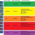
Location of the tumor is one of the most important factors affecting the outcome of treatment of NMSCs. Certain sites of the head and neck region are more likely to recur and metastasize. Swanson illustrated high-risk locations of face as an “H” zone, because of the higher recurrence rate and the functional and cosmetic importance ( Fig. 1 ). Recurrences are most commonly seen on nose, cheek, auricular area, periocular area, scalp, and forehead. SCCs located on ear, temple, forehead, and anterior scalp that drain to parotid gland and lower lip are associated with higher incidence of metastases.
Size
Lesion size is also an important prognostic risk factor for NMSCs and larger tumors have higher recurrence rates. The degree of subclinical extension was unpredictable in tumors 2 cm or larger in diameter when compared with tumors less than 2 cm in diameter. The horizontal diameter of BCCs is an important factor for tumor invasion; a tumor with larger diameters invades more deeply. The lesion size of SCC was found to be associated with recurrence and depth of invasion. Furthermore, lesion size 4 cm or greater was one of the most significant factors that diminished disease-specific survival. In another study, tumor size greater than 2 cm was found to be an independent risk factor for recurrence-free survival. Cherpelis and colleagues showed that tumor size significantly correlated with metastasis, with increasing risk for tumors larger than 2 cm in size, and they proposed that invasive SCCs that are smaller than 1 cm in size may metastasize, but it is uncommon.
Histologic Subtype
Histologic subtypes of NMSCs are a predictor of risk of recurrence and metastasis. The most potent predictor of recurrence for BCCs was found to be aggressive histologic type of the tumor, besides positive excision margins. Any histologic variant of BCC may be locally aggressive and metastasize, but metatypical and basosquamous, morpheaform, infiltrating, and micronodular subtypes of BCC are more prone to exhibit an aggressive behavior. Infiltrative, morpheic, and micronodular subtypes of BCC show locally aggressive behavior and invade more deeply than nodular BCCs. On eyelid, 29.41% of patients with fibrous or sclerozing BCC, referred to as infiltrative, superficial multicentric, or morpheo growth pattern, recurred, whereas only 2.38% of nodular BCCs recurred.
Clinical behaviors of subtypes of SCCs also vary widely, although all squamoid neoplasms are commonly referred as generic SCC. Desmoplastic growth pattern of SCC is an independent risk factor for local recurrence. Cassarino and colleagues made a clinicopathologic classification of cutaneous SCC according to metastatic rate in which tumors were separated as low risk (≤2% metastatic rate); intermediate risk (3%–10% metastatic rate); high risk (>10% metastatic rate); and intermediate behavior (insufficient data to determine the metastatic risk) ( Table 1 ).
| Low-Risk SCC Types I | Intermediate-Risk Types | High-Risk Tumors | Intermediate Category |
|---|---|---|---|
| SCC arising in actinic keratoses Human papilloma virus–associated SCC, Tricholemmal carcinoma Spindle cell SCC | Adenoid (acantholytic) SCC Intraepidermal epithelioma with invasion Lymphoepithelioma-like carcinoma of the skin | Invasive Bowen disease Adenosquamous carcinoma Malignant proliferating pillar tumor Desmoplastic SCC De novo SCC SCC arising in association with predisposing factors | Clear cell SCC Signet ring cell SCC Papillary SCC Pigmented SCC Follicular SCC SCC arising from benign sweat gland cysts Squamoid eccrine ductal carcinoma |
Degree of Differentiation
Differentiation of the tumor is also an important prognostic criterion for cutaneous SCC. Poorly differentiated SCCs tend to present more locally aggressive behavior and demonstrate increased recurrence rates compared with well-differentiated SCCs. There is also a higher propensity to develop regional metastases in poorly differentiated SCCs.
Tumor Thickness and Depth of Invasion
Tumor thickness or depth of invasion is another important prognostic predictor of SCCs. Deep invasion diminishes disease-specific survival in cutaneous SCCs. Brantsch and colleagues found tumor thickness of SCC greater than 6 mm to be an independent risk factor for local recurrence and proposed to divide SCC into three main risk categories for metastasis according to tumor thickness: (1) no detectable risk (≤2 mm tumor thickness); (2) low risk (2–6 mm tumor thickness); and (3) high risk (>6 mm tumor thickness). Metastatic SCCs are more likely to be deeper (Clark level V). Depth of invasion greater than 4 mm is associated with an increasing risk of metastases.
Perineural and Lymphovascular Invasion
Perineural invasion diminishes disease-specific survival in cutaneous SCCs and was found to be an independent risk factor for overall and recurrence-free survival of cutaneous SCC of the head and neck. Presence of lymphovascular invasion may also increase the risk of developing nodal metastases.
Immunosuppression
SCC of skin behaves more aggressive in patients who are immunosuppressed than in patients who are immunocompetent, and has higher metastatic potential. Although the recurrence rate of SCC and BCC was found to be similar after renal transplantation, 48% and 42.8%, respectively, any role of immunosuppression on aggressive behavior that has been shown for SCC has not been proved for BCC.
Recurrent Tumor
Recurrent tumors have more extensive subclinical spread. The incidence of incomplete excision of BCC was 3.7% in primary lesions, but it was 13.1% in the recurrent lesions.
Site of Chronic Wound and Burn Scar
SCCs arising at the site of chronic wound or burn scar have three times diminished 5-year recurrence-free survival compared with SCCs as a whole (28% vs 74%).
Risk factors of recurrence and metastases
Location
Location of the tumor is one of the most important factors affecting the outcome of treatment of NMSCs. Certain sites of the head and neck region are more likely to recur and metastasize. Swanson illustrated high-risk locations of face as an “H” zone, because of the higher recurrence rate and the functional and cosmetic importance ( Fig. 1 ). Recurrences are most commonly seen on nose, cheek, auricular area, periocular area, scalp, and forehead. SCCs located on ear, temple, forehead, and anterior scalp that drain to parotid gland and lower lip are associated with higher incidence of metastases.
Size
Lesion size is also an important prognostic risk factor for NMSCs and larger tumors have higher recurrence rates. The degree of subclinical extension was unpredictable in tumors 2 cm or larger in diameter when compared with tumors less than 2 cm in diameter. The horizontal diameter of BCCs is an important factor for tumor invasion; a tumor with larger diameters invades more deeply. The lesion size of SCC was found to be associated with recurrence and depth of invasion. Furthermore, lesion size 4 cm or greater was one of the most significant factors that diminished disease-specific survival. In another study, tumor size greater than 2 cm was found to be an independent risk factor for recurrence-free survival. Cherpelis and colleagues showed that tumor size significantly correlated with metastasis, with increasing risk for tumors larger than 2 cm in size, and they proposed that invasive SCCs that are smaller than 1 cm in size may metastasize, but it is uncommon.
Histologic Subtype
Histologic subtypes of NMSCs are a predictor of risk of recurrence and metastasis. The most potent predictor of recurrence for BCCs was found to be aggressive histologic type of the tumor, besides positive excision margins. Any histologic variant of BCC may be locally aggressive and metastasize, but metatypical and basosquamous, morpheaform, infiltrating, and micronodular subtypes of BCC are more prone to exhibit an aggressive behavior. Infiltrative, morpheic, and micronodular subtypes of BCC show locally aggressive behavior and invade more deeply than nodular BCCs. On eyelid, 29.41% of patients with fibrous or sclerozing BCC, referred to as infiltrative, superficial multicentric, or morpheo growth pattern, recurred, whereas only 2.38% of nodular BCCs recurred.
Clinical behaviors of subtypes of SCCs also vary widely, although all squamoid neoplasms are commonly referred as generic SCC. Desmoplastic growth pattern of SCC is an independent risk factor for local recurrence. Cassarino and colleagues made a clinicopathologic classification of cutaneous SCC according to metastatic rate in which tumors were separated as low risk (≤2% metastatic rate); intermediate risk (3%–10% metastatic rate); high risk (>10% metastatic rate); and intermediate behavior (insufficient data to determine the metastatic risk) ( Table 1 ).
| Low-Risk SCC Types I | Intermediate-Risk Types | High-Risk Tumors | Intermediate Category |
|---|---|---|---|
| SCC arising in actinic keratoses Human papilloma virus–associated SCC, Tricholemmal carcinoma Spindle cell SCC | Adenoid (acantholytic) SCC Intraepidermal epithelioma with invasion Lymphoepithelioma-like carcinoma of the skin | Invasive Bowen disease Adenosquamous carcinoma Malignant proliferating pillar tumor Desmoplastic SCC De novo SCC SCC arising in association with predisposing factors | Clear cell SCC Signet ring cell SCC Papillary SCC Pigmented SCC Follicular SCC SCC arising from benign sweat gland cysts Squamoid eccrine ductal carcinoma |
Degree of Differentiation
Differentiation of the tumor is also an important prognostic criterion for cutaneous SCC. Poorly differentiated SCCs tend to present more locally aggressive behavior and demonstrate increased recurrence rates compared with well-differentiated SCCs. There is also a higher propensity to develop regional metastases in poorly differentiated SCCs.
Tumor Thickness and Depth of Invasion
Tumor thickness or depth of invasion is another important prognostic predictor of SCCs. Deep invasion diminishes disease-specific survival in cutaneous SCCs. Brantsch and colleagues found tumor thickness of SCC greater than 6 mm to be an independent risk factor for local recurrence and proposed to divide SCC into three main risk categories for metastasis according to tumor thickness: (1) no detectable risk (≤2 mm tumor thickness); (2) low risk (2–6 mm tumor thickness); and (3) high risk (>6 mm tumor thickness). Metastatic SCCs are more likely to be deeper (Clark level V). Depth of invasion greater than 4 mm is associated with an increasing risk of metastases.
Perineural and Lymphovascular Invasion
Perineural invasion diminishes disease-specific survival in cutaneous SCCs and was found to be an independent risk factor for overall and recurrence-free survival of cutaneous SCC of the head and neck. Presence of lymphovascular invasion may also increase the risk of developing nodal metastases.
Immunosuppression
SCC of skin behaves more aggressive in patients who are immunosuppressed than in patients who are immunocompetent, and has higher metastatic potential. Although the recurrence rate of SCC and BCC was found to be similar after renal transplantation, 48% and 42.8%, respectively, any role of immunosuppression on aggressive behavior that has been shown for SCC has not been proved for BCC.
Recurrent Tumor
Recurrent tumors have more extensive subclinical spread. The incidence of incomplete excision of BCC was 3.7% in primary lesions, but it was 13.1% in the recurrent lesions.
Site of Chronic Wound and Burn Scar
SCCs arising at the site of chronic wound or burn scar have three times diminished 5-year recurrence-free survival compared with SCCs as a whole (28% vs 74%).
Surgical treatment options
There are three margin concepts to differentiate in the treatment of NMSCs: (1) clinical margin, (2) surgical margin, and (3) pathologic margin ( Fig. 2 ). Clinical margin defines the visible borders of a tumor. However, the clinical margin of a tumor does not necessarily refer to histopathologic borders of the tumor, namely pathologic margin. Tumor may have some amount of subclinical extension that cannot be identified visibly. Therefore, excised specimen must incorporate sufficient amount of normal tissue around the tumor to prevent recurrences. The preferred excision margin of a tumor with some amount of normal-appearing tissue defines the surgical margin. However, one must also avoid removing excessive tissue to spare normal tissue in order not to disturb cosmesis and function. Therefore, determining surgical margins of a tumor is crucial in the surgical treatment of NMSC.
Identification of lesions with extensive subclinical spread preoperatively helps surgeon to determine the optimal surgical procedure and surgical margin. Batra and Kelley performed a retrospective study to develop a scale to predict extensive subclinical spread of NMSCs. Analyses of 1095 BCCs and SCCs revealed that the significant predictors of extensive subclinical spread of NMSCs are basosquamous, morpheaform, nodular, and recurrent subtypes located on the nose; morpheaform BCC on the cheek; any tumor on the eyelid, temple, or ear helix; any tumor on the neck in men; recurrent BCC in men; and preoperative size less than 10 mm.
The surgical procedures used in the treatment of NMSCs aim to remove the tumor with a tumor-free surrounding tissue. This may be achieved by either surgical excision with standard margins ; surgical excision with frozen-section margin assessment ; surgical excision with delayed repair (staged surgery) ; or Mohs micrographic surgery (MMS) . The comparison of these options regarding advantages and disadvantages is summarized in Table 2 .
| Surgical Excision with Standard Margin | Surgical Excision with Frozen Section | Surgical Excision with Delay Repair | Mohs Micrographic Surgery |
|---|---|---|---|
| Relies on clinical margins | Relies on vertical frozen section assessment | Relies on permanent section assessment | Relies on horizontal frozen section assessment |
| Subtotal margin control (changes depending on the type of sectioning) | Subtotal margin control (changes depending on the type of sectioning) | Subtotal margin control (changes depending on the type of sectioning) | 100% microscopically controlled margin |
| No special training and facilities required | Requires pathologist intraoperatively | No special training and facilities required | Special training and facilities required |
| Results in larger defect size | Results in larger defect size | Results in larger defect size (smaller when narrow margin is used) | Results in smaller defect size |
| Shorter operation time | Time-consuming operation | Shorter operation time (only for the excision) | Time-consuming operation |
| Re-excision may be required | Re-excision may be required (less than surgical excision and surgical excision with delay repair) | Re-excision may be required | No re-excision after operation |
| Reconstruction at the time of excision | Reconstruction at the time of excision | Reconstruction at a second stage | Reconstruction at the time of excision (if another reconstructive surgeon is not required) |
| Complicated reconstructions cannot be performed safely | Complicated reconstructions can be performed more safely | Complicated reconstructions can be performed most safely | Complicated reconstructions can be performed most safely |
| Least expense | Less expense | Increased expense | Increased expense |
| Relatively worse outcome | Better outcome | Best outcome | Best outcome |
Surgical Excision with Standard Margins
This traditional surgical treatment is based on the removal of tumor relying on the clinical margins. Therefore, this approach is preferred for the treatment of tumors with well-defined clinical margins. Surgical excision with standard margins is indicated mostly in the treatment of low-risk NMSCs.
In most cases, tumors can be excised under local anesthesia. Identifying the clinical margin of the tumor preoperatively is crucial. The lesion is carefully inspected under optimal lighting ideally with magnification and palpated to determine the exact clinical margin of the tumor. The clinical margin, the assessed outline of the tumor, is dotted with ink or fine-tipped marking pen, and then the surgical margin is marked according to preferred peripheral clearance margin. Preoperative margin detection by digital dermoscopy may obtain better results to achieve safe margins. Curettage before excision of NMSC may also be used to delineate margins of the tumor. Curettage before excision decreased the surgical failure rate for BCC by 24%, but did not decreased the rate for SCC. It is advised to use a measurement of scale for marking the surgical margins. If the lesions are marked using only the naked eye, there is a tendency to underestimate surgical margins for large lesions and to overestimate for small lesions. It was shown that marking surgical margins with aid of magnification reduces the incidence of incomplete resection. Marking the relaxed skin tension lines is beneficial to obtain the orientation of incision to hide within a normal wrinkle.
Local anesthetic agent infiltration must be performed after assessing the clinical margin of the tumor in order not to impair the assessment. The lesion is excised by adhering exactly to the edge of the drawn surgical margin. The tumor is excised down into the subcutaneous fat, or adjacent involved anatomic layer ( Fig. 3 ). Surgical excision provides a single specimen to evaluate the involvement of peripheral and deep margins by the tumor. The excised specimen should be marked for orientation during histopathologic assessment of the surgical margins by either sutures or different colors of tissue dyes. Safety of margins is checked by the pathologist postoperatively with permanent sectioning.
Wolf and Zitelli showed that a minimum margin of 4 mm was necessary to totally eradicate well-defined bordered, primary BCCs with a diameter less than 2 cm, in more than 95% of cases. Significant correlation is present between the size of the lesion and margin involvement; incomplete excision is seen almost three times more in tumors greater than 2 cm in diameter.
Narrow-margin elliptical excision with surgical margins up to 3 mm was found to be only 80% effective in achieving tumor-free margins. However, Bisson and colleagues compared the surgical margins and histologic margins of BCCs and found that in cases where the tumor was larger than they had anticipated, the discrepancy was never greater than 3 mm. They concluded that 3 mm surgical margin was sufficient for the complete resection of the tumor. Griffiths and colleagues propounded that 1- to 2-mm histologic margins may generally be considered adequate and to achieve such histologic clearance, clinical margins of 2 to 3 mm are required to allow tissue shrinkage. As the surgical margins are reduced, the incidence of incomplete excision increases. Gulleth and colleagues showed that increasing the size of margins decreased recurrence rates linearly until it reached 5 mm, which had the lowest recurrence rate. There was no significant change in the rate of tumor-free margins between 3- and 5-mm surgical margins, but recurrence rates decreased.
Minimal margins of excision around the clinical borders of SCC were recommended to be 4 mm margins in low-risk tumors, whereas at least a 6-mm margin for high-risk tumors was proposed. Thomas and coworkers showed that a surgical margin of 4 mm achieved an optimal excision beyond one microscopic high-power field in 96% of BCCs and in 97% of SCCs. They proposed to excise NMSCs less than 2 cm in diameter with a 4-mm margin of normal-appearing skin under magnification.
Unlu and colleagues excised BCCs located on head and neck according to either “inspected healthy margins” visible on outer margins of the erythema or induration area surrounding the tumor or “safety margins,” 2-mm margin for tumors less than 1 cm in size and 4-mm margin for tumors greater than 1 cm in size. Comparisons revealed that incomplete excision rates with inspected healthy margins and safety margins were similar for tumors less than 1 cm and greater than 1 cm in size. They recommended excising just what was seen, especially at the perioral, periorbital, and nasal regions where morbidity might be a problem.
Surgical excision with preferred margins is a good treatment option, especially for small NMSCs ( Fig. 4 ). It shortens the operation time and costs less compared with other surgical options; however, multiple re-excisions may be needed, so the cost increases. Surgical margins should be determined according to pathologic characteristics, size, and location of the tumor. Surgical margin of 3 to 4 mm seems sufficient for most of the tumors. Safety margins of SCCs should be at least 4 to 6 mm. When the tumor is larger than 2 cm in diameter, has poorly defined borders, and high-risk to recur or metastases, it is better to prefer other treatment options. Otherwise, surgical margins should be increased. Because of the possibility of re-excisions, simple excision with permanent section margin assessment is usually preferred for lesions that can be repaired by primary closure.