Histopathologic examination is the gold standard for the diagnosis of skin cancer. Because analysis of molecular parameters such as nucleic acids and DNA are also gaining importance in diagnosis, prognosis, and therapy, an understanding of the molecular mechanisms underlying the pathogenesis of nonmelanoma skin cancer of the head and neck is of growing importance for the diagnostician and surgeon alike. This article presents a description of the effect on cells and impact on DNA of ultraviolet radiation, with a discussion of squamous cell and basal cell carcinoma in terms of the effects of genetic pathways and apoptosis.
- •
The main cause of squamous and basal skin cancer is exposure to ultraviolet (UV) radiation.
- •
Ultraviolet radiation, consistent of a small UV-B and complete UV-A band, is responsible for the biological effects on human skin.
- •
Destruction of repair mechanisms or activation of oncogenes may lead to carcinogenesis. Pathways involving p53, tumor supressor gene, apoptosis, transforming growth factor β, platelet-derivated growth factor, telomerase enzyme and hedgehog signaling system may participate in this process.
- •
Better understanding and increased awareness of molecular mechanisms underlying nonmelanoma skin cancer may aid in development of innovative diagnostic, therapeutic, and preventive measures.
Introduction
Cancer is a cellular disease characterized by a transformed cell population with abnormal cell growth. Malignant transformation is an irreversible transition of one cell that leads to the formation of a cancer. Histopathologic examination is the gold standard for the diagnosis of skin cancer. In the future, analysis of molecular parameters such as nucleic acids and DNA will also gain importance for diagnosis, prognosis, and therapy. Understanding the molecular mechanisms underlying the pathogenesis of nonmelanoma skin cancer of the head and neck of growing importance for the diagnostician and surgeon alike.
Ultraviolet radiation
The main cause of skin cancer is exposure to ultraviolet (UV) radiation. The solar UV spectrum consists of UVC (wavelengths below 280 nm), UVB (280–315 nm), and UVA bands (315–400 nm). The predominant part of the short-wave, high-energy, and destructive UV spectrum cannot reach the Earth’s surface: the ozone layer in the outer Earth atmosphere absorbs the shorter wavelengths up to 310 nm (UVC and main part of UVB radiation). The remaining transmitted UV spectrum, that is, a small UVB and the complete UVA band, is responsible for biological effects on human skin.
The consistently increasing incidence of melanocytic and nonmelanocytic skin tumors is associated with recreational sun exposure. Epidemiologic data indicate that excessive or cumulative sunlight exposure takes place years before the cancer occurs. The most important protection strategies against UV damage in human skin consist of melanin synthesis and active DNA-repair mechanisms. DNA is the major target of direct and indirect UV damage.
Animal models have demonstrated that UVB is more likely than UVA to induce skin cancer. UV-induced DNA photoproducts are able to cause specific mutations (UV-signature) in susceptible genes for squamous cell carcinoma (SCC) and basal cell carcinoma (BCC). In SCC, development of UV-signature mutations in the p53 tumor suppressor gene are the most common event, as precancerous lesions reveal fewer than 80% and SCCs more than 90% UV-specific p53 mutations.
UV light does not penetrate the body any deeper than the skin and is absorbed by the different skin layers in a wavelength-dependent manner. UVB is almost completely absorbed by the epidermis; only 10% to 20% of UVB energy reaches the epidermal stratum basale and papillary dermis. UVA penetrates deeper into the dermis and deposits 30% to 50% of its energy into the papillary dermis. These absorption characteristics of human skin explain why UVB effects occur predominantly in the epidermis (development of skin cancer) whereas UVA effects occur in the dermis (solar elastosis, skin aging). UV radiation also displays immunosuppressive effects and is able to generate tolerance against immunogenetic skin tumors. Thus UV is considered to be a “double-edged sword,” causing skin cancer by DNA damage on the one hand and enabling tumor escape from immune surveillance on the other.
UV-induced carcinogenesis in squamous cell carcinoma
The precursor cell of SCC and its precancerous progenitor, the actinic keratosis (AK), is assumed to be the interfollicular epidermal basal keratinocyte. AK and SCC are strongly related to UV exposure because 90% occur at predilection sites where the skin is regularly exposed to sun damage:
- •
Head
- •
Forehead
- •
Nose
- •
Ears
- •
Back of hands
The lifetime risk of developing an SCC correlates very closely with the individual cumulative UV dose. De Gruijl demonstrated a wavelength dependency of SCC induction effectiveness by means of hairless mice. SCC were generated in AK by UV with UV-action spectrum peaks at 300 nm in the UVB band that continuously decreased through the UVA band, with a smaller peak at 380 nm. Similar findings were observed in the murine model. When these findings were applied to estimates of human skin tolerance, the relative efficacy to induce SCC at 300 nm UVB irradiation was 1000-fold higher than UVA.
UVA predominantly causes photosensitizer-mediated oxidative DNA damage. Because in this part of the UVA spectrum (347–400 nm) direct DNA damage is not observed, it can be assumed that the indirect oxidative UVA effects contribute to the mutagenic potential of UVA in this wavelength range. UVB is even more powerful than UVA in its ability to induce AK and SCC, and is considered to be the predominant carcinogen.
The most important protein involved in early UV-induced carcinogenesis of SCC appears to be the tumor suppressor p53. p53 is an essential and well-defined transcription factor regulating cell-cycle control and apoptosis. UV radiation helps to stabilize transcriptional activity and protein degradation. Specific p53 mutations can be found in 75% to 80% of AK and in more than 90% of cutaneous SCC.
UV-induced carcinogenesis in basal cell carcinoma
BCC is the most common malignancy in Caucasians, with a worldwide increasing incidence. Exposure to UV radiation is assumed to be the main causative pathogenic factor for BCCs as well, but the precise relation between amount, timing, and pattern of UV exposure and BCC risk is still unknown. Compared with SCC, the correlation of UV with basal cell carcinogenesis is far less obvious, and epidemiologic data are not completely in line with the impact of cumulative UV dose. Some studies reveal a link between cumulative UV dose and BCC risk, although the relative risks are small, with odds ratios of 1.0 to 1.5.
BCCs predominantly affect :
- •
Seborrheic central parts of the face: root of nose, nasal ala, nasolabial fold
- •
Head
- •
Trunk
- •
Lower limbs
The BCC precursor cell is less well defined than that of SCC and is hypothesized to stem from interfollicular epidermal basal keratinocytes with retained basal morphology, from the follicular outer root sheath or sebaceous gland derived keratinocytes. The deeper anatomic localization of the BCC cells originating in hair follicles and sebaceous glands may be one explanation for differences in the carcinogenesis of BCC and SCC.
BCCs are predominantly sporadic but also appear in persons suffering from rare hereditary disorders such as nevoid BCC syndrome (NBCCS; Gorlin syndrome) or xeroderma pigmentosum (XP).
The most common inherited disorder associated with BCCs is NBCCS. NBCCS is an autosomal dominant neurocutaneous disorder. Affected individuals have a wide range of developmental anomalies, including skeletal abnormalities, craniofacial dysmorphism, and macrocephaly. Multiple early-onset BCCs are a hallmark of the syndrome that also includes features such as odontogenic keratocysts, palmar and plantar pits, and calcification of the falx cerebri. In addition to BCCs, patients have an increased incidence of medulloblastomas, meningiomas, and ovarian and cardiac fibromas. Germline mutations in the PTCH (patched) gene are found in the majority of patients with this syndrome.
XP is caused by inherited defects in nucleotide excision repair (NER) genes, resulting in a complex skin pathology including lentigines, epidermal hyperplasias, BCCs, SCCs, and cutaneous melanomas triggered by exposure to the sun. This disease is an exaggerated response to UV light, with the early onset of a vastly increased number of skin lesions that would otherwise occur in small numbers late in life. Because of the deficient DNA repair, the rate at which skin cancers develop in young XP patients is increased at least 1000-fold.
The hedgehog signaling pathway
The hedgehog pathway is a fundamental signal transduction pathway in embryogenesis that is responsible for the proper development of various organs, including:
- •
Neural tube
- •
Axial skeleton
- •
Limbs
- •
Lungs
- •
Teeth
- •
Skin
- •
Hair
Consequently, germline mutations in genes encoding members of the hedgehog pathway result in human diseases characterized by tumor predisposition and/or developmental defects.
The Hh gene, encoding a secret ligand, was originally identified in screens for mutations that disrupt segment polarity in the fruit-fly Drosophila . The gene has 3 vertebrate homologues, of which sonic Hh (Shh) is the most widely expressed. The Hh ligand binds to the membrane receptor PTCH1. On binding, PTCH1-mediated inhibition of the transmembrane protein, smoothened (SMOH), is relieved, allowing the latter to transduce a signal into the cell. This process results in altered activity of the Gli family of transcription factors. In fact, the binding of Hh to PTCH1 switches on Gli-mediated expression of pathway target genes such as PTCH1 and Gli1.
In the absence of hedgehog, Ptc inhibits signal transduction by repressing SMOH activity. Binding of hedgehog to Ptc abrogates Ptc-mediated inhibition of SMOH, and the signal is transduced from the cytoplasm into the nucleus via a large, microtubulin-associated protein complex. Although vertebrate homologues have been identified for most of the Drosophila hedgehog pathway genes, the vertebrate pathway seems to be more complex and is far less well understood. There are 3 different hedgehog proteins identified in vertebrates :
- 1.
Desert hedgehog (Dhh)
- 2.
Indian hedgehog (Ihh)
- 3.
Sonic hedgehog (Shh)
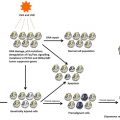
The most striking evidence that Hh pathway deregulation is an early event in BCC formation comes from in vivo transgenic model systems. In these studies the Hh pathway was deregulated in both epidermal cells of transgenic mice and transgenic, reconstituted human skin. Tumors that were indistinguishable from BCC developed within weeks, in the absence of mutation in other genes such as HRAS and TP53. Based on these studies, current evidence shows that Hh pathway deregulation alone can rapidly generate BCC directly from normal keratinocytes. This finding may explain why, in contrast to melanoma and SCC, BCC has no apparent precursor lesion.
Related posts:

Stay updated, free articles. Join our Telegram channel

Full access? Get Clinical Tree


