Nipple–areola reconstruction

CHAPTER 58 Nipple–areola reconstruction


History


Nipple–areola reconstruction represents the final stage of breast reconstruction, whereby a reconstructed breast mound is transformed into a breast with maximal realism when compared with the patient’s opposite breast. Essentially all post-mastectomy patients are fraught with distress brought on by the diagnosis of breast cancer and they suffer a severe alteration of body image and the resultant adverse psychological consequences. Nipple–areola reconstruction is an extremely vital part of breast reconstruction in the sense that it visually transforms the breast mound into a breast. The combination of nipple projection, areola color patch symmetry, and areola projection provides the reconstructed breast with the maximum degree of realism that any breast reconstruction can ever achieve. The evolution of nipple reconstruction techniques has enabled much progress toward this goal of realism.


Numbers of flap designs for nipple reconstruction have been well-described in the literature. There are several reasons why preferred techniques have changed throughout the years; most notably, these include a focus by surgeons on simplicity, reliability, and a reduction in the donor site. Several techniques have fallen into complete disfavor. Nipple banking (i.e. saving the native nipple for later transfer) is an absolutely inappropriate method for nipple reconstruction, as the risk of transferring cancerous cells back to the reconstructed breast has been previously documented. In the past, authors have advocated free grafting of composite tissue from sites such as the auricle, asserting that nipple projection is much better preserved over a long-term period. Here again, however, donor-site morbidity and a steep learning curve seem to underscore the unreasonable cost implicit in using such methods when other, more simple options for nipple reconstruction exist. Composite grafts from distant donor sites have also been previously used for nipple reconstruction but have not been nearly as successful.


All nipple reconstructions lose projection because of the natural processes of contraction inherent with wound healing. Postoperative care is fundamental for satisfactory results, and includes avoiding dressings that apply direct pressure on the nipple. A number of studies indicate that this loss of projection may be substantial, and therefore it is a common practice to overbuild the nipple relative to the opposite nipple projection in anticipation of decreased long-term nipple projection.


Reconstruction of the breast mound has consistently improved with multiple techniques that are selected on the basis of the extent of the defect and the patient’s and surgeon’s preferences. Nipple–areola reconstruction has also made significant strides in regard to multiple surgical options. Since 1944, when Adams described the reconstruction of the nipple–areola complex (NAC) with the nipple–areola graft, the techniques for NAC reconstruction have improved with grafts of a variety of donor areas and local flaps. The most popular techniques include the skate flap and the star flap. With improved techniques, nipple–areola reconstruction has become more popular in the past two decades as many patients consider the idea of a reconstructed breast without the NAC undesirable.


Anatomy


Nipple–areola anatomy is remarkably variable in dimension, texture, and color across ethnic groups and among individuals. Moreover, an appreciable difference often exists in the two nipple–areola complexes even in the same patient. The presence of an elevated structure in the center of a pigmented area on the breast mound usually represents a nipple, yet wide variability exists as to what constitutes the normal dimensions of the complex. In general, an aesthetically balanced B–C cup breast has an areola diameter of 4.2–5 cm, with the nipple diameter and projection or height equal to one-third to one-fourth of the areola diameter.


The central position of the nipple cylinder in the areola also has significant variability, ranging from one-fourth to one-half of the radius off-center.


Nipple projection results from the primary location of the mammary ducts in the central portion of the nipple complex. This arrangement produces a semi-rigid structure with a significantly more fibrotic element than the soft and pliable surrounding areola. The contractile properties of the areola also contribute to the gradual change in nipple projection obtained with direct or neural stimuli.


The NAC is ideally located at the most prominent point of the breast or above the level of the projected inframammary crease. Average projection of the nipple is 5 mm. The areola projects slightly and has an average diameter of 35 to 45 mm. The areola texture can be smooth or rough at the location of the Montgomery tubercles. There is a wide variation in color, shape, size, and projection of the NAC as a result of race, the aging process, hormonal changes (pregnancy and menopause), and variation in weight.


Technical steps


The goal of this final stage of reconstruction is to transform the reconstructed mound into an attractive, natural, pleasing breast. This is a complex task especially in unilateral reconstructions as the opposite breast is soft, mobile, and ptotic. All patients need to be educated regarding the need of further revision to achieve symmetry and some may come close but never be completely symmetrical. Therefore during the decision making, the authors involve the patient to confirm the proposed NAC position and size, since ultimately patient satisfaction and acceptance are directly related to the patient’s sense of involvement and decision concerning her surgery.


In general, the authors like to delay the nipple–areola reconstructions as too many variables have an effect against achieving the best result. Nipple reconstruction should be performed as a final stage, after adjustments to the flap or implant and contralateral breast are completed. It is usually performed 2 to 3 months after the final breast shape is achieved. If subsequent surgical procedures are planned for the opposite breast for symmetry with the reconstructed breast, it is preferable to delay nipple reconstruction until the opposite breast surgery is performed. Some prefer to perform the nipple reconstruction and the contralateral matching surgery simultaneously. This again can be a complication with the multiple variables that are present in cases of a reduction mammaplasty or a mastopexy for the matching procedure. As the contralateral breast regains its final shape, the NAC may be lower than the reconstructed site; therefore a contralateral procedure should be performed first followed by NAC reconstruction as this may give the surgeon the opportunity to revise the opposite site again to achieve symmetry.


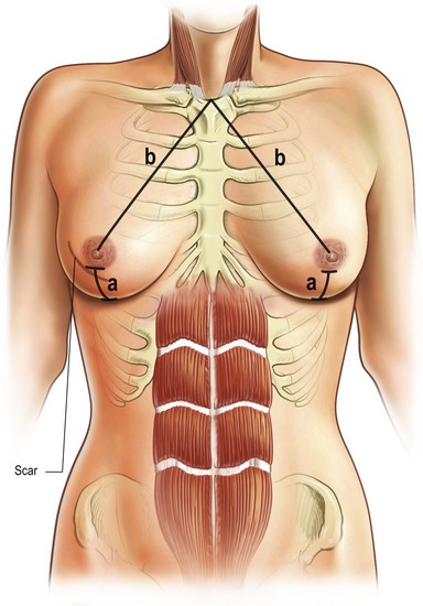
When the patient has agreed to proceed with nipple–areola reconstruction, the patient and the plastic surgeon must determine the optimal position. Specific identifiable landmarks help to determine the proper nipple areolar position visually and geometrically. These include the level of the nipple areola, the triangles with the sternal notch and the umbilicus and the nipple areolar position relative to each breast and to the inframammary fold (Fig. 58.1).


The nipple–areola position is determined visually with the patient in a standing position and the shoulder relaxed without abduction. In addition to measurement, visually the NAC is centered on the breast at the point of maximal convexity and projection, symmetric with the opposite NAC. This is why simultaneous contralateral surgery is not recommended as final shape and NAC position of the contralateral breast will change again.


On the other hand in a bilateral reconstruction, the nipple–areola complex should be positioned along the breast meridian starting from the mid-clavicular point and extending to the breast fold to a point 11 cm from the midline. Taking measurements with the patient in the sitting position, arms at sides; measurements are taken of the chest to determine nipple–areola distances from inframammary line, sternal notch, mid-clavicular line, and midline adjacent to the fourth intercostal space. Once again the NAC should be placed at the most projecting point of the breast mound, approximately 21–23 cm from the sternal notch and 5–7 cm from the inframammary fold. These dimensions are guides and averages, and each patient should be individualized and tailored to each patient’s breast dimensions. The final position should be confirmed visually and this may not always correlate to the measurements.


After the location is determined, the patient is asked to look in the mirror and approve the selected position. This will give the opportunity to the patient to select and ascertain the desired location just prior to surgery. Some also recommend taking a digital photograph and showing it to the patient.


Nipple–areola reconstruction is usually performed as a delayed procedure and can be either under local or general anesthesia and it is done on an outpatient basis. Several technical options are available for nipple and areolar creation. The method selected depends on the size and color of the opposite nipple and areola, the type of breast mound on which it is placed and the patient’s and surgeon’s preference (Table 58.1).

Only gold members can continue reading. Log In or Register to continue

Stay updated, free articles. Join our Telegram channel

Mar 4, 2016 | Posted by in Aesthetic plastic surgery | Comments Off on Nipple–areola reconstruction

Full access? Get Clinical Tree

Get Clinical Tree app for offline access