Fig. 5.1
Mucoepidermoid carcinoma. An ulcerated nodule on the forehead of an old patient
Pathology
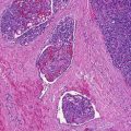
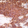
MEC is usually a dermal-based nodular, nonencapsulated solid, and/or cystic neoplasm without continuity with the overlying epidermis (Fig. 5.2). It is characterized by proliferation of squamous, intermediate, mucin-secreting columnar and/or clear cells with gland formation in varying proportions (Figs. 5.3 and 5.4). On the basis of the histopathologic features, including pattern of growth, nuclear atypia and cellular differentiation, presence of mucous cells, and glands formation, this tumor is subdivided in low, intermediate, and high grade. Peritumoral fibrosis is a common feature. Mucin stains such as mucicarmine and Alcian blue highlight the mucin-secreting cells (Fig. 5.5) and the content of glandular and cystic spaces, while immunohistochemistry for CEA, EMA, (Fig. 5.6) and CK7 results strongly positive in the mucinous and glandular cells. The immunohistochemical staining for p63 gives positive result in primary cutaneous MEC and is helpful in differentiating cutaneous metastatic MEC.
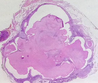
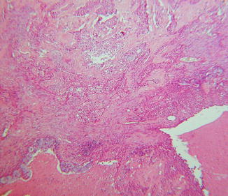
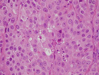
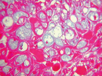
Fig. 5.2
Mucoepidermoid carcinoma. A dermal-based epithelial neoplasm with solid-cystic component in the absence of epidermal connection
Fig. 5.3
Mucoepidermoid carcinoma. An atypical squamous dermal proliferation with ductal structures
Fig. 5.4
Mucoepidermoid carcinoma. The proliferation is made of atypical epidermoid, clear cells, and mucinous cells
Fig. 5.5


Mucoepidermoid carcinoma. Mucin-secreting cells are stained by Alcian blue stain
Related posts:


Stay updated, free articles. Join our Telegram channel
Full access? Get Clinical Tree
