Author
Study design
Patients (n)
Local recurrence (%)
NAC recurrence (%)
Median follow up (month)
Gerber et al. [43]
Prospective
112
5.4 % NSM, 8.2 % MRM (P = 0.6)
0.9 %
59
Petit et al. [33]
Prospective
27
0.00 %
0
6
Caruso et al. [30]
Prospective
50
2 %
2 %
66
Sacchini et al. [15]
Retrospective
192
3 %
0
24.6
Petit et al. [12]
Prospective
106
1 %
0
13
Benediktsson and Perbeck [42]
Prospective
216
20.8 %
0
156
Regolo et al. [32]
Retrospective
102
0
0
16
Petit et al. [43]
Prospective
579
2.40 %
0
19
Crowe et al. [31]
Prospective
149
1.30 %
0
41
Sookhan et al. [45]
Retrospective
18
0
0
10.8
Voltura et al. [48]
Retrospective
51
5.9
0
18
Garwood et al. [37]
Prospective
102
0.6 %
0
13
Gerber et al. [46]
Prospective
112*
11.7 % NSM, 10.4 % SSM, 11.5 % MRM (P = 0.974)
0.9 %
101
Garcia-Etienne et al. [41]
Retrospective
42 NSM
0
0
10
Paepke et al. [47]
Retrospective
96
2 %
0
34
De Alcantara Filho et al. [50]
Prospective
353
0
0
10.4
Petit et al. [51]
Prospective
934
4 %
1.18 %
50
3.3 Cancerous Involvement of Non-areola Skin
Because the areola is in continuity with the breast skin, it would seem reasonable to consider that those factors favoring occult involvement of breast skin in general would similarly apply to the areola.
The involvement of the skin in areas different from those of the nipple areola complex, occurs in most cases for direct infiltration of the tumor, which invades the overlying skin. In this sense, the risk factor determinant is related to the size of the tumor at its original site. Occult lesions in the skin may be present in a very small percentage (0.5–1.3 %) and are, however, related to the detection of neoplastic emboli in the skin. But even in this case, these lesions are accompanied by clinically visible skin infiltration and increased size of the primary lesion [28].
3.4 Paget’s Disease of the Areola
In 1995, van der Putte and co-workers [29] reported the first case of Paget’s disease with involvement of the areola, the nipple sparing. The patient had a slowly growing lesion of the areola present for at least 10 years at diagnosis ranged from 1.5 to 2 cm. There was no cancer found underlying and the disease was limited to the areola. Serial histological sections of the areola showed 39 glands, some of which were simple tubules, while others branched off into lobules. Groups of cells similar to clear Paget’s cells were noted in seven areas outside of the areola. The patient has undergone a partial mastectomy, and, at the time of publication, were free of disease although the follow-up was only 2 years.
This limited reporting that acquires the characters for the anecdotal skimpiness cases, demonstrates the very low incidence of Paget’s disease with only the areola involvement. Nevertheless, should always be taken into account.
3.5 Nipple’s Viability after Nipple Sparing Mastectomy
The incidence of necrosis of the nipple as a result of the conservation of the nipple areola complex, varies from 0 to 48 % in the literature, with the most series of greatest relevance by number that relates to the incidence of <10 % (Fig. 3.1). The comparison between the studies is difficult because many of these recruit a limited number of cases and lacks an adequate score for defining the severity of ischemic injury to the tissue level. Furthermore, we must consider the negative effect on the cosmetic results, determined by the removal of a nipple necrotic days or weeks after the initial surgery and patient satisfaction resulting from such unexpected event. Finally, do not forget that this complication involves the risk of implant loss as a result of infection that may arise. These consequences are rarely mentioned in other published series. It is likely that necrosis nipple is affected by patient factors and surgical technique. Komorowski et al. [39] refers that the age of 45 years had a significant impact on the risk of necrosis (odds ratio 4.51, 95 % confidence interval (CI) 0.83–0.98, P < 0.050), and Garwood [40] and co-workers demonstrated that smoking is a risk factor (odds ratio 7.9, 95 % CI 1.80–35.3, P = 0.006). Overall, however, the effect of age, smoking, vascular diseases, and diabetes have not been reported in all studies.
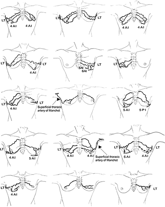
Fig. 3.1
The main sources of blood supply to the nipple areola complex (NAC) and various patterns of supply. Perforating arteries of the internal thoracic artery indicated by numbers on the sternum. Branches from the lateral thoracic artery (LT) and branches from the anterior intercostal arteries (AI). Branches from the posterior intercostal artery (PI) (From Petrus V. van Deventer, Aesth. Plast. Surg. 27:393–398, 2004, Springer Ed.)
In this context, the additional element that can be highlighted, is the appearance of surgical technique that affects on the viability of the nipple, as reported for the note just above.
Compared to the nipple Sparing with standard technique, the minimally invasive video-assisted mastectomy nipple sparing offers the advantage of not requiring additional periareolar incisions to complete emptying of the glandular tissue. In a comprehensive descriptive report, Garwood et al. [40] showed that incisions extending around more than 30 % of the areolar circumference were an independent risk factor for necrosis. Furthermore, by comparing consecutive cohorts, they demonstrated that changing the incision (among other measures) contributed to a decrease in nipple necrosis rates from 20 to 5 % (P = 0·003).
This more conservative approach, does not provide to discontinue the vascular supply of the areola and nipple better preserving its vascularization and its vitality. In this sense it is still difficult to bring consistent data showing that advantage. To confirm this, we must await the completion of numerous case studies that can be compared with the standard approach Table 3.2.
Table 3.2
Features of the primary breast cancer reported to increase risk of occult nipple-areola involvement
Feature |
|---|
<2 cm from areola |
Dimpling (tethering) from areola |
Tumors >4 cm |
Multicentric tumors |
Multifocal tumors
Related posts:Stay updated, free articles. Join our Telegram channel
Full access? Get Clinical Tree
 Get Clinical Tree app for offline access

|