Subungual hyperkeratosis and onycholysis
Psoriasis
Contact dermatitis
Nail bed lichen planus
Onycholysis
Traumas
Yellow nail syndrome
Nail bed tumors (warts, exostosis, squamous cell carcinoma, melanoma)
Subungual hyperkeratosis
Pachyonychia congenita
Nail Psoriasis
Epidemiology
Clinical Features
Nail psoriasis presents clinically with pitting, onycholysis with erythematous border, salmon patches, subungual hyperkeratosis, and splinter hemorrhages, among other nail abnormalities [3].
The most common change seen in nail psoriasis is pitting, in which small depressions of less than 1 millimeter diameter are seen on the surface of the nail plate, due to the presence of parakeratotic cells [3]. This is the result of psoriatic lesions, which consist of clusters of parakeratotic cells in the proximal nail matrix. These clusters disrupt normal keratinization and eventually are removed as the nail grows outward, leaving depressions on the nail surface (Fig. 14.1).
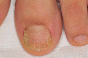
Fig. 14.1
Nail psoriasis. The presence of pitting suggests diagnosis
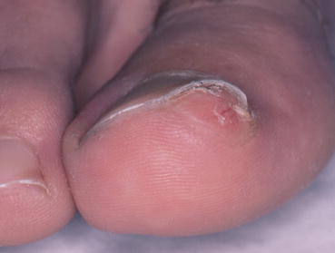
A salmon-colored patch may be seen beneath the nail plate, resembling an oil drop. The red-yellow discoloration is the result of parakeratotic lesions within the nail bed that are visible through the overlying nail plate. This “oil drop” sign is the most diagnostic finding of nail psoriasis. Onycholysis occurs when the parakeratotic lesions involve the hyponychium. Air enters the space between the nail bed and overlying nail plate and may cause white discoloration [1]. Psoriatic onycholysis is typically surrounded by an erythematous border.
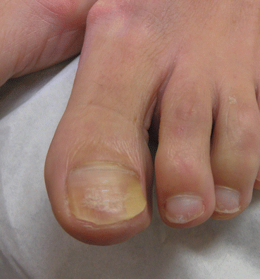
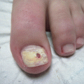
Subungual hyperkeratosis results from deposition and accumulation of desquamated cells underneath the nail plate. This leads to detachment of the nail plate from the nail bed. The degree of this detachment depends upon the level of psoriatic activity present. Subungual hyperkeratosis in nail psoriasis is typically characterized by a silvery-white discoloration as opposed to the typical yellow, greasy appearance seen in onychomycosis, but this is not always the case [1, 4] (Fig. 14.2).
Fig. 14.2
Nail psoriasis presenting with subungual hyperkeratosis, onycholysis, and yellow discoloration
Splinter hemorrhages are less commonly seen and appear as linear, thin, deep, red to black lines in the distal nail. These occur in the dermis of the nail bed, when small capillaries rupture into the linearly oriented epidermal-dermal ridges. Splinter hemorrhages are not only associated with psoriasis, as they can present in a number of other medical conditions, particularly trauma, as observed in 20 % of cases. They are also seen in onychomycosis and even in healthy individuals with inherently delicate capillaries [4].
Lichen Planus
Epidemiology
Lichen planus is a chronic inflammatory disease with unknown etiology that affects the skin, hair, nails, and mucous membranes [1, 6]. The prevalence of lichen planus is not known, but is estimated to be less than 1 %. It affects women and men equally. It can occur at any age, but the majority of cases are in patients between 30 and 60 years old [7]. Lichen planus involves the nails in about 10 % of cases and can be limited to the nails only [8].
Clinical Features
Lichen planus most commonly involves the nail matrix causing longitudinal grooves and fissures, as well as progressive thinning and distal splitting of the nail plate [8]. Nail matrix destruction causes dorsal pterygium, in which the proximal nail fold adheres to the nail bed [1, 6, 9].
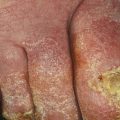
However, lichen planus can also affect the nail bed causing subungual hyperkeratosis and onycholysis. In the toenails it often causes yellow discoloration and thickening (yellow nail syndrome-like presentations) (Fig. 14.3). In general, patients also present signs of nail matrix involvement that suggest correct diagnosis.
Fig. 14.3
Nail bed lichen planus. Nails are thickened and yellow in color; note onychorrhexis that suggests correct diagnosis
Subungual Tumors
Warts
Epidemiology
Periungual warts are most commonly seen in children and teenagers, occurring most frequently in those ranging from 12 to 16 years old. There is increased incidence in those who bite their nails, who suck their fingers, and who work in a wet environment. Often, warts disappear spontaneously [10].
Clinical Features
Periungual warts are usually caused by HPV genotypes 1, 2, and 4. They affect fingernails more often than toenails and appear as hyperkeratotic papules with a rough surface (Fig. 14.4). Pathologically, they are characterized as having sharply demarcated hyperplasia with acanthosis, papillomatosis, and hyperkeratosis with areas of parakeratosis [10].
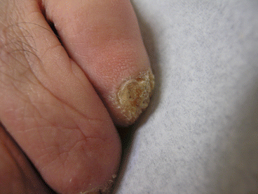
Fig. 14.4
Periungual wart
Initially, the warts are small in size, shiny, smooth, and translucent. Weeks to months later, they grow in size and appear rough, dirty, brownish black in color, and horny [11].
When located in the proximal nail fold, warts produce periungual hyperkeratosis forming a hyperkeratotic cuticle. Subungual warts raise the nail plate causing onycholysis and appear as a subungual nodular lesions. They may also produce a longitudinal band of onycholysis with splinter hemorrhages due to linear growth underneath the nail plate. Warts in the hyponychium of the toenails may cause distal thickening.
Exostosis
Epidemiology
Squamous Cell Carcinoma
Epidemiology
Squamous cell carcinoma of the nail is rare [15]. It is most common in the fifth and sixth decades of life and tends to have a male predominance [16].
The etiology of subungual squamous cell carcinoma is unclear, although repeated trauma, chronic infection, radiation, tar, arsenic exposure, UV radiation, immunosuppression, and HPV infection may each play a role. HPV infection is particularly relevant as it is present in 60–90 % of cases [15].
Clinical Features
Subungual squamous cell carcinoma most frequently presents on the hands rather than the feet [17]. It most often involves only one digit, with the thumb and hallux being the most common [15]. The right index and middle fingers are also commonly affected [18].
Patients most commonly present with a wartlike appearance of the nail bed with nail dystrophy [15]. Nail pigmentation with longitudinal melanonychia is common (Fig. 14.6) [18].
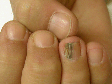
Fig. 14.6
Squamous cell carcinoma presenting with longitudinal melanonychia
Subungual Melanoma
Epidemiology
Nail melanoma is not common, as it accounts for only about 1–3 % of cutaneous melanomas diagnosed in the general population [19, 20]. Nail melanomas are seen more often in patients 50–70 years old and more often in men than women [19, 20]. Darker-skinned individuals are more commonly affected with this subtype of melanoma. Up to 75 % of cutaneous melanomas are localized in the nail in Africans, 10 % in Japanese, and 25 % in Chinese populations [20].
Clinical Features
Nail melanomas originate either from the nail matrix, or the nail bed (subungual melanomas), and may involve other parts of the nail unit such as the proximal nail fold and hyponychium. Nail matrix melanoma presents as bands of longitudinal brown-black discolorations of the nail plate (longitudinal melanonychia) [20]. The pigmented band is usually wider than 3 mm and has dishomogeneous color and blurred lateral margins (Fig. 14.7) [19, 20]. Nail bed melanoma appears as a pigmented or nonpigmented subungual nodule that initially causes nail plate detachment. It gradually enlarges, eventually leading to nail plate destruction [19]. Ulceration, pain, inflammation, discharge, and surrounding discoloration are common [20].
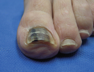
Fig. 14.7
Nail matrix melanoma presenting with longitudinal melanonychia
Hutchinson sign is a characteristic feature of invasive subungual melanoma. It is defined as extension of the dark pigment into the lateral or proximal periungual folds [1].
Nail melanomas are more often found in the hands than in the feet and most commonly in the thumb and hallux [19]. Although UV radiation is a well-known risk factor for cutaneous melanoma, it is unable to penetrate the nail plate and therefore is not a risk factor for subungual melanoma [19]. Instead, direct trauma to the nail is frequently reported although there is lack of evidence to form a direct correlation [20].
Contact Dermatitis
Epidemiology
Contact dermatitis can be allergic or irritant and causes inflammation of the skin due to chemical damage. It can occur at any age and is more prevalent in women and manual laborers [1].
Clinical Features
Contact dermatitis frequently affects the nail bed as chemicals penetrate through the thin onychodermal band causing subungual inflammation with onycholysis and subungual hyperkeratosis. This can be very severe in patients with contact allergy to acrylic nails (Fig. 14.8). Splinter hemorrhages are also common.
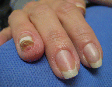
Fig. 14.8
Contact dermatitis due to acrylic nails
Diagnosis is suggested by the presence of periungual erythema and scaling as well as Beau’s lines.
Traumatic Onycholysis
Epidemiology
Trauma of the nail unit is a common injury that can mimic onychomycosis. Trauma can be due to footwear, mechanical injury, or athletics. Traumatic nail lesions can be observed in patients of any race, sex, or age and is very commonly misdiagnosed and treated as onychomycosis.
Clinical Features
Signs of trauma include onycholysis, subungual hyperkeratosis, abnormalities of the nail plate, changes in the hyponychium, ingrown nails, paronychia, and onychomadesis [1]. Traumatic onycholysis usually affects the big toes and is common in athletes and in women wearing high-heel shoes. It affects the lateral aspect of the toenail when caused by overlapping of the second toe or the distal nail [21] (Fig. 14.9).